MONITORING THE STORY BEHIND THE STORY Human error

MONITORING “THE STORY BEHIND THE STORY”


Human error…. . . ASA Status
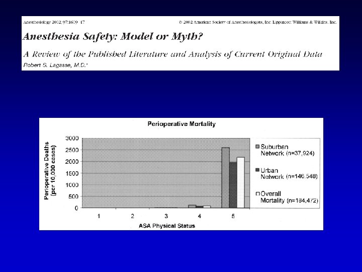

Arbous MS, Grobbee DE, et al. Anaesthesia 2001; 56: 1141 -53 869483 anesthesia 769 pts died within 24 hours after anesthesia 42 pts comatose Inadequate monitoring Postop monitoring: → 10% anesthesia related deaths inadequately 8% pts unavailable 5% pts

Arbous MS, Grobbee DE, et al. Anaesthesia 2001; 56: 1141 -53

“Vital Signs” Monitoring Guidelines Cardiovascular Respiratory Others

ASA standards for basic anesthetic monitoring 2001 Standard 1: Qualified anesthesia personnel shall be present in the room throughout the conduct of all general anesthetics, regional anesthetics and monitored anesthesia care Standard 2: During all anesthetics, the patient's oxygenation, ventilation, circulation, and temperature shall be continually evaluated

ASA standards for basic anesthetic monitoring 2001 Oxygenation: Oxygen analyzer for inspired gases-Observation of the patient Pulse oximetry Ventilation: Auscultation of breath sounds-Observation of the patient Observation of the reservoir bag Capnography (Carbon dioxide monitoring) Circulation: Continuous ECG display Heart rate and BP recorded every 5 minutes Evaluation of circulation Auscultation of heart sounds Palpation of pulse Pulse plethysmography Pulse oximetry Intrarterial pressure tracing Temperature: Monitor temperature when changes are intended, anticipated, or suspected

Buhre W and Rossaint R. The Lancet 2003; 362: 1839 -46

Buhre W and Rossaint R. The Lancet 2003; 362: 1839 -46 Monitoring recommendations of the Association of Anesthetists of Great Britain and Ireland

Buhre W and Rossaint R. The Lancet 2003; 362: 1839 -46

Buhre W and Rossaint R. The Lancet 2003; 362: 1839 -46

Buhre W and Rossaint R. The Lancet 2003; 362: 1839 -46 “There is growing evidence that no single monitoring device can improve outcome in the OR or in the ICU.

ECG blood pressure pulse oximetry capnography + anesthetic gas concentrations + Fi. O 2 temperature

Immediately available Anesthesiology 2002; 96: 742 -52

Periop monitoring Guidelines Cardiovascular Respiratory Others

The electricity of the heart

What to expect from the ECG • Essential monitor • Rate, rhythm, propagation of the excitation wave, heart position, muscle hypertrophy, regional ischemia • NO information about pump function

Lead Selection • Lead II is the same as standard lead two as seen in a 12 lead ECG. • It is the most common monitoring lead. • It is not the optimal monitoring lead.

Lead Selection • V 5 = the best lead to detect ST-T change

The shape of the ECG P T Q R S

Normal ECG

ECG interpretation Rate Rhythm Intervals QRS complexes ST segments & T waves

Intraoperative Monitoring invasive - invasive AP • Swan Ganz cath • Pi. CCO System • Advanced PAC: SVO 2, CCO, REF, EDV …………………TEE

Intraoperative Monitoring invasive

Intraoperative Monitoring invasive 8’ 3’’ + oscill 2 -4’’
 • Pros – Healthy patients")
NIBP vs. Arterial Cannulation • NIBP (auscultatory / oscillometric) • Pros – Healthy patients – Short case • Cons – Bladder cuff size – Flow dependent – Motion – Interruption of IV infusion – Injury – Cuff deflation rate – Hydrostatic errors • Arterial Cannulation • Pros – Continuous BP – Sick patients – Difficult cases – ABG monitoring • Cons – Nerve dysfunction – Thrombosis / Ischemia – Hematoma formation – Infection – Hydrostatic errors

Art Line Complications • • Thrombus formation Arterial laceration Hematoma Loss of distal perfusion to hand…ouch! Nerve dysfunction from dissection Infection Errors in monitoring Failed attempt. Always consider failure as a potential complication.

Arterial Waveform Evaluation • Tf – Foot – Onset of ejection – Systole • T 1 - First Shoulder – Peak flow • T 2 - Second Shoulder – Peak pressure • • Ti – dichotic notch End of ejection Closure of aortic valve Precedes the onset of diastole • Tt – Pulse Duration

Arterial Line Tracing

Central Line Indications • Peripheral venous access is required for: – Administration of fluids – Administration of drugs • Central venous access is required for: – Parenteral nutrition – Anticipated Inotropic medication infusion – Anticipated large volume resuscitation – Monitoring of central venous pressure (CVP) – Cardiac pacing – Difficult peripheral access

Central Line Contraindications • • Patient refusal? Severe Coagulopathy Bundle Branch Blocks relative contraindication Infection at site Previous failed attempts at specific site Hematoma Unusual anatomy

Central Line Techniques • Sterile techniques should be used for all central line cannulation • Surgical scrub with Sterile gown and gloves • Sterile prep of skin and surgical drapes. • Local anesthetic should be used for central catheters in awake patients • Success may be improved by using ultrasound guidance • Techniques of gaining access include: – – Catheter over needle Catheter through needle Seldinger technique Surgical cut-down is surgical technique as last resort.

Anatomy of Central Assess • Internal jugular vein – – Right sided access preferred. Why? Apical pleura does not rise as high on right and avoids thoracic duct Patient positioned head down In the low approach triangle formed by two heads of sternomastoid and clavicle identified – Cannula aimed down and lateral towards ipsilateral nipple • Subclavian vein – – – Usually approached from below clavicle Patient positioned head down Needle inserted below junction of medial 2/3 and lateral 1/3 of the clavicle Needle aimed towards suprasternal notch Passes immediately behind clavicle Vein encountered after 4 -5 cm

Normal CVP Waveform

Waveform Interpretation • + a wave : This wave is due to the increased atrial pressure during right atrial contraction. It correlates with the P wave on an EKG. • + c wave : This wave is caused by a slight elevation of the tricuspid valve into the right atrium during early ventricular contraction. It correlates with the end of the QRS segment on an EKG. • - x descent : This wave is probably caused by the downward movement of the ventricle during systolic contraction. It occurs before the T wave on an EKG.

Waveform Interpretation • + v wave : This wave arises from the pressure produced when the blood filling the right atrium comes up against a closed tricuspid valve. It occurs as the T wave is ending on an EKG. • - y descent : This wave is produced by the tricuspid valve opening in diastole with blood flowing into the right ventricle. It occurs before the P wave on an EKG.

Periop monitoring Guidelines Cardiovascular Respiratory Others
 Stethoscope Used with placement of the endotracheal (ET) tube Will hear")
Monitoring Devices (Types) Stethoscope Used with placement of the endotracheal (ET) tube Will hear breath sounds clearly with the delivery of oxygen into the ET tube with correct placement Can use in placement of nasogastric (NG) tube
 Fi-Fe volatile an. Peep Sp.")
Respiratory monitoring Fi -Fe. O 2 Aw. P (peak-plateau) Fi-Fe volatile an. Peep Sp. O 2 compliance Et. CO 2 P/V e flow slope
 Fi-Fe volatile an. Peep Sp. O 2")
Fi -Fe. O 2 Aw. P (peak-plateau) Fi-Fe volatile an. Peep Sp. O 2 compliance Et. CO 2 P/V e flow slope

PULSE OXIMETRY

During observation in the recovery room, the incidence of hypoxemia in the pulse oximetry group was 1. 5 -3 time less.
 Fi-Fe volatile an. Peep Sp. O 2")
Fi -Fe. O 2 Aw. P (peak-plateau) Fi-Fe volatile an. Peep Sp. O 2 compliance Et. CO 2 P/V e flow slope

Et. CO 2 CAPNOGRPHY mm. Hg 80400 Time in sec

Tetevossian RG, Wo CC, Shoemaker WC. 48 pts whit blunt and hemodynamic instability Crit Care Med 2000; 28: 2248 -53

Tetevossian RG, Wo CC, Shoemaker WC. Crit Care Med 2000; 28: 2248 -53 Ptc. O 2 and Pt. CO 2 early indicators of tissue hypoxia, subclinical hypovolemia, and hemodynamic shock in ER severely ill patients. Ptc-gas values reflect local skin perfusion during normal conditions and in period of circulatory dysfunction and shock.

Periop monitoring Guidelines Cardiovascular Respiratory Others: Temperature Depth of GA NMT Glycemia Lactate

Kurz A, Sessler I, Lenhardt R. NEJM 1996; 334: 1209 -15 80 pts elective colon surgery • Normotermia 37± 0. 3 °C • Ipotermia 34. 4± 0. 4 °C “Intraoperative core temperatures approximately 2°C below normal triple the incidence of wound infection and prolong hospitalization by about 20%. ”

TEMPERATURE

Periop monitoring Guidelines Cardiovascular Respiratory Others: Temperature Depth of GA NMT Glycemia Lactate

BIS…. . PSA 4000 and AEP

BIS HYPNOSIS STATE 100 Awake / moderate sedation Mild hypnosis state 70 Low probability of awareness < 70 Moderate hypnosis state 60 Not awareness <60 Deep hypnosis state 40 0 EEG suppression
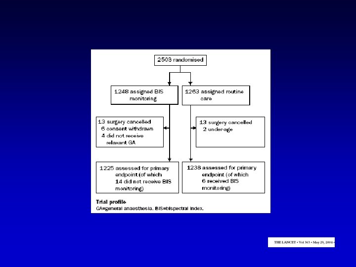

13 AWARENESS: 11 CTRL 2 BIS

49 Possible AWARENESS: 27 CTRL 22 BIS
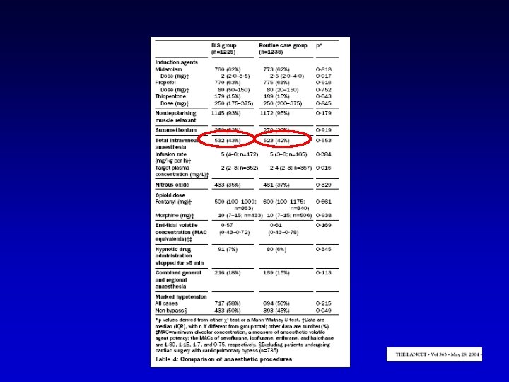

Conclusions: BIS cost = 16 USD To prevent 1 case of awerenwss: 2200 USD

Periop monitoring Guidelines Cardiovascular Respiratory Others: Temperature Depth of GA NMT Glycemia Lactate

NMT MONITORING IN ANESTHESIA TOF WATCH Reversal: M. R. Chelant

Residual neuromuscolar block is a risk factor for postoperative pulmonary complication. Berg H, Viby-Mogensen J, Roed J et al. Acta Anaesthesiol Scand 1997; 41: 1095 -03 691 pz PANC-ATR-VEC Intraop TOF ogni 12 sec Postop TOF+Meccanomiografia Postop Pulm Complications ch generale maggiore >durata <T°C >età PANC se TOF
: 1042 -7")
526 pz VEC-ROC-ATR No reveral Anesthesiology 2003; 98(5): 1042 -7

• 1 hour 12 -19 nmol/kg/min ROC c. i. in guinea pigs • after 30 min Org 25969 c. i 50 nmol/kg/min o NS Anesthesiology 2003; 99(3): 632 -7

Periop monitoring Guidelines Cardiovascular Respiratory Others: Temperature Depth of GA NMT Glycemia Lactate

GLYCEMIC CONTROL Splachnic perfusion

RESPONSE TO HYPERGLYCEMIA Splachnic perfusion

Anesthesiology 2003; 98: 774 -779

Anesthesiology 2003; 98: 774 -779

Anesthesiology 2003; 98: 774 -779

Periop monitoring Guidelines Cardiovascular Respiratory Others: Temperature Depth of GA NMT Glycemia Lactate

“Lactate metabolism: a new paradigm for the third millenium” Tissue hypoperfusion, hypoxia and resulting anaerobic glycolysis are probably not the only causes of increased La- production during shock Lactate plays an important role as intermediatory in numerous metabolic process, a mobile fuel for aerobic metabolism, perhaps a mediator of redox state among various compartment both within and between cells Gladden JB. J Physiol 2004; 558: 5 -30

“Lactate metabolism: a new paradigm for the third millenium” Lactate can no longer be considered the usual suspect for metabolic crimes, but is instead a central player in cellular, regional and whole body metabolism” Gladden JB. J Physiol 2004; 558: 5 -30

Anesth Analg 2002; 95: 294 -8

CONCLUSIONS

COMPUTERIZED CHART KEEPING IS AN ESSENTIAL PART OF MONITORING

DATA CAN ACCESSED BY: On line Trends Snap Shots

“Vital signs” monitoring: When ? type of surgery and pt condition Where ? OR and/or ICU / PACU / Ward How ? ……. . “several” techniques Why ? to manage…. to improve outcome

ASA News Letter 2002

Conclusion: YOU ALL ARE EXHAUSTED
- Slides: 82