Molecular mechanism of muscle contraction Sliding Filament theory


- Slides: 19

Molecular mechanism of muscle contraction Sliding Filament theory Huxley, A. F. Niedergerke, R. , Huxley H. E. , Hanson, J.

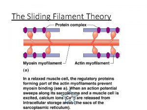
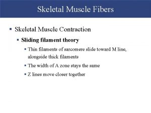
Molecular mechanism of muscle contraction • Most accepted theory at present “ Sliding filament theory” • Proposes that a muscle shortens or lengthens because the myofibrillar filaments slide past each other without actually changing their length. • The molecular motor to drive this shortening process is the action of the Myosin cross bridges, which cyclically bind or attach, rotate and detach from the actin filaments with energy provided by ATP hydrolysis.

Sliding Filament Theory

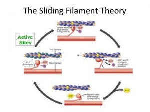
I Sliding Filament Theory Calcium Myosin Filament Actin Filament ATP ADP +P

Sliding filament theory • • When muscle is in the relaxed state Ca ion conc. in the cytosol is low. At this point actin and myosin filaments lie along each other in the sarcomere. The Myosin head at this point is in a high energy condition “cocked up” with ADP and inorganic phosphate bound to it Active sites on the G actin molecules are covered by the troponin tropomyosin complex.

II Sliding Filament Theory Actin Filament Myosin Filament ADP +P Myosin head cocked up

Sliding filament theory • Action potentials in the T tubule cause the release of Ca ions from SR into the muscle cytosol • Ca binds Troponin C. A conformational change is induced in the Troponin weakens the bond between it and Actin. • This allows tropomyosin to move laterally and expose the active sites on G Actin. • The cocked up myosin molecule rapidly binds to the Actin: this link is a “cross bridge”

III Sliding Filament Theory Calcium binds to Actin ADP +P

IV Sliding Filament Theory Calcium opens binding sites

V. Sliding Filament Theory Cross bridge forms, Connecting Myosin to Actin

Sliding Filament Theory • Myosin head then undergoes a conformational change causing a “rachet action” and pulls the actin filament to the centre of the sarcomere. • ADP and Pi are released by this process • This is called the “power stroke” which causes the sliding action

VI. Sliding Filament Theory Conformational Changes Myosin head: Actin Moves~ “power stroke” Release of ADP+P

Sliding filament theory • An ATP binds to the Actomyosin complex • This causes the affinity of myosin for actin to decrease • The myosin head changes its position to close around the ATP and hydrolyze it. • This change in conformation of the Myosin head releases the myosin from the actin.

VII. Sliding Filament Theory New ATP binds

IX. Sliding Filament Theory ADP +P ATP hydrolysed Myosin returns to cocked up position Fresh cycle starts

Sliding filament theory • Cycling continues until cytosolic Ca levels remain high • One Ca ion releases one Troponin which covers 7 active sites. • All myosin molecules do not move simultaneously but sequentially like oars on a boat and cause the myosin slide along the Actin filament

Muscle relaxed Calcium pumped out of cytosol: active sites covered ADP +P

Muscle energetics • Energy currency for muscle contraction is ATP • Hydrolysis of ATP by Myosin ATPase energizes cross bridges prior to cycling. Binding of ATP to myosin dissociates cross bridges bound to actin allowing the bridges to repeat their cycle of activity. Hydrolysis of ATP by Ca –ATPase provides energy for active transport of Ca into sarcoplasmic reticulum thus ending the contraction and allowing the muscle fiber to relax. • •

Rigor mortis • A condition of the muscles seen after death • ATP not available • Cycle stops at the point of formation of actomyosin complexes • “permanent actomyosin” complexes formed • Leads to a state of rigor • Ends with denaturation of protiens
 Smooth muscle fibres are fusiform and syncytial
 Sliding filament theory
 Crash course sliding filament theory
 Sliding filament theory
 Winding filament theory
 Skeletal striations
 Unipennate muscle
 Winding filament theory
 Sliding filament
 Sliding filament model
 Define autorhythmicity
 Is atp needed for muscle contraction
 3 phases of muscle contraction
 Types of muscle contraction
 Isotonic vs isometric contraction
 Muscle twitch
 Muscle twitch
 Muscle contraction
 Wave summation
 Mussels anatomy
