Molecular Biology of the Cell Fifth Edition The

Molecular Biology of the Cell Fifth Edition The Cell Cycle

Fig 17 -1 The cell cycle. Figure 17 -1 Molecular Biology of the Cell (© Garland Science 2008)

Fig 17 -2 The major events of the cell cycle. Figure 17 -2 Molecular Biology of the Cell (© Garland Science 2008)

Fig 17 -4 The four phase of the cell cycle. Figure 17 -4 Molecular Biology of the Cell (© Garland Science 2008)

Fig 17 -5 A comparison of the cell cycles of fission yeasts and budding yeasts. Figure 17 -5 Molecular Biology of the Cell (© Garland Science 2008)

Fig 17 -6 The behavior of a temperature-sensitive Cdc mutant. Figure 17 -6 Molecular Biology of the Cell (© Garland Science 2008)

Fig 17 -6 The morphology of budding yeast cells arrested by a Cdc mutation. Figure 17 -7 Molecular Biology of the Cell (© Garland Science 2008)

Fig 17 -8 A mature Xenopus egg, ready for fertilization. Figure 17 -8 Molecular Biology of the Cell (© Garland Science 2008)

Fig 17 -9 Oocyte growth and egg cleavage in Xenopus. Figure 17 -9 Molecular Biology of the Cell (© Garland Science 2008)

Fig 17 -10 Studying the cell cycle in a cell-free system. Figure 17 -10 Molecular Biology of the Cell (© Garland Science 2008)

Fig 17 -11 Mammalian cells proliferating in culture. Figure 17 -11 Molecular Biology of the Cell (© Garland Science 2008)

Fig 17 -13 Analysis of DNA content with a flow cytometer. Figure 17 -13 Molecular Biology of the Cell (© Garland Science 2008)
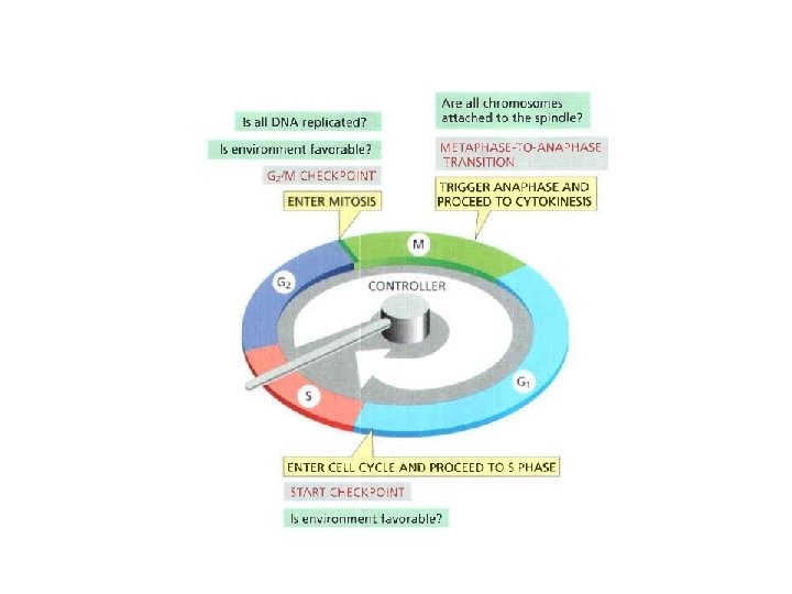

Fig 17 -15 Two key components of the cell-cycle control system. Figure 17 -15 Molecular Biology of the Cell (© Garland Science 2008)

Fig 17 -16 Cyclin-Cdk complex of the cell-cycle control system. Figure 17 -16 Molecular Biology of the Cell (© Garland Science 2008)
")
Table 17 -1 Molecular Biology of the Cell (© Garland Science 2008)

Fig 17 -17 The structural basis of Cdk activation. Figure 17 -17 Molecular Biology of the Cell (© Garland Science 2008)

Fig 17 -18 The regulation of Cdk activity by inhibitory phosphorylation. Figure 17 -18 Molecular Biology of the Cell (© Garland Science 2008)

Fig 17 -19 The inhibition of a cyclin-Cdk complex by a CKI Figure 17 -19 Molecular Biology of the Cell (© Garland Science 2008)

Fig 17 -20 The control of proteolysis by APC/C and SFC during the cell cycle. Figure 17 -20 a Molecular Biology of the Cell (© Garland Science 2008)
")
Table 17 -2 Molecular Biology of the Cell (© Garland Science 2008)

Fig 17 -21 An overview of the cell-cycle control system. Figure 17 -21 Molecular Biology of the Cell (© Garland Science 2008)

Fig 17 -22 Control of chromosome duplication. Figure 17 -22 Molecular Biology of the Cell (© Garland Science 2008)

Fig 17 -23 Control of the initiation of DNA replication. Figure 17 -23 Molecular Biology of the Cell (© Garland Science 2008)

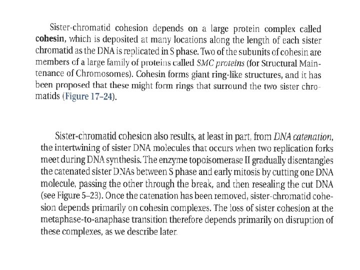
Fig 17 -24 Cohesin Figure 17 -24 Molecular Biology of the Cell (© Garland Science 2008)
")
Figure 17 -25 Molecular Biology of the Cell (© Garland Science 2008)

Fig 17 -26 The mitotic chromosome. Figure 17 -26 Molecular Biology of the Cell (© Garland Science 2008)

Fig 17 -27 Condensin. Figure 17 -27 Molecular Biology of the Cell (© Garland Science 2008)
")
Figure 17 -28 Molecular Biology of the Cell (© Garland Science 2008)

Fig 17 -29 The centrosome. Figure 17 -29 Molecular Biology of the Cell (© Garland Science 2008)

Fig 17 -30 Major motor proteins of the spindle. Figure 17 -30 Molecular Biology of the Cell (© Garland Science 2008)

Fig 17 -31 Centriole replication. Figure 17 -31 Molecular Biology of the Cell (© Garland Science 2008)

Fig 17 -34 Spindle self-organization by motor proteins. Figure 17 -34 Molecular Biology of the Cell (© Garland Science 2008)

Fig 17 -35 Bipolar spindle assembly without centrosome in parthenogenetic embryos of the insect Sciara (or fungus gnat). Figure 17 -35 Molecular Biology of the Cell (© Garland Science 2008)

Fig 17 -36 The kinetochore. Figure 17 -36 a, b Molecular Biology of the Cell (© Garland Science 2008)

Fig 17 -37 A microtubule attachment site in a kinetochore.

Fig 17 -38 The capture of centrosome microtubules by kinetochores. Figure 17 -38 Molecular Biology of the Cell (© Garland Science 2008)
")
Figure 17 -39 Molecular Biology of the Cell (© Garland Science 2008)

Fig 17 -40 How depolymerization may pull the kinetochore toward the spindle pole. Figure 17 -40 Molecular Biology of the Cell (© Garland Science 2008)

Fig 17 -41 Microtubule flux in the metaphase spindle. Figure 17 -41 a Molecular Biology of the Cell (© Garland Science 2008)

Fig 17 -42 How opposing forces may drive chromosomes to the metaphase plate. Figure 17 -42 a Molecular Biology of the Cell (© Garland Science 2008)

Fig 17 -43 Sister-chromatid separation at anaphase. Figure 17 -43 Molecular Biology of the Cell (© Garland Science 2008)

Fig 17 -44 The initiation of sister-chromatid separation by the APC/C. Figure 17 -44 Molecular Biology of the Cell (© Garland Science 2008)
")
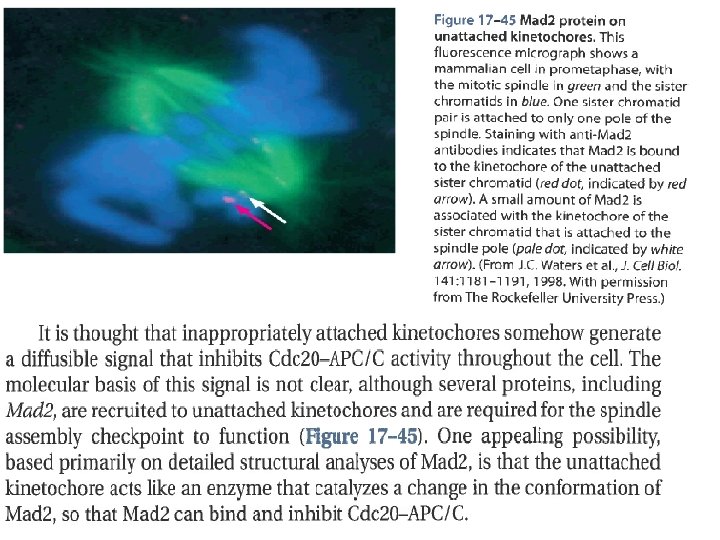
Figure 17 -46 Molecular Biology of the Cell (© Garland Science 2008)

Fig 17 -47 Comparison of meiosis and the mitotic cell cycle. Figure 17 -47 Molecular Biology of the Cell (© Garland Science 2008)

Fig 17 -48 A crossover between homologs. Figure 17 -48 Molecular Biology of the Cell (© Garland Science 2008)

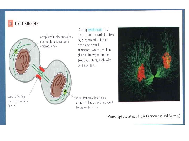
Fig 17 -49 Cytokinesis. Figure 17 -49 a Molecular Biology of the Cell (© Garland Science 2008)

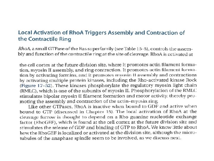
Fig 17 -50 The contractile ring. Figure 17 -50 a Molecular Biology of the Cell (© Garland Science 2008)

Fig 17 -51 The midbody. Figure 17 -51 a Molecular Biology of the Cell (© Garland Science 2008)
")
Figure 17 -52 Molecular Biology of the Cell (© Garland Science 2008)
")
Figure 17 -53 Molecular Biology of the Cell (© Garland Science 2008)

Fig 17 -54 An experiment demonstrating the influence of the position of microtubule asters on the subsequent plane of cleavage in a large egg cell. Figure 17 -54 Molecular Biology of the Cell (© Garland Science 2008)
")
Figure 17 -55 Molecular Biology of the Cell (© Garland Science 2008)

Fig 17 -55 Cytokinesis in a plant cell in telophase. Figure 17 -56 Molecular Biology of the Cell (© Garland Science 2008)

Fig 17 -57 The special features of cytokinesis in a higher plant cell. Figure 17 -57 Molecular Biology of the Cell (© Garland Science 2008)

Fig 17 -58 An asymmetric cell division segregating cytoplasmic components to only one daughter cell. Figure 17 -58 Molecular Biology of the Cell (© Garland Science 2008)
")
Figure 17 -59 a Molecular Biology of the Cell (© Garland Science 2008)
")
Figure 17 -60 Molecular Biology of the Cell (© Garland Science 2008)
")
Figure 17 -61 Molecular Biology of the Cell (© Garland Science 2008)
")
Figure 17 -62 Molecular Biology of the Cell (© Garland Science 2008)
")
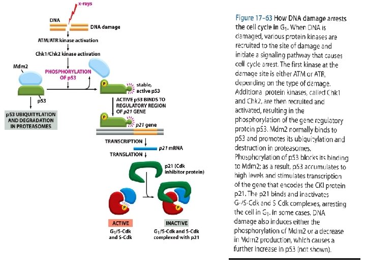
Figure 17 -64 Molecular Biology of the Cell (© Garland Science 2008)

Fig 17 -66 Potential mechanisms for coordinating cell growth and division. Figure 17 -66 Molecular Biology of the Cell (© Garland Science 2008)
- Slides: 75