Molecular Biology Lecture 1 2 Dr Jehad AlShuneigat

Molecular Biology Lecture 1 & 2 Dr Jehad Al-Shuneigat

Molecular Biology syllabus • • • Introduction Significance of molecular biology and genetics in biology and medicine Living systems and experimental models Chromosomes, genes, and DNA and RNA structures General characteristics of DNA and RNA structures Chemical structure of DNA-protein interaction and binding Structure of chromosomes Chromosomal structure and chromatin in prokaryotes and eukaryotes Chromosomal karyotyping and Chromosomal Disorders DNA replication in prokaryotes and eukaryotes RNA and gene transcription Types and structure of RNA in prokaryote and eukaryotes Transcription in prokaryote and eukaryotes Post-transcriptional regulation Translation in prokaryote and eukaryotes - Post-translational regulation, Eukaryotic Pre-m. RNA Processing, RNA Splicing

• Genetic Testing Newborn screening, Presymptomatic testing, Prenatal testing • Cancer Causes, genetics, p 53, treatment • Stem cell: Types and properties - Gene therapy: Introduction, uses and risk - Signal transduction: General principles and examples Examples of signaling pathways • Gene Regulation (Operon system) • DNA mutations and chromosomal anomalies Types of chromosomal anomalies Examples of genetic diseases Types of DNA mutation • Mechanisms of DNA repair

List of laboratories 1. Extraction of genomic DNA from bacteria 2. Concept of restriction endonucleases and DNA cloning introduction (Lecture) 3. Restriction endonucleases Lab 4. Gel electrophoresis & results analysis 5. PCR introduction (Lecture) 6. PCR Lab and Gel electrophoresis of amplified PCR & results analysis

Introduction • Molecular biology is the study of biological molecules and the molecular basis of structure and function in living organisms. • The field of molecular biology arose from the meeting point of work of geneticists, biochemists, physicists, and structural chemists on a common problem: the structure and function of the gene. • The goal of molecular biology is to understand the cell growth, division, specialization, movement, and interaction in terms of various molecules that are responsible for them.
 of Molecular Biology It estimated that humans has 30, 000 genes")
Central Idea (Dogma) of Molecular Biology It estimated that humans has 30, 000 genes It is estimated that each gene codes for at least 4 proteins which can be explained by splicing. Genetic code is the sequence of nucleotides in DNA that determines the sequence of amino acids in proteins.

Models of biological system • Viruses Head Tail DNA Tail fiber • Bacteria prokaryote, agar (Agar is a complex polysaccharide derived from red algae), cell division, prototroph, auxotroph • Yeast eukaryotes • Animal cells cell culture, cancer cell culture, stem cells • Plant cells

Genetics and Medicine • Most disease result from environmental influences interacting with the individual genetic makeup {genetic predisposition (sometimes also called genetic susceptibility)}. • e. g. , high blood pressure, diabetes mellitus, psychiatric disorders • More than 3000 defined human genetic diseases are known. • Genetically determined diseases are not a marginal group. • The total estimated frequency of genetically determined diseases of different categories in the general population is about 3. 5– 5. 0%. • Genetic counselling is generally offered prior to marriage or conception, in order to predict the likelihood of conceiving an affected child, during pregnancy. • Gene therapy is used to correct defective genes that cause disease not yet an active current therapy.
 Gene RNA Proteins")
Introduction • • • Genome Chromosome DNA (Watson-Crick model) Gene RNA Proteins

Nucleic Acids • Nucleic acids occur in two forms: 1 - Deoxyribo. Nucleic Acid (DNA) 2 - Ribo. Nucleic Acid (RNA). • Both DNA and RNA are polymers of nucleotides. • DNA is the molecule of heredity in all cells except some viruses where RNA is the molecule of heredity. • RNA molecules are synthesized on DNA templates and participate in protein synthesis in the cytoplasm.
. • Has a diameter of about")
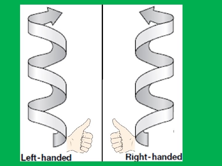
DNA • Right-handed antiparallel doublestranded helix (spiral structure). • Has a diameter of about 20 A 0 (2 nm). • Makes complete turn every 34 A 0 (3. 4 nm) • The distance between adjacent nucleotide is 3. 4 A 0. • The bases of DNA are flat and stacked above one another.

The nitrogen bases are hydrophobic therefore to avoid water they are stacked in the center, while the sugar and phosphates are hydrophilic stay outside. To prevent water from reaching nitrogen bases DNA twists to reduce space between the nitrogenous bases and the phosphate and sugar strands.

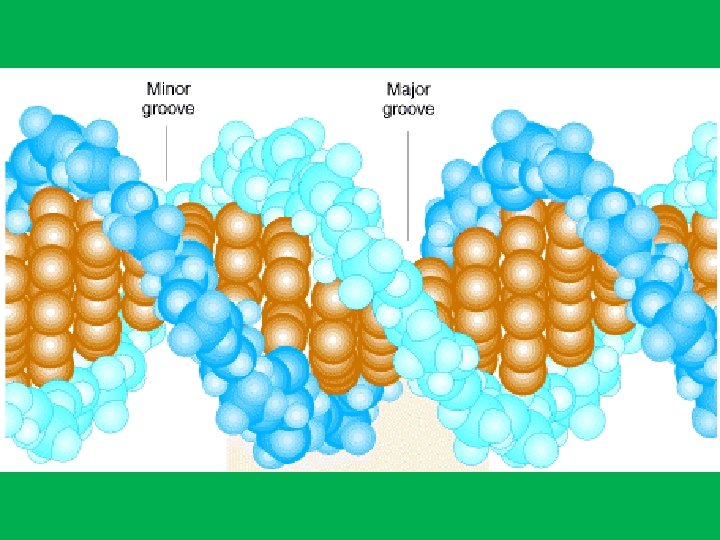
• Why DNA is spiral? • The distance between two sugars or phosphate is about double that of the thickness of the nitrogen bases which thus leaves a hole between the bases. • How DNA gets rid of the ‘holes’? • Each base pair is twisted about 360 to the next base pair and this keeps the distance between the sugars and phosphate constant and at the same time get ride of the holes. • The twisting of the two strand around one another form a double helix with a minor groove about 12 A 0 across and a major groove about 22 A 0.

RNA • Single strand of nucleotides • There are three classes of RNA based on their functions: • 1) transfer RNA (t. RNA); • 2) messenger RNA (m. RNA); • 3) ribosomal RNA (r. RNA).

• Chemically RNA differs from DNA in two respects: • 1 - RNA contains ribose sugar instead of deoxyribose. • 2 - RNA has uracil (U) instead of thymine (T).
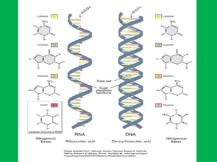
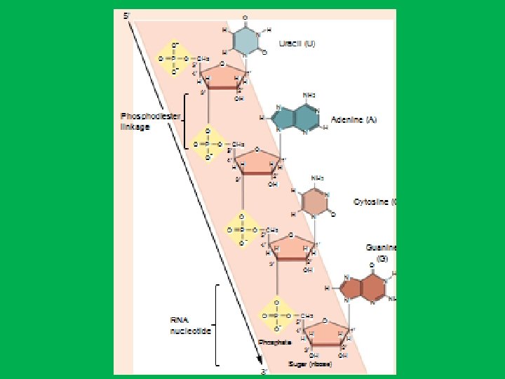

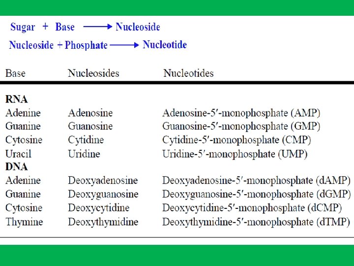
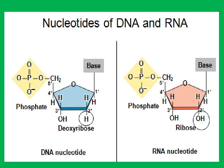
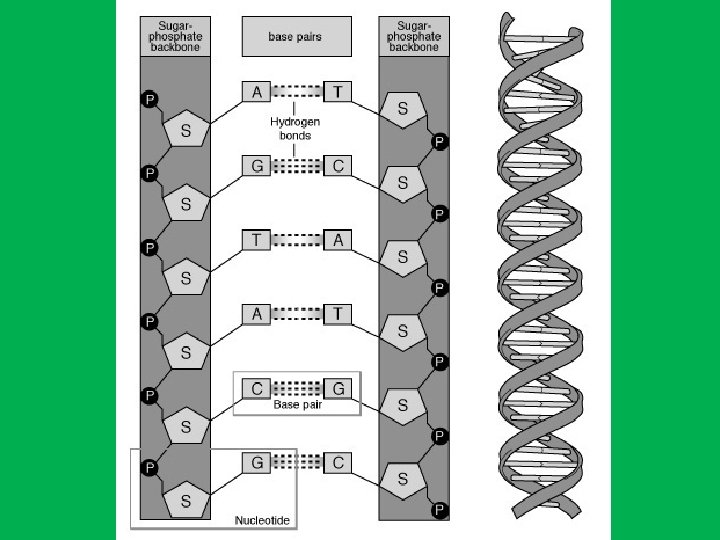
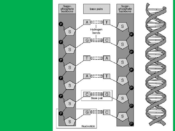
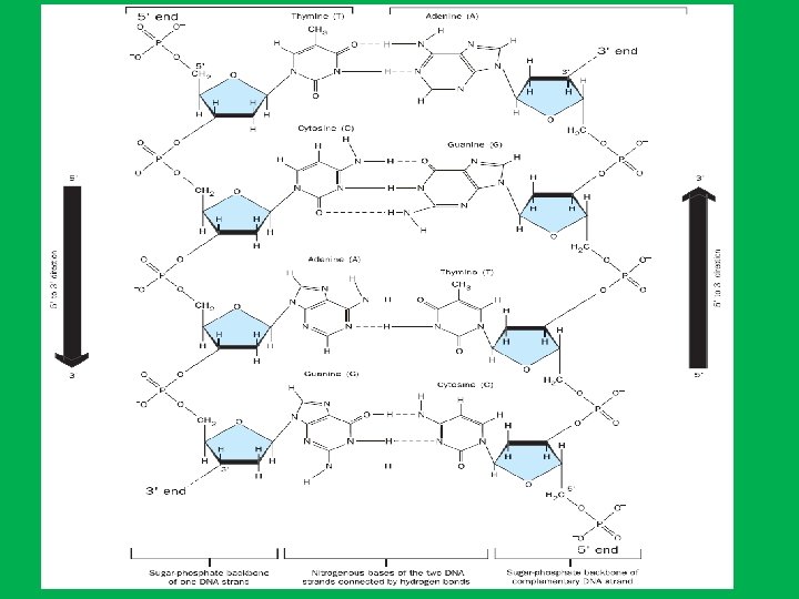
Nucleotides • DNA and RNA are long, unbranched polymer of nucleotides • A nucleotide is made up of: • • 1 - Sugar (deoxyribose in DNA and ribose in RNA) 2 - Phosphate 3 - Nitrogen base. In DNA nitrogen bases are adenine (A), guanine (G), cytosine (C), and thymine (T), while in RNA adenine (A), guanine (G), cytosine (C), and uracil (U)). • Nucleotides are named after the nitrogen bases present, • The genetic information is stored in the sequence of bases • Nucleosides are formed by joining a nitrogenous base to a sugar

Nucleotide
 & Deoxyribose (C 5 H")
1 - Ribose (C 5 H 10 O 5) & Deoxyribose (C 5 H 10 O 4) Sugar • The carbon atoms on the sugar ring are numbered 1’ (one prime) to 5’ to distinguish them from atoms in the bases. The ribose in DNA differs from that of RNA by the absence of oxygen at the carbon atom number 2 and is thus 2 -deoxy-β-D-ribose in DNA while in RNA β-D-ribose. prime

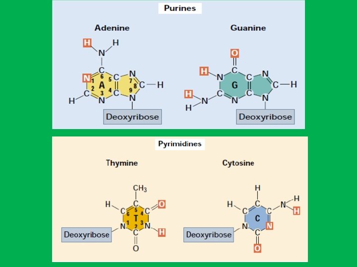
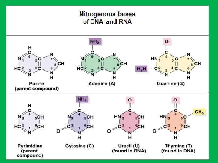
• 2 - Nitrogen bases • Because of their nitrogen content and basic qualities (hydrophobic) they are known as nitrogenous bases. • The organic bases are of two general types: purines and pyrimidines. A. Purines • The purines are adenine (A) and guanine (G). • Each purine consists of a six-sided ring attached to a five-sided ring B. Pyrimidines • The pyrimidines are cytosine (C), thymine (T), and uracil (U). • Each pyrimidine consists of a six-sided ring only. • Thymine is found primarily in DNA and uracil is found only in RNA. • Uracil differs from thymine by lacking a methyl group on its C 5

5 5 Nitrogen bases are hydrophobic with a hydrophilic edge. These hydrophilic edges are the -C=O and -N-H groups that can act as H-bond acceptors and donors. The hydrophilic edges of the bases interact in a very specific way A=T and G≡C, and thus this will leave the nitrogen bases as pure hydrophobic in the centre of the DNA.
")
• 3 - Phosphate group • A phosphoric acid (H 3 PO 4) is bounded to one oxygen by a double bond and three hydroxyl groups (–OH). • Two of the hydroxyl groups can form covalent bonds, phosphodiester bonds, with the sugar hydroxyl groups by splitting out water. • The third -OH group on the phosphate is free and dissociates a hydrogen ion (H+ ions) and leaving negatively charged oxygens at physiologic p. H. • In this form, the structure is referred to as phosphate. • Therefore, phosphate is a negatively charged which gives the polymer its acidic property and promoting their attraction to positively charged histone proteins that partially neutralized this negative charges • because of the phosphate charges both DNA and RNA are negatively charged.

Phosphoric acid H PO 3 4

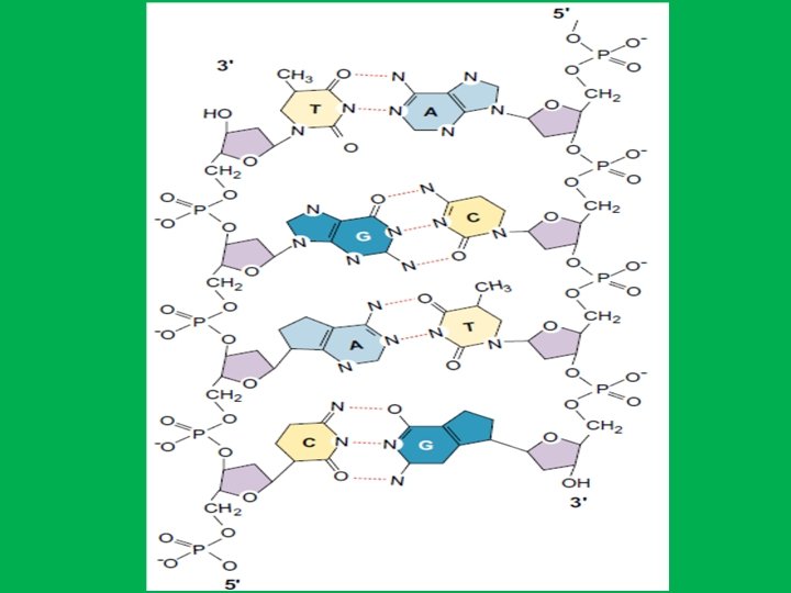
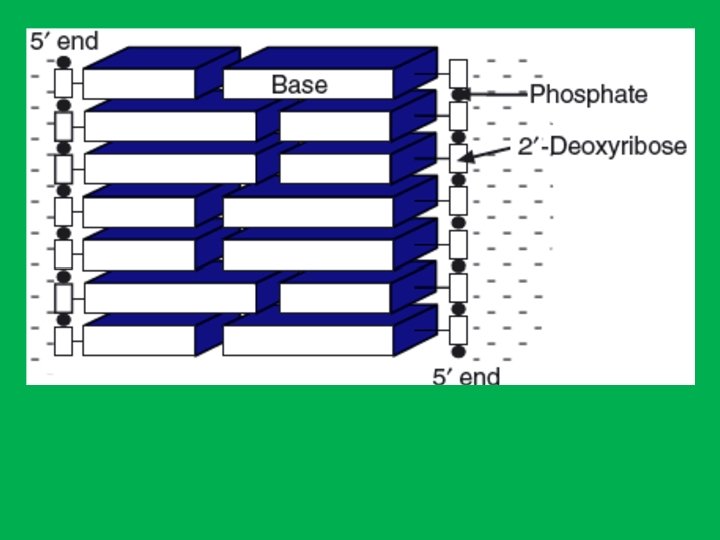
• The nitrogen atom in position 9 of a purine or in position 1 of a pyrimidine is bound by N-glycosidic bond to the carbon number 1 of the sugar. • On the other hand, the phosphate group links the 3’ end of one nucleotide to the 5’ end of the next nucleotide through phosphodiester bonds. • This gives the sugar-phosphate backbone directionality a 5’ end and 3’ end. • In DNA the direction of the nucleotides in one strand is opposite to their direction in the other strand thus called antiparallel. • The 5' end has a terminal phosphate group and the 3' end has a terminal hydroxyl group. • Sequences are written and read in the 5’ to 3’ direction (from left to right); for example, the sequence AUG is assumed to be (5’)AUG(3’)

In a nucleotide, the nitrogen atom in position 9 of a purine or in position 1 of a pyrimidine is bound to the carbon number 1 of the sugar by N-glycosidic bond.

Covalent bonds in DNA N-glycosidic bond
 specifically binds to thymine (T)")
Base Pairs and bonds in DNA • Adenine (A) specifically binds to thymine (T) by two hydrogen bonds (A=T) and • Cytosine (C) specifically binds to guanine (G) by three hydrogen bonds (C≡G) • The human genome contains approximately 3 billion of these base pairs, which reside in the 23 pairs of chromosomes within the nucleus of all our cells. Chargaff's Rule: in any sample of doublestranded DNA, the amount of adenine equals the amount of thymine, the amount of guanine equals the amount of cytosine, and the total amount of purines equals the total amount of pyrimidines.

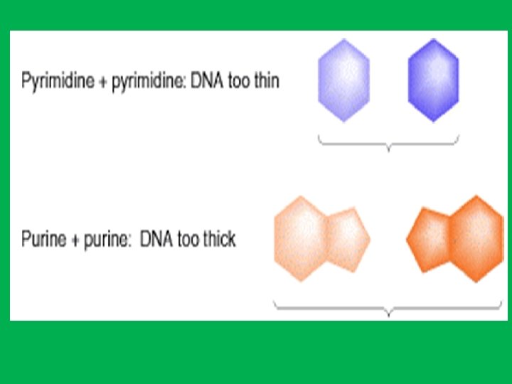
• The A-T and G-C bonds are the most stable of the various base-pairs on energetic grounds. • These two base pairs have essentially identical dimensions within the double helix structure of DNA and thus avoiding partnership of purine-purine which will be too wide or pyrimidine-pyrimidine which is too narrow. This means that these are also the only two base pairs which will properly fit into the double helix that is the outside diameter of the double helix will be uniform over its length • Hydrogen bonds are weak they can be broken and rejoined relatively easily

Suppose we tried to pair an adenine with a cytosine. 1 - Then we would have a hydrogen bond acceptor (N 1 of adenine) lying opposite a hydrogen bond acceptor (N 3 of cytosine). 2 - Likewise, two hydrogen bond donors, the NH 2 groups at C 6 of adenine and C 4 of cytosine, would lie opposite each other. • Thus, an A: C base pair would be unstable

The DNA Double Helix Is a Stable Structure First, Hydrogen bond • The two strands of DNA are held together by H-bonds • Polar atoms in the sugar–phosphate backbone form external H bonds with surrounding water molecules. Second, the negatively charged phosphate groups are all situated on the exterior surface of the helix in such a way that they have minimal effect on one another and the phosphates interact with positively charged (magnesium, potassium, or sodium) ions and with positively charged histone proteins. Third, Stacking interactions which involve hydrophobic interaction and Van der Waals forces in the core of the helix. Base stacking helps to minimize contact of the bases with water, and base-stacking interactions are very important in stabilizing the three-dimensional structure of nucleic acids.

Significance of chemical differences between DNA and RNA 1 - DNA contains thymine instead of uracil Deamination of cytosine forms uracil Thymine has methyl group to distinguish itself from uracil

2 - DNA contains 2 -deoxyribose sugar instead of ribose sugar. • The absence of the 2’-hydroxyl group in DNA further increases its resistance to hydrolysis.

Heat and alkali effect on nucleic acid • In DNA both alkali and heat cause • The two strands of the DNA helix to separate (denature). • It does not break the phosphodiester bonds. • In RNA alkali causes the cleavage of the phosphodiester bonds. • Therefore, alkali is used to remove RNA from DNA and to separate DNA strands before, or after, electrophoresis on polyacrylamide or agarose gels. • The separation of DNA strands is called melting Tm. • The Tm is defined as the temperature in degrees Celsius, at which 50% of all molecules of a given DNA sequence are hybridized into a double strand, and 50% are present as single strands. • If the temperature is slowly decreased, complementary single strands can realign and base-pair, re-forming a double helix essentially identical to the original DNA. This process is known as renaturation, reannealing, or hybridization.
 N 7 atom of the purine ring and the C")
Grooves (space between strands) N 7 atom of the purine ring and the C 5 atom of the pyrimidine ring face out into the major groove 22 Å T A 12 Å

Minor groove Major groove 46

Chemical Groups Exposed in the Major and Minor Grooves from the Edges of the Base Pairs. The letters in red identify hydrogen bond acceptors (A), hydrogen bond donors (D), nonpolar hydrogens (H), and methyl groups (M).

different types of helical structure • • • DNA can adopt 3 forms including, A DNA right-handed has 11 base pairs per turn B DNA right-handed has 10 base pairs per turn Z DNA left-handed has 12 base pairs per turn Under physiological conditions, most of the DNA in a bacterial or eukaryotic genome is of the BDNA.

A DNA Major groove proportions B DNA Extremely narrow but very deep Wide and with intermediate depth Exist when less water is present. Exist in normal physiological conditions Z DNA Flattened out on helix surface Exist when particular base sequences are present, such as stretches of alternating C and G sequences ZDNA may play a role in regulating gene transcription Helical sense Diameter Right handed 26 A 0 Right handed 20 A 0 Left handed 18 A 0

Each structure shown here has 36 base pairs
 High affinity binding to a specific site.")
DNA-protein interaction and binding Regulatory proteins (1) High affinity binding to a specific site. (2) Only small regions of the protein make direct contact with DNA. (3) The protein-DNA interactions are maintained by hydrogen bonds and van der Waals forces. (4) Proteins with the helix-turn-helix or leucine zipper motifs form symmetric dimers, and their respective DNA binding sites are symmetric palindromes (sequence of units that can be read the same way in either direction). In proteins with the zinc finger motif, the binding site is repeated two to nine times. ACCTAGGT palindromic TGGATCCA
 and")
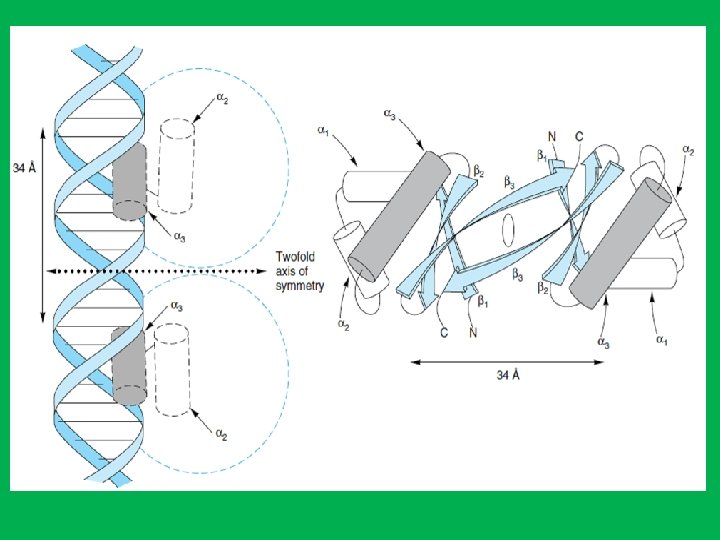
The Helix-Turn-Helix Motif • Consists of three antiparallel β sheets (β 1–β 3) and three α helices (α 1–α 3). • The helix-turn-helix motif is formed because the α 3 and α 2 helices are held at about 90 degrees to each other by a turn of four amino acids. • The α 3 helices form the DNA recognition surface of many proteins, and the rest stabilize structures. • The DNA recognition domain interacts with 5 bp. • Two monomers associate through the antiparallel β 3 sheets to form a dimmer that has a twofold axis of symmetry. • The average diameter of an α helix is 1. 2 nm, which fit in the major groove in the B form of DNA.
 binding to DNA")
• Structure of receptor (dimer) binding to DNA

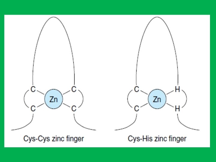
The Zinc Finger Motif • Zinc finger motifs: zinc bound at four positions with either four cysteine, or two cysteine and two histidine in a sequence of approximately 20 amino acids. • The zinc is required to maintain the tertiary structure of this domain. • Each zinc finger contacts about 5 bp of DNA. • Zinc finger motifs consist of an α-helix and a β- sheet. The nucleotide recognition signal (contained within the α-helix) of at least one zinc finger binds to a specific sequence of bases in the major groove of DNA.
 Motif • Leucine zippers also function as dimers to regulate")
The Leucine Zipper (LZ) Motif • Leucine zippers also function as dimers to regulate gene transcription. • The leucine zipper motif is an α-helix of 30 to 40 amino acid residues • LZ contains a leucine (nonpolar hydrophobic aa) every seven amino acids, positioned so that they align on the same side of the helix. • Two helices dimerize so that the leucines of one helix align with the other helix through hydrophobic interactions to form a coiled coil. • The portions of the dimer adjacent to the zipper bind the DNA through basic amino acid residues (arginine and lysine) that bind to the negatively charged phosphate groups. This DNA binding portion of the molecule also contains a nucleotide recognition signal.

Alpha helix makes one full turn every 3. 6 residues

• Helix-loop-helix • It consists of a short a helix connected by a flexible loop of amino acids to a second longer a helix. (loop is a number of aa their function is to connect two strands and change direction) • A highly basic set of amino acids in one of the helices binds to the DNA. • They also function as dimers that is similar to leucine zipper proteins. • The dimerization region consists of a portion of the DNA-gripping helix and a loop to another helix.

Helix loop helix
 • Mitochondrial DNA is a double strand circular that is")
Mitochondria DNA (mt. DNA) • Mitochondrial DNA is a double strand circular that is not covered with histone and lacks introns. Exons code for polypeptide, whereas introns do not • The mt. DNA contains 37 genes, 13 of these genes provide instructions for making enzymes involved in oxidative phosphorylation. The remaining genes provide instructions for making transfer RNA (t. RNA) and ribosomal RNA (r. RNA). • mt. DNA is only inherited from our mother. The mitochondria in mammalian sperm are usually destroyed by the egg cell after fertilization and are present at the base of the sperm's tail, which is usually lost during fertilization. • mt. DNA also does not recombine; there is no shuffling of genes from one generation to the other, as there is with nuclear gen. • On the other hand Y chromosome is inherited from the father it didn’t get recombined or shuffled (mutations occasionally occur) when passed from father to son.

• Why Study mt. DNA? • There are many diseases caused by mutations in mt. DNA. Mutations in mt. DNA increase the production of potentially harmful molecules called reactive oxygen species (ROS) which is the cause of many diseases including cancer. Tumour development in most of cases is associated with mutation to mitochondrial DNA. • Because the mitochondria produce energy in cells, symptoms of mitochondrial diseases often involve degeneration or functional failure of tissue. For example, mt. DNA mutations have been identified in some forms of diabetes, deafness, and certain inherited heart diseases. In addition, mutations in mt. DNA are able to accumulate throughout an individual's lifetime due to lack of repair mechanism. Evidence suggests that the mt. DNA mutations contribute to the progression of Parkinson's and Alzheimer's disease. • In addition to the critical cellular energy-related functions, mitochondrial genes are useful to evolutionary biologists because of their maternal inheritance. By studying patterns of mutations, scientists are able to reconstruct patterns of migration and evolution within and between species. For example, mt. DNA analysis has been used to trace the migration of people from Asia to North and South America.
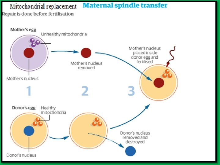
 • Used to avoid transmission of")
Three-parents baby • Mitochondrial replacement IVF (mt. IVF) • Used to avoid transmission of mitochondrial disease. • The technique 1. Take the nucleus of mother egg 2. Insert it into a donor egg that its nucleus removed but still contains mitochondrial DNA 3. Fertilize this hybrid egg with father sperm. • Religious and ethical objections
- Slides: 66