
Molecular Biology I Replication Transcription 1 STRUCTURE OF
Molecular Biology - I Replication Transcription 1
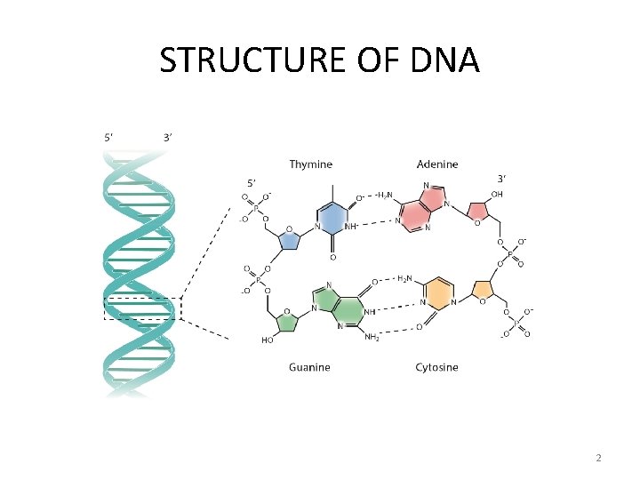
STRUCTURE OF DNA 2
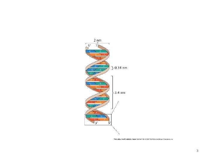
3
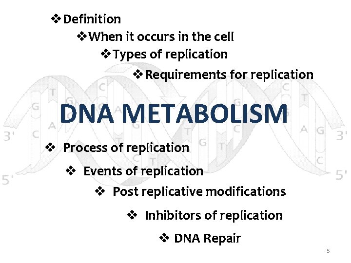
v. Definition v. When it occurs in the cell v. Types of replication v. Requirements for replication DNA METABOLISM v Process of replication v Events of replication v Post replicative modifications v Inhibitors of replication v DNA Repair 5
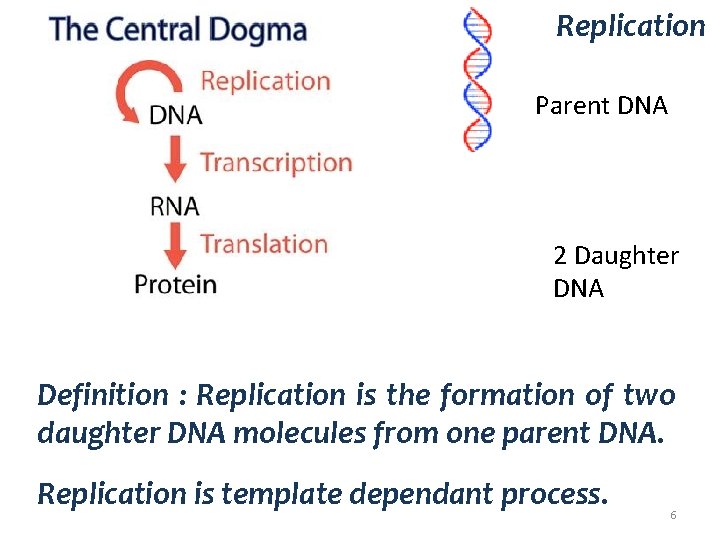
Replication Parent DNA 2 Daughter DNA Definition : Replication is the formation of two daughter DNA molecules from one parent DNA. Replication is template dependant process. 6

Replication occurs during “S” Phase of the cell cycle. Cell Cycle Each chromosome now consists of two sister chromatids. 7
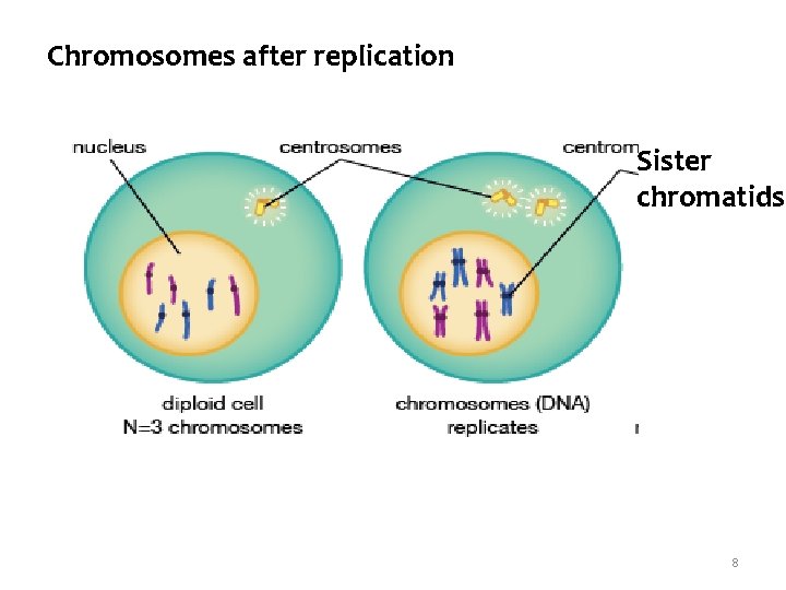
Chromosomes after replication Sister chromatids 8
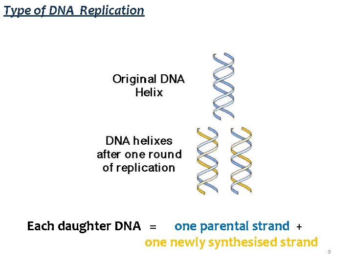
Type of DNA Replication Each daughter DNA = one parental strand + one newly synthesised strand 9
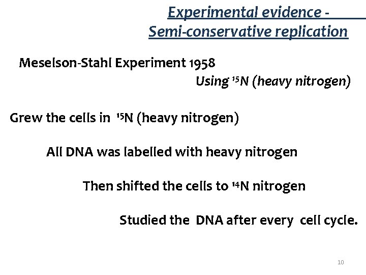
Experimental evidence - Semi-conservative replication Meselson-Stahl Experiment 1958 Using 15 N (heavy nitrogen) Grew the cells in 15 N (heavy nitrogen) All DNA was labelled with heavy nitrogen Then shifted the cells to 14 N nitrogen Studied the DNA after every cell cycle. 10
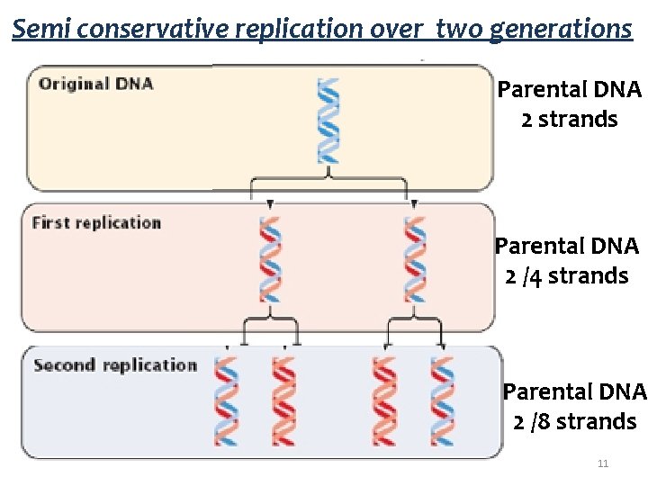
Semi conservative replication over two generations Parental DNA 2 strands Parental DNA 2 /4 strands Parental DNA 2 /8 strands 11
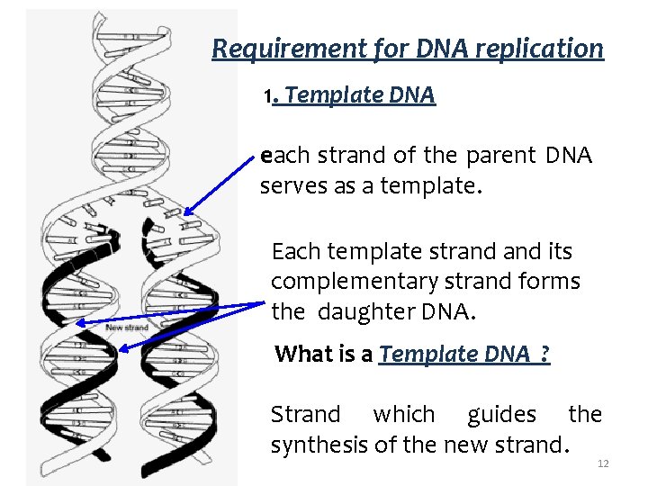
Requirement for DNA replication 1. Template DNA each strand of the parent DNA serves as a template. Each template strand its complementary strand forms the daughter DNA. What is a Template DNA ? Strand which guides the synthesis of the new strand. 12
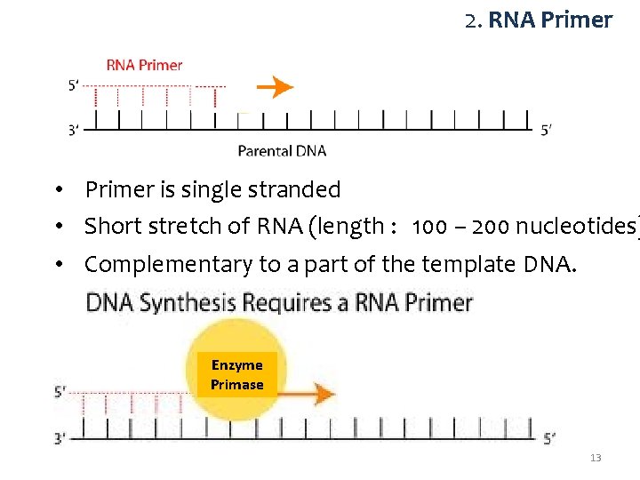
2. RNA Primer • Primer is single stranded • Short stretch of RNA (length : 100 – 200 nucleotides) • Complementary to a part of the template DNA. Enzyme Primase 13
3. Substrates Ø Substrates for DNA synthesis. Deoxy adenosine triphosphate (d. ATPs) Deoxy guanosine triphosphate (d. GTPs) Deoxy cytidine triphosphate (d. CTPs) Deoxy thymidine triphosphate (d. TTPs) Ø Substrates for RNA Primer synthesis. The four Ribonucleoside triphosphates (NTPs) – ATP, GTP, UTP, CTP 14
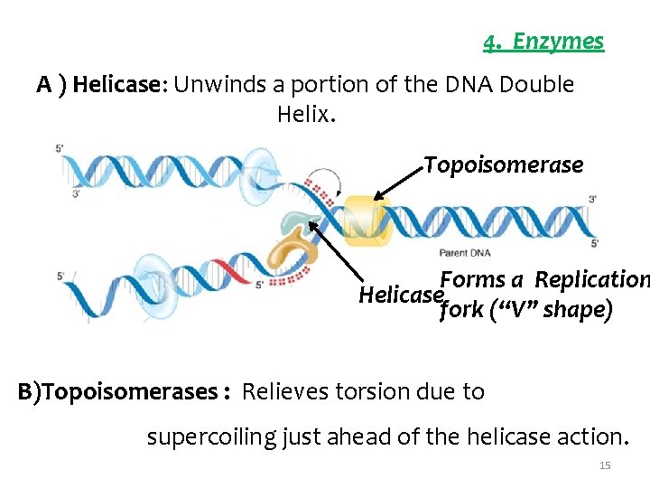
4. Enzymes A ) Helicase: Unwinds a portion of the DNA Double Helix. Topoisomerase Forms a Replication Helicase fork (“V” shape) B)Topoisomerases : Relieves torsion due to supercoiling just ahead of the helicase action. 15
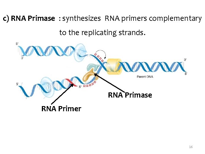
c) RNA Primase : synthesizes RNA primers complementary to the replicating strands. RNA Primase RNA Primer 16
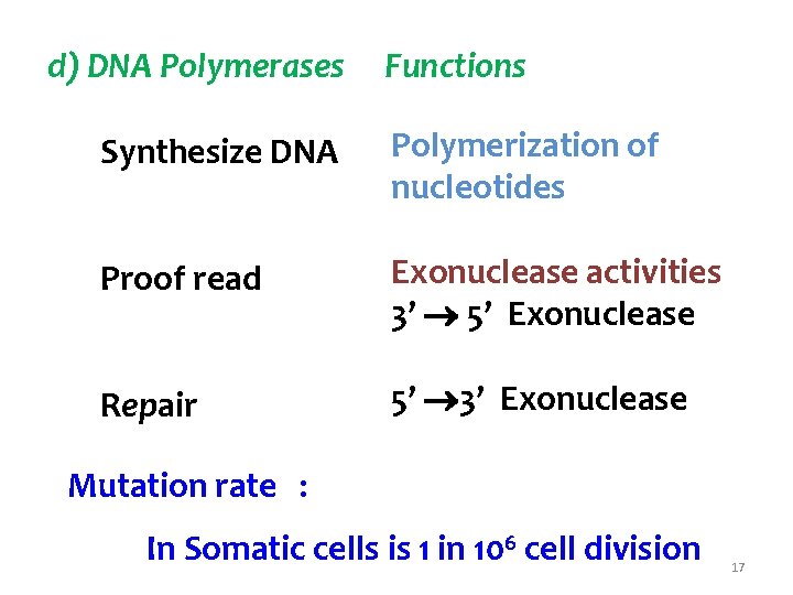
d) DNA Polymerases Functions Synthesize DNA Polymerization of nucleotides Proof read Exonuclease activities 3’ 5’ Exonuclease Repair 5’ 3’ Exonuclease Mutation rate : In Somatic cells is 1 in 106 cell division 17
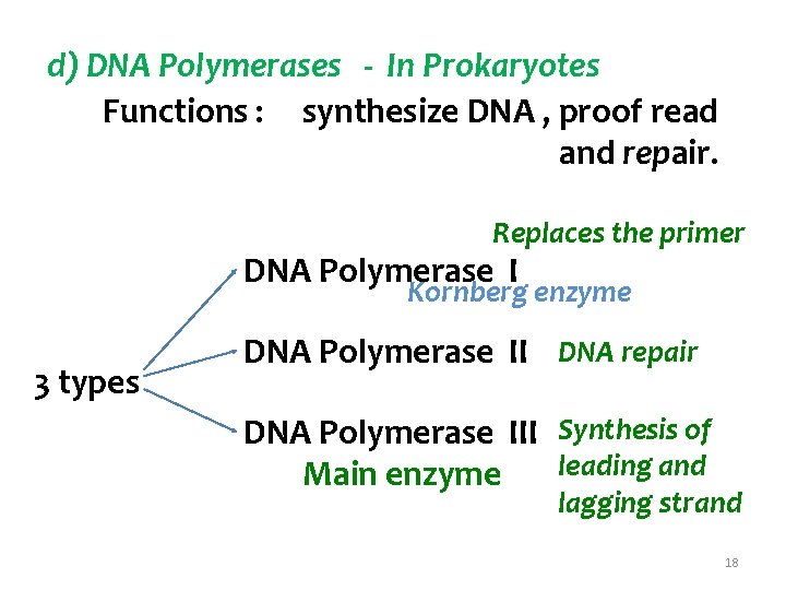
d) DNA Polymerases - In Prokaryotes Functions : synthesize DNA , proof read and repair. Replaces the primer DNA Polymerase I Kornberg enzyme 3 types DNA Polymerase II DNA repair DNA Polymerase III Synthesis of leading and Main enzyme lagging strand 18
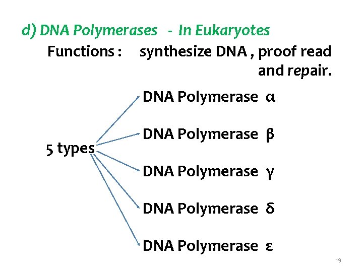
d) DNA Polymerases - In Eukaryotes Functions : synthesize DNA , proof read and repair. DNA Polymerase α 5 types DNA Polymerase β DNA Polymerase γ DNA Polymerase δ DNA Polymerase ε 19
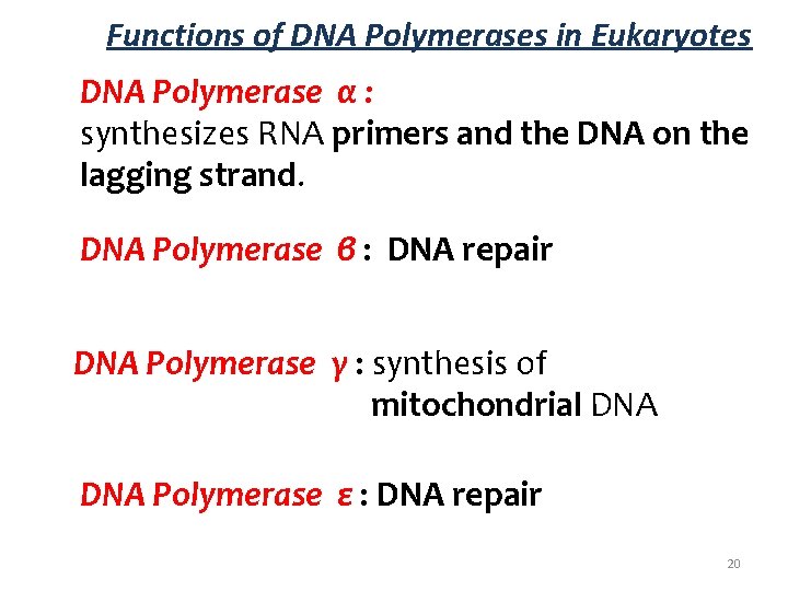
Functions of DNA Polymerases in Eukaryotes DNA Polymerase α : synthesizes RNA primers and the DNA on the lagging strand. DNA Polymerase β : DNA repair DNA Polymerase γ : synthesis of mitochondrial DNA Polymerase ε : DNA repair 20
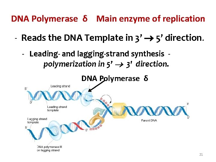
DNA Polymerase δ Main enzyme of replication - Reads the DNA Template in 3’ 5’ direction. - Leading- and lagging-strand synthesis polymerization in 5’ 3' direction. DNA Polymerase δ 21
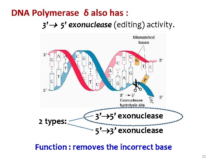
DNA Polymerase δ also has : 3' 5' exonuclease (editing) activity. 2 types: 3’ 5’ exonuclease 5’ 3’ exonuclease Function : removes the incorrect base 22
DNA Polymerase δ Cannot initiate DNA synthesis. Can only add d. NTP’s to 3’ OH group of a already existing DNA / RNA primer. Mechanism of DNA replication: d. NTP is brought as per base pair rule. Phosphodiester bond formation occurs. Rate of Replication Prokayotes : 1000 nucleotides / sec Eukayotes : 100 nucleotides / sec 23
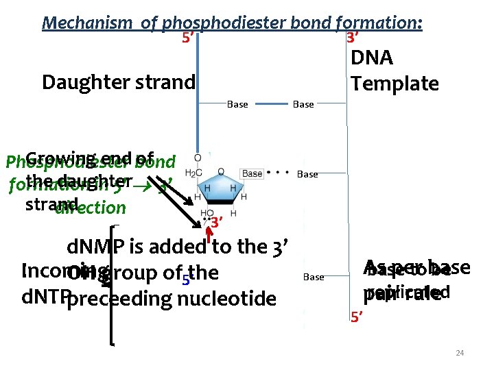
Mechanism of phosphodiester bond formation: 5’ 3’ DNA Template Daughter strand Base Growing end of Phosphodiester bond the daughter 3’ formation in 5’ strand direction Base 3’ d. NMP is added to the 3’ Incoming OH group of 5’the d. NTPpreceeding nucleotide Base As per base tobase be replicated pair rule 5’ 24
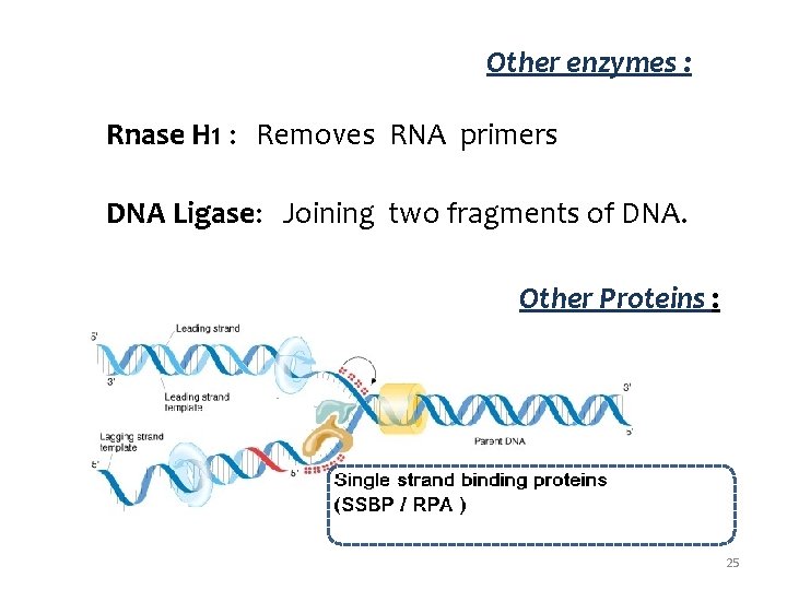
Other enzymes : Rnase H 1 : Removes RNA primers DNA Ligase: Joining two fragments of DNA. Other Proteins : 25
Enzymes involved in DNA replication: Helicase : Unwinds DNA helix Topoisomerase I and II : Removes supercoiling Primase : Synthesizes RNA primers DNA Pol : Polymerizes nucleotides to form the complementary strand. Rnase H 1 : Removes RNA primers DNA Ligase: Joining fragments of DNA. 26
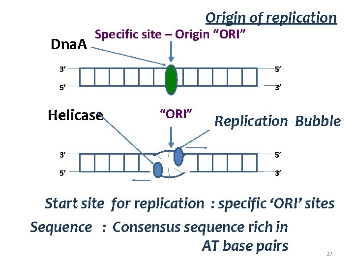
Origin of replication Dna. A Specific site – Origin “ORI” 3’ 5’ 5’ 3’ Helicase “ORI” Replication Bubble 3’ 5’ 5’ 3’ Start site for replication : specific ‘ORI’ sites Sequence : Consensus sequence rich in AT base pairs 27
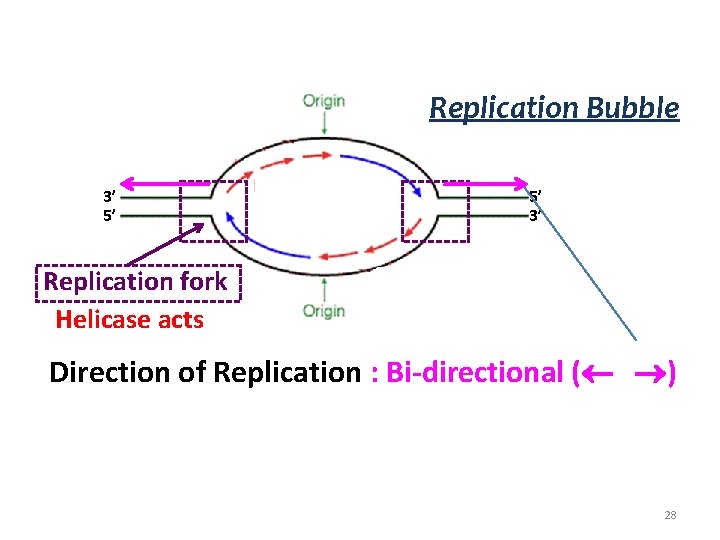
Replication Bubble 3’ 5’ 5’ 3’ Replication fork Helicase acts Direction of Replication : Bi-directional ( ) 28
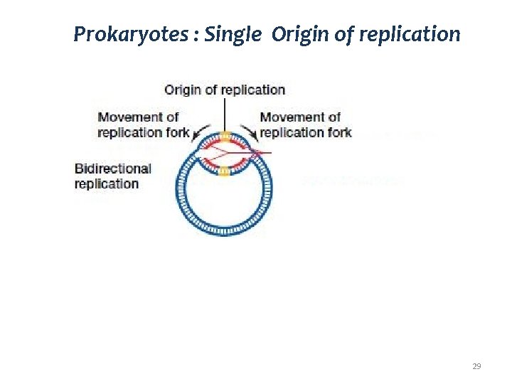
Prokaryotes : Single Origin of replication 29
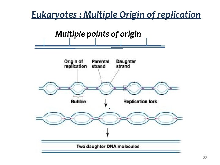
Eukaryotes : Multiple Origin of replication Multiple points of origin 30

Leading strand lagging strand DNA Polymerase δ can read the template in 3' 5' direction only. synthesize the daughter strand 5' 3' direction only. 5’ 3’ Leading strand ds. DNA 3’ 5’ 3’ DNA Polymerase δ Replicating fork Lagging strand Okazaki fragments 5’ 31
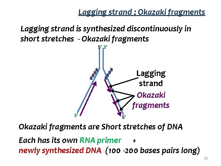
Lagging strand ; Okazaki fragments Lagging strand is synthesized discontinuously in short stretches - Okazaki fragments 5’ 3’ Lagging strand Okazaki fragments 3’ 5’ Okazaki fragments are Short stretches of DNA Each has its own RNA primer + newly synthesized DNA (100 -200 bases pairs long) 32
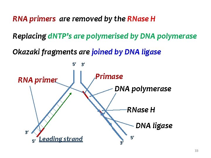
RNA primers are removed by the RNase H Replacing d. NTP’s are polymerised by DNA polymerase Okazaki fragments are joined by DNA ligase 5’ 3’ RNA primer Primase DNA polymerase RNase H DNA ligase 3’ 5’ Leading strand 3’ 5’ 33
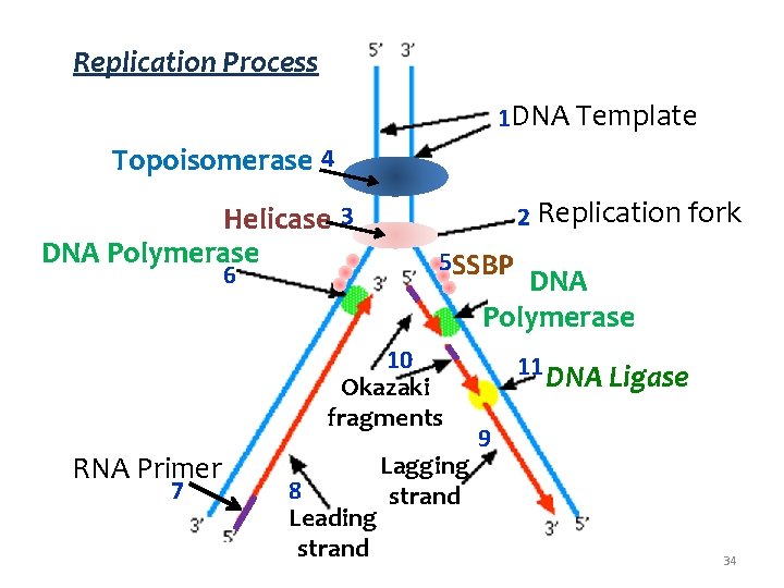
Replication Process 1 DNA Template Topoisomerase 4 Helicase 3 DNA Polymerase 6 2 Replication fork 5 SSBP DNA Polymerase 10 Okazaki fragments RNA Primer 7 8 Leading strand Lagging strand 11 DNA Ligase 9 34
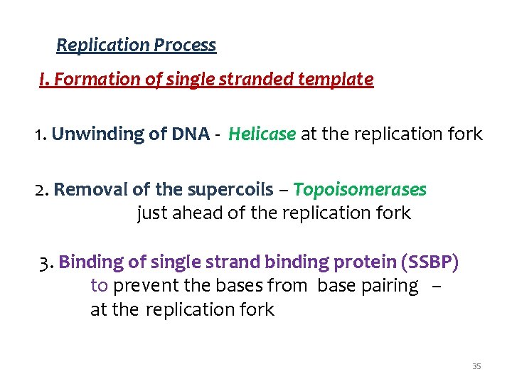
Replication Process I. Formation of single stranded template 1. Unwinding of DNA - Helicase at the replication fork 2. Removal of the supercoils – Topoisomerases just ahead of the replication fork 3. Binding of single strand binding protein (SSBP) to prevent the bases from base pairing – at the replication fork 35
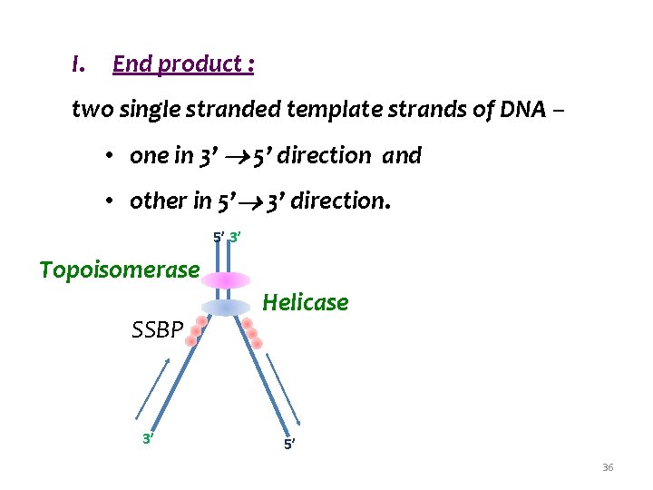
I. End product : two single stranded template strands of DNA – • one in 3’ 5’ direction and • other in 5’ 3’ direction. 5’ 3’ Topoisomerase SSBP 3’ Helicase 5’ 36
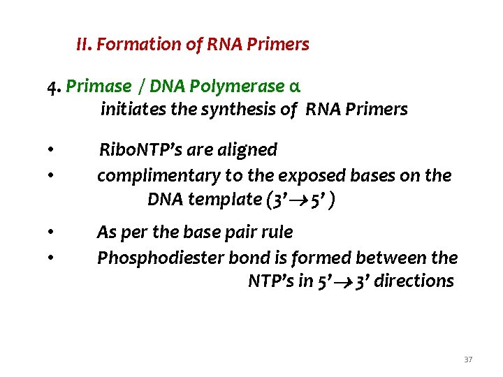
II. Formation of RNA Primers 4. Primase / DNA Polymerase α initiates the synthesis of RNA Primers • Ribo. NTP’s are aligned • complimentary to the exposed bases on the DNA template (3’ 5’ ) • • As per the base pair rule Phosphodiester bond is formed between the NTP’s in 5’ 3’ directions 37
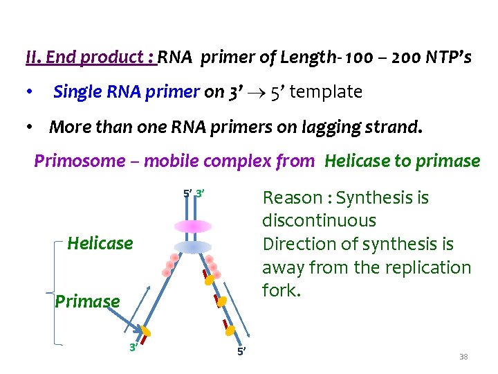
II. End product : RNA primer of Length- 100 – 200 NTP’s • Single RNA primer on 3’ 5’ template • More than one RNA primers on lagging strand. Primosome – mobile complex from Helicase to primase Reason : Synthesis is discontinuous Direction of synthesis is away from the replication fork. 5’ 3’ Helicase Primase 3’ 5’ 38
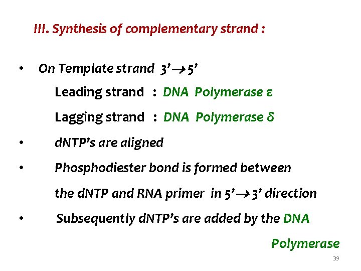
III. Synthesis of complementary strand : • On Template strand 3’ 5’ Leading strand : DNA Polymerase ε Lagging strand : DNA Polymerase δ • d. NTP’s are aligned • Phosphodiester bond is formed between the d. NTP and RNA primer in 5’ 3’ direction • Subsequently d. NTP’s are added by the DNA Polymerase 39
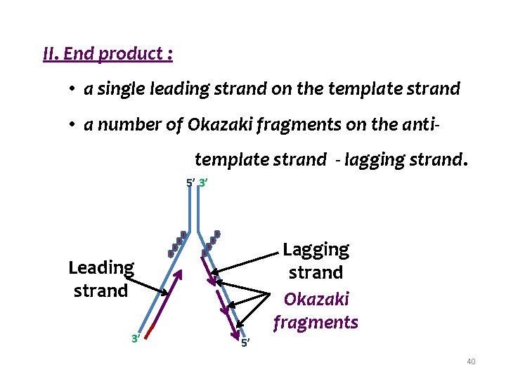
II. End product : • a single leading strand on the template strand • a number of Okazaki fragments on the antitemplate strand - lagging strand. 5’ 3’ Lagging strand Okazaki fragments Leading strand 3’ 5’ 40
IV. Removal of RNA Primers, filling the gaps, joining the fragments On the lagging strand - • Removal of RNA Primers - 3’ 5’ exonuclease activity of DNA polymerase δ / RNase H • Filling the gaps DNA polymerase δ • Joining the fragments DNA Ligase 41
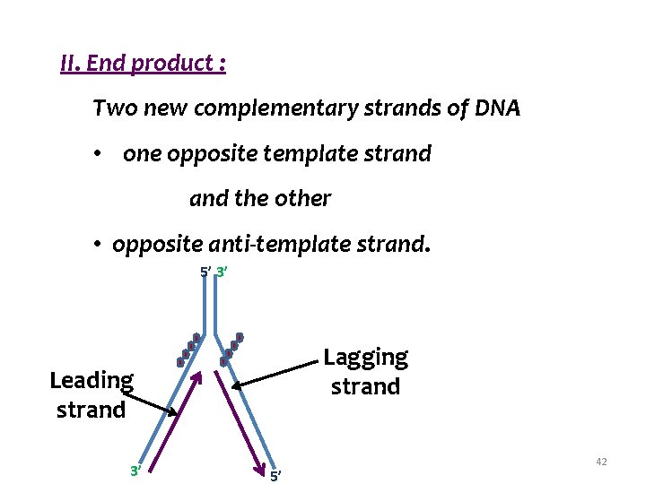
II. End product : Two new complementary strands of DNA • one opposite template strand and the other • opposite anti-template strand. 5’ 3’ Lagging strand Leading strand 3’ 5’ 42
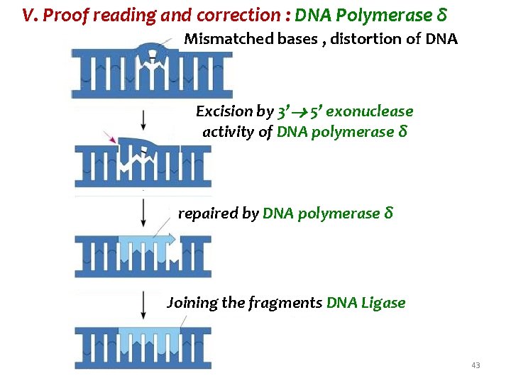
V. Proof reading and correction : DNA Polymerase δ Mismatched bases , distortion of DNA Excision by 3’ 5’ exonuclease activity of DNA polymerase δ repaired by DNA polymerase δ Joining the fragments DNA Ligase 43
V. Proof reading and correction : DNA Polymerase δ Mismatched base pairs are • excised by - 3’ 5’ exonuclease activity of DNA polymerase δ • replaced by the correct base and polymerised by DNA polymerase δ • Joining the fragments DNA Ligase 44
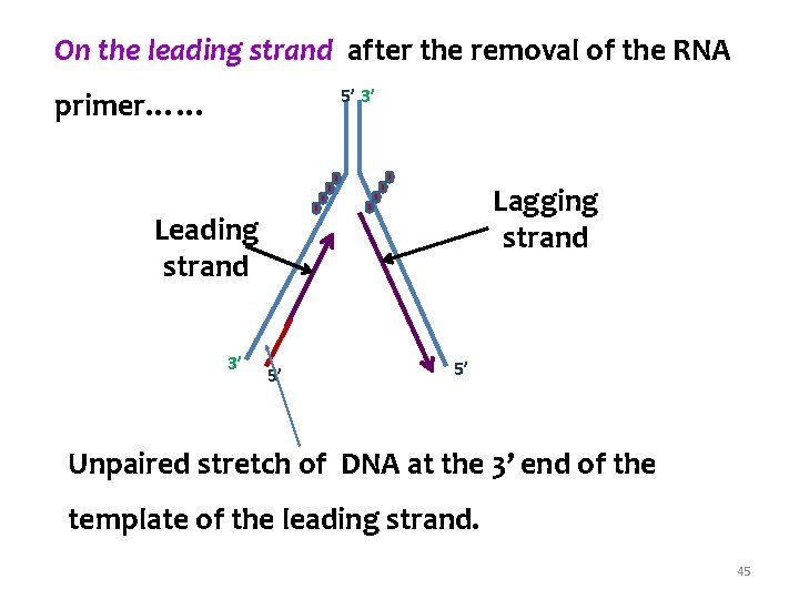
On the leading strand after the removal of the RNA 5’ 3’ primer…… Lagging strand Leading strand 3’ 5’ 5’ Unpaired stretch of DNA at the 3’ end of the template of the leading strand. 45
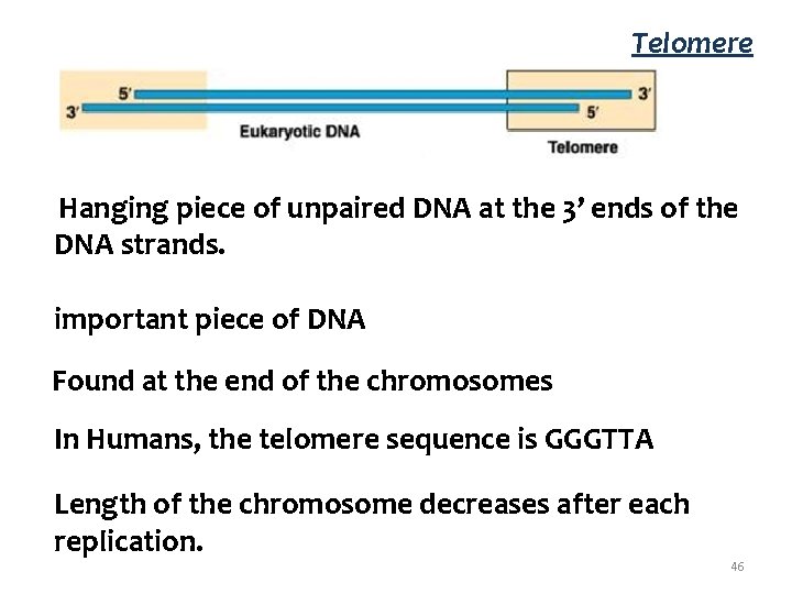
Telomere Hanging piece of unpaired DNA at the 3’ ends of the DNA strands. important piece of DNA Found at the end of the chromosomes In Humans, the telomere sequence is GGGTTA Length of the chromosome decreases after each replication. 46
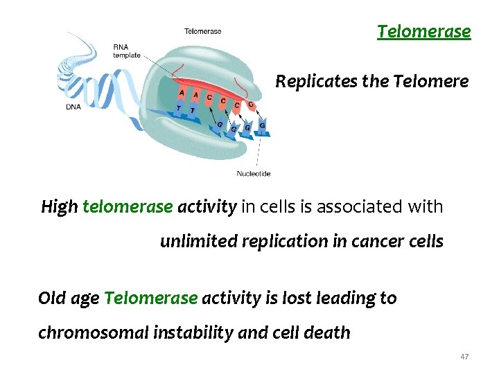
Telomerase Replicates the Telomere High telomerase activity in cells is associated with unlimited replication in cancer cells Old age Telomerase activity is lost leading to chromosomal instability and cell death 47
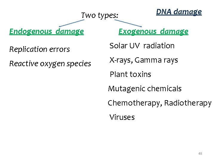
DNA damage Two types: Endogenous damage Exogenous damage Replication errors Solar UV radiation Reactive oxygen species X-rays, Gamma rays Plant toxins Mutagenic chemicals Chemotherapy, Radiotherapy Viruses 48
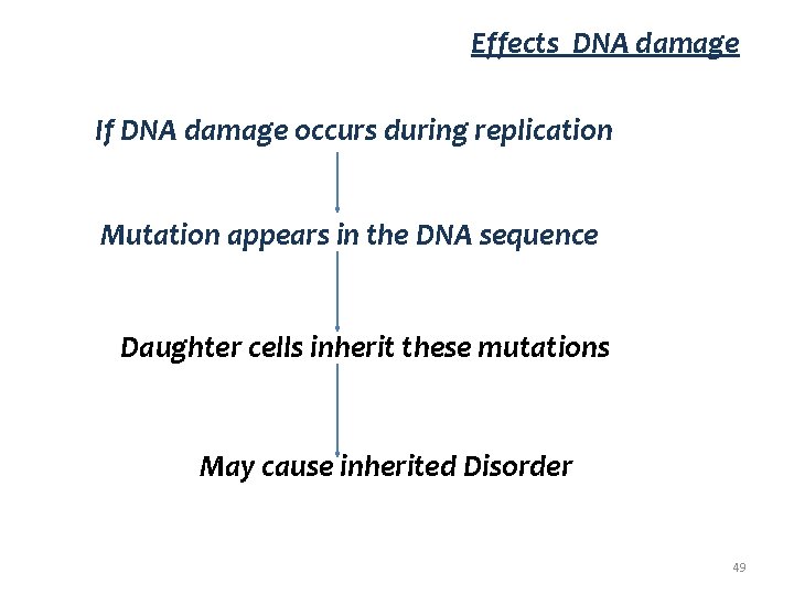
Effects DNA damage If DNA damage occurs during replication Mutation appears in the DNA sequence Daughter cells inherit these mutations May cause inherited Disorder 49
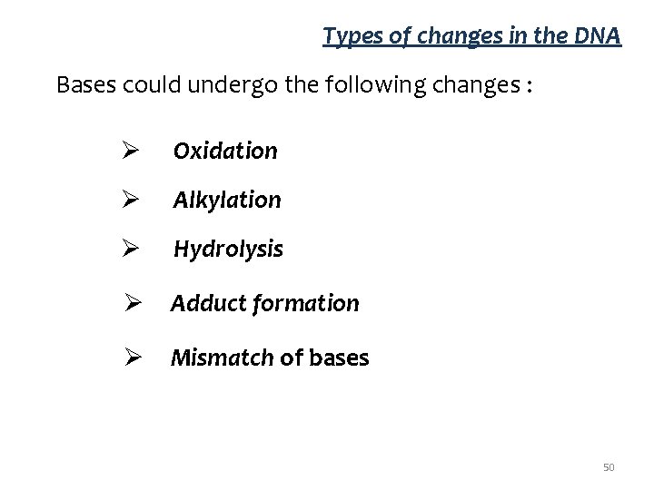
Types of changes in the DNA Bases could undergo the following changes : Ø Oxidation Ø Alkylation Ø Hydrolysis Ø Adduct formation Ø Mismatch of bases 50
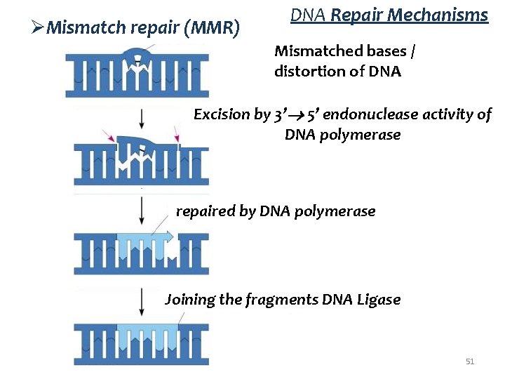
ØMismatch repair (MMR) DNA Repair Mechanisms Mismatched bases / distortion of DNA Excision by 3’ 5’ endonuclease activity of DNA polymerase repaired by DNA polymerase Joining the fragments DNA Ligase 51
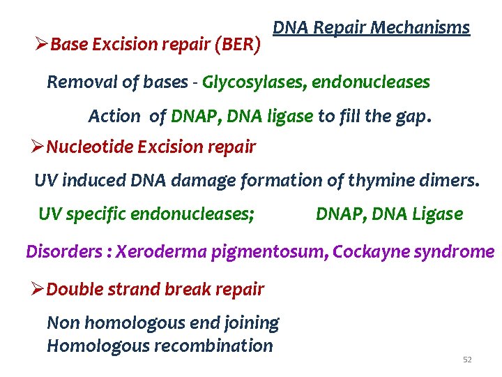
ØBase Excision repair (BER) DNA Repair Mechanisms Removal of bases - Glycosylases, endonucleases Action of DNAP, DNA ligase to fill the gap. ØNucleotide Excision repair UV induced DNA damage formation of thymine dimers. UV specific endonucleases; DNAP, DNA Ligase Disorders : Xeroderma pigmentosum, Cockayne syndrome ØDouble strand break repair Non homologous end joining Homologous recombination 52

53
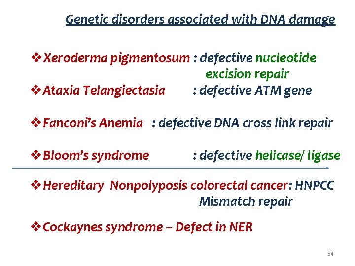
Genetic disorders associated with DNA damage v. Xeroderma pigmentosum : defective nucleotide excision repair v. Ataxia Telangiectasia : defective ATM gene v. Fanconi’s Anemia : defective DNA cross link repair v. Bloom’s syndrome : defective helicase/ ligase v. Hereditary Nonpolyposis colorectal cancer: HNPCC Mismatch repair v. Cockaynes syndrome – Defect in NER 54
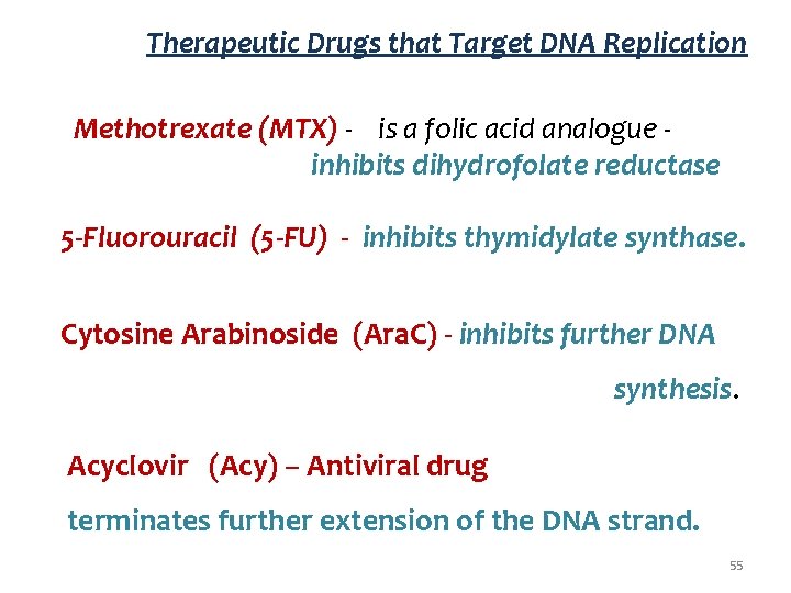
Therapeutic Drugs that Target DNA Replication Methotrexate (MTX) - is a folic acid analogue inhibits dihydrofolate reductase 5 -Fluorouracil (5 -FU) - inhibits thymidylate synthase. Cytosine Arabinoside (Ara. C) - inhibits further DNA synthesis. Acyclovir (Acy) – Antiviral drug terminates further extension of the DNA strand. 55
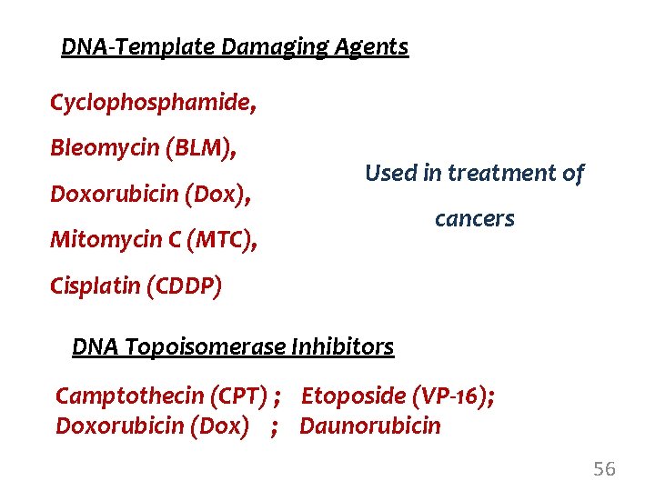
DNA-Template Damaging Agents Cyclophosphamide, Bleomycin (BLM), Doxorubicin (Dox), Used in treatment of Mitomycin C (MTC), cancers Cisplatin (CDDP) DNA Topoisomerase Inhibitors Camptothecin (CPT) ; Etoposide (VP-16); Doxorubicin (Dox) ; Daunorubicin 56
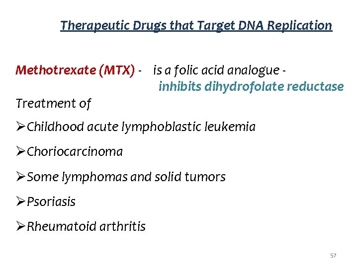
Therapeutic Drugs that Target DNA Replication Methotrexate (MTX) - is a folic acid analogue inhibits dihydrofolate reductase Treatment of ØChildhood acute lymphoblastic leukemia ØChoriocarcinoma ØSome lymphomas and solid tumors ØPsoriasis ØRheumatoid arthritis 57
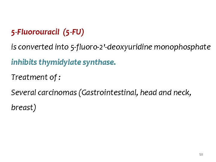
5 -Fluorouracil (5 -FU) is converted into 5 -fluoro-2'-deoxyuridine monophosphate inhibits thymidylate synthase. Treatment of : Several carcinomas (Gastrointestinal, head and neck, breast) 58
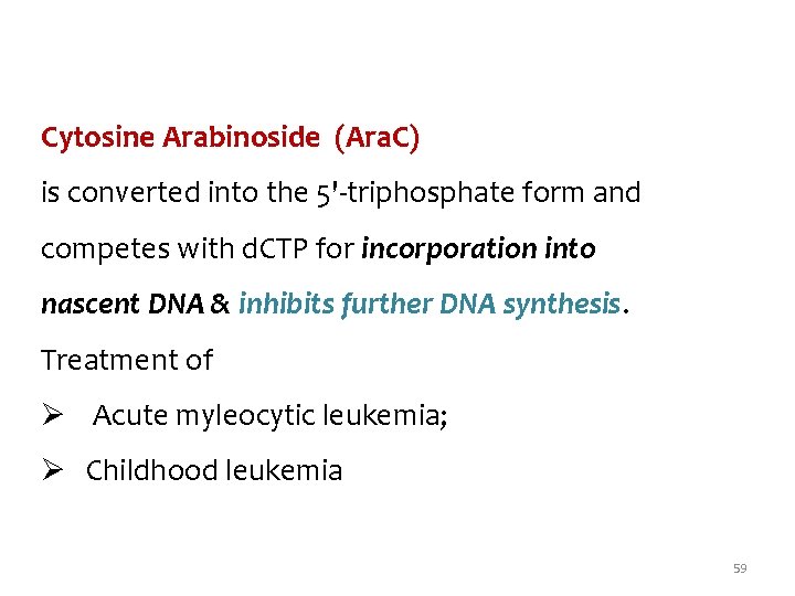
Cytosine Arabinoside (Ara. C) is converted into the 5'-triphosphate form and competes with d. CTP for incorporation into nascent DNA & inhibits further DNA synthesis. Treatment of Ø Acute myleocytic leukemia; Ø Childhood leukemia 59
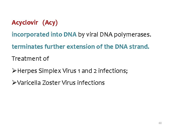
Acyclovir (Acy) incorporated into DNA by viral DNA polymerases. terminates further extension of the DNA strand. Treatment of ØHerpes Simplex Virus 1 and 2 infections; ØVaricella Zoster Virus infections 60
- Slides: 59