Molecular Biology Biol 480 Lecture 4 January 16

Molecular Biology Biol 480 Lecture 4 January 16, 2019

Announcements/Assignments • Comments on ”Decoding Watson”? – Pay attention to the history of DNA structure discovery--- • New articles to read

Where we… • Prions. Insoluble particles composed of a protein called Pr. P. The protein of prions folds very differently compared to the normal form of this protein, despite being encoded by the same gene. The mis-folded Pr. P proteins stick together forming prions which damage nerve cells. Misfolded Pr. P proteins induce the mis-folding of normal proteins. They cause a type of protein replication without DNA

Where we…? • Describe/sketch DNA as a chemical—what are its components and characteristics? • Describe/sketch DNA as a strand—how are nucleotides linked together? • Describe/sketch DNA as a double helix

The Watson-Crick Model B-form DNA A=angstrom= 0. 1 nm 3. 4 nm . 34 nm Figure 10. 14
 is the most common form of DNA")
• B-DNA (also called Watson-Crick form) is the most common form of DNA in cells, but not the only structure that has been described using X-ray crystallography. • Helical direction, # of bases in a turn of helix, helix diameter, shape of backbone can vary

• DNA can change shape due to dehydration, presence of ions, and specific base sequences. • For example, Z-DNA forms readily when DNA contains G-C-G-C…. In order for base pairing to occur, the sugar-phosphate backbone makes sharp turns (zig-zag) pattern in a left turning helix.

• A-DNA forms when DNA is not surrounded by water. It is a right handed helix, but wider, due to the increased number of bases in each turn of helix. • Both A and Z-DNA are thought to exist in cells, under certain conditions or in certain regions of a chromosome, and may even affect gene expression

A-DNA B-DNA z-DNA

• Let’s think about base-pairs in DNA. The structure is entirely the result of the stability of the interaction between A and T and between G and C. • The bond itself, is not covalent, but noncovalent hydrogen bond.

H-bonds • Weak electrostatic interactions between a covalently bonded hydrogen atom and a highly electronegative atom such as nitrogen or oxygen. • The hydrogen is slightly + charged and the O and N slightly negatively charged due to their unequal pull on electrons in their covalent bonds.
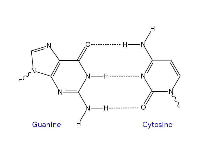

• Only the pairings of A and T and G and C set up bond lengths and alignment to produce a stable molecule—the helix described by W and C.

Individually H-bonds are weak, but in polymers their collective strength is great. Question. How many H-bonds are in a DNA molecule with 10 nucleotides, and the sequence of one strand 5’ ATTCGGCTAA 3’?

• H-bonds are not only important in DNA. – Water! The life-giving properties of water are the result of the extensive formation of H-bonds. – H-bonds are important in other macromolecules, especially in forming protein structure.

Characteristics of DNA • A basic characteristic of DNA was revealed by spectrophotgraphy. It was shown early on in the search for the genetic material that nucleic acids absorb light in the UV range. • The bases of DNA and RNA absorb light at wavelength 260 nm. You can detect and quantify DNA and RNA solutions by their optical density using 260 nm.

• In a future lab you will quantify and analyze the purity of DNA you isolate using UV spectrophotgraphy.

How do we study DNA? • Many of the techniques used to study DNA involve analysis of specific DNA sequence and on its complementary strand nature. For example, DNA can vary quite a bit in the amount of repetitive sequence interspersed between protein coding genes.

Range of repetitive DNA sequence • Single copy---a sequence that appears one time in all the DNA of the organism. Many genes are single copy. • Middle repetitive— 10 -10, 000 copies • Highly repetitive-- > 106 copies

• Repetitive DNA can be found in multiple positions on many or all chromosomes of an organism. Called interspersed repetitive DNA. There are relatively short repeats known as Short Interspersed Elements or SINEs and relatively long sequences called Long Interspersed Elements known as LINEs.

• We will talk more of these features of DNA in chapter 12. (next)

• In my genetics class we do a lab in which we analyze a specific spot in our own genome (on chromosome 16) for the presence of absence of a SINE called an Alu repeat. – Some people have an Alu repeat in this region, whereas some people don’t. And some of us have the repeat on one chromosome 16 and not on the other. Because of the variability in this region, it is know as a gentic polymorphism.

• Repetitive DNA can also be found in the genome in multiple copies repeated over and over in the same region. – These are known as tandem repeats – Tandem repeats can vary in length from 2 -4 nucleotides (called microsatellite repeats) – Longer tandem repeats (10 -60 nucleotides) are called minisatellite repeats.

• Spectrophotometry can be used to analyze the amount and type of repetitive DNA in an organism. • The analysis takes advantage of the complementary base-pairing of the DNA strands.

• As you know, DNA strands are held together by multiple H-bonds between specific bases on opposite strands. – The bonds can be broken by various means including high heat and high p. H. The process of disrupting the base-pairing of 2 DNA strands is called denaturing.

• Likewise, DNA that is carefully, purposely denatured can be renatured or reanneal---if conditions are reversed, the DNA can reform its H-bonds and complementary base-pairs can form. • You. Tube: https: //www. youtube. com/watch? v=k 7 Wov. Eb. Xf. Q
 at 260 nm")
• As DNA denatures, its absorption of light (optical density—OD) at 260 nm increases. A sample of DNA which is heated to near 100 degrees C will show an increase in absorption until all the DNA is single stranded.

Also…see Figure 10. 16 • Sigmoidal curve showing denaturing of DNA The melting temperature (Tm) is the temp for a given sample of DNA in which half is SS and half is DS.

Likewise… • If you cool the DNA slowly you can allow the bases to reform and the DNA renatures or reassociates into double strands. • As renaturation occurs, the OD decreases until all the DNA is double stranded.
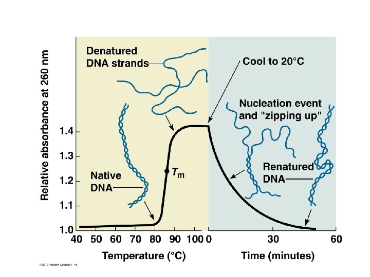

• Renaturation/reassociation kinetics tells you something about the DNA sequence. – What might slow or speed the reassociation of DNA strands? (figure 10 -16)

Using reassociation kinetics to analyze repetitive sequence in DNA • DNA from different organisms can be analyzed using DNA denaturing and reassociation kinetics. 1. Isolate all the DNA from cells of an organism 2. Denature/melt DNA with heat 3. Cool DNA to allow reassocation of DNA strands 4. At the same time, measure OD 260 over time.

Time OD 260

Think of the curve as a comparison of prokaryotic and eukaryotic DNA • Make some conclusions from this experiment. • Start with basic observations, then look more deeply at the data.

• The more repeats in the genome of an organism, the faster the renaturation. Why? • Renaturation occurs in phases or fractions. • Be able to interpret a reassocation curve.
- Slides: 35