Molecular Biology Biol 480 Lecture 26 April 3

Molecular Biology Biol 480 Lecture 26 April 3 rd , 2019
 meeting • Cool talk")
Announcements/Assignments – Attended the American Association for cancer research (AACR) meeting • Cool talk on alternative splicing! Different ancestry groups. . i. e. West Africans and descendants show different splicing forms of specific genes than other ancestry groups. Relevant in the risk of developing certain cancers and in the effectiveness of certain treatments. – Exam 3—returned Friday

Where we? Translation • Protein primary, secondary, tertiary, quaternary structure. • Protein domains • Protein motifs • Shared motifs in different proteins suggests exons have been moved/shuffled throughout evolutionary history.

Functional Domains • Domain: portion of a protein that folds into stable, unique conformations independent of the rest of the molecule • • • Domains give different functional capabilities Proteins can contain one to several domains Thought to have arisen as a result of “exon shuffling” (see upcoming slides)

LDL Receptor Exons Figure 14. 22

Some common protein motifs

How did unique proteins come to have common domains? Exon Shuffling… It’s thought that new proteins have been made over time by DNA recombination and movement of DNA in the genome. In a sense, just reusing a limited number of protein coding sequences in different ways…. read more on your own. http: //en. wikipedia. org/wiki/Exon_shuffling#His tory

Protein folding process • Some proteins require other proteins to fold properly. Because of this tendency for proteins to fold right away, the end terminus may fold up and be unable to form proper structure with a more C-terminal region. • Proteins called chaperones function to sort of prevent some folding of a newly made protein until more of the amino acids are available.

• Some chaperones also serve to protect proteins from unfolding if a cell is exposed to denaturing conditions. • For example, special chaperone proteins called heat-shock proteins, serve to protect a cell from short exposures to increased heat.

• Without these heat shock proteins some proteins would denature and likely stick together with even small increases in heat. • http: //en. wikipedia. org/wiki/Heat_shock_prot ein

Moving to chapter 15 • If we have a good handle on the genetic code, and protein synthesis, and on the intimate connection between amino acid sequence and folded structure and on the link between protein structure and function… …We can pretty easily link DNA alterations--mutations, to genetic variation and natural selection, and to genetic diseases.
 is")
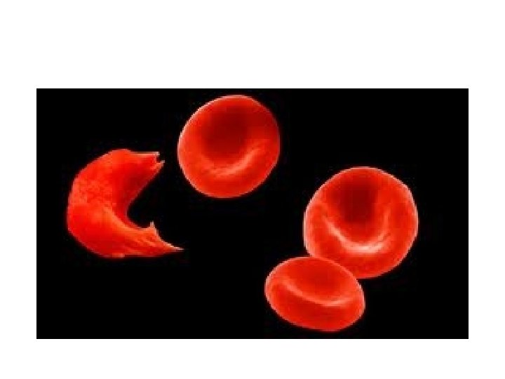
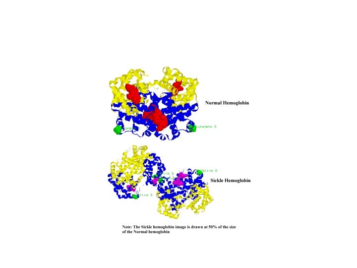
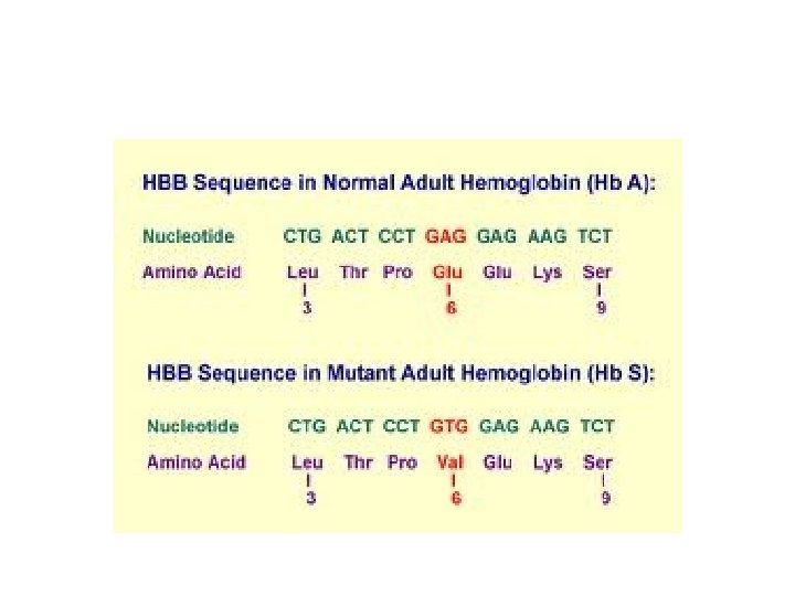
• The classic example (and the first disease linked definitively to mutation) is sickle cell anemia. • This disease is the result of abnormal hemoglobin—the oxygen-carrying protein in our red blood cells.

Review…linking classical with molecular genetics • To begin will need to think somewhat mechanically about mutations. – Mutations are permanent changes in DNA – DNA is organized into chromosomes. – Chromosomes are located in cells… – Diploid organisms have 2 copies of each gene

Classification…location of mutation • Somatic mutations---occur in DNA of any cell in the body, other than germ cells (egg or sperm) • Germline mutations occur in DNA of germ cells/gametes/egg and sperm. • Autosomal mutations occur within genes on chromosomes that do not have genes that determine sex development. • Sex-linked mutations occur within genes on chromosomes that do have genes that determine sex development.

Recall…in diploid organisms… Depending on the function of the protein encoded by the gene…mutations can be: • Dominant---giving a phenotypic change if one copy of the gene is mutated • Recessive---having no effect unless both copies of the gene have mutations.

Dominant mutations… • Thinking in terms of the protein encoded… – The mutant phenotype may be the result of haploinsufficiency. One normal copy of the gene may be unable to produce enough of the protein for normal function of the cell. – The mutant phenotype may be the result of a dominant-negative effect. The abnormal form of the protein may inhibit the activity of the normal form of the protein.
 because a")
• Recessive mutations---no noticeable effect (if only one allele is mutated) because a normal copy of a gene provides a normal copy of the protein. There seems to be no difference between one and 2 copies of the normal protein. • Only when both alleles are mutated, and no normal form of the protein is produced is there noticeable effect on the cell/organism.

• Example? Instances occur where a region of a chromosome is lost---if this occurs in both copies of the chromosome, BOTH copies of a gene are lost---no protein encoded by this gene can be made. • (go beyond thinking of a recessive allele as a small letter!)

At the molecular level… • Point mutation—a change in one nucleotide in a gene. Also known as a base substitution. – 3 outcomes in terms of the protein produced. • Substitution changes the triplet code to encode a different amino acid= missense mutation • Substitution introduces a stop codon in the protein code=nonsense mutation • Substitution changes the triplet code to a different code, but which encodes the same amino acid or a functionally similar amino acid= silent mutation

• Nucleotide substitutions are sometimes classified by the change made. – If a purine is substituted for another purine or pyrimidine for a pyrimidine, it is called a transition mutation – If a purine is substituted for a pyrimidine or vice versa, it is called a transversion mutation

• Frameshift mutation– insertion or deletion of 1 or 2 nucleotides, which completely changes the reading frame of the protein. • Figure 15 -1 -- • Why are frameshift mutations generally more serious than point mutations?

Be able to describe this as a molecular change…not just an English sentence… Figure 16. 1

Functional classifications of mutations • Loss of function mutations---as expected, these types of mutations completely eliminate the normal function of the protein encoded by the gene. • These can result from point mutations, frameshift mutations, large deletions of DNA including the gene. • Sometimes called null mutations

• Loss of function mutations can be dominant or recessive---see earlier slides---and read more!

• Gain of function mutations. As the name implies, these mutations change the protein in a way that gives in a new activity or so that it is made in excess and this greatly enhances its effect in a cell. • These mutations are usually dominant.

How do mutations arise? • 2 primary ways: – Mistakes by DNA polymerase, during replication. • Insertion of a non-complementary base to the template (with a failure to correct) • DNA polymerase slippage or stuttering that ultimately misses some nucleotides or duplicates some region of the DNA – Base changes which occur in existing DNA

Some common base modifications • Tautomeric shifts – DNA bases can exist in 2 forms, isomers, that differ only in the position of one proton. These different forms are known as tautomers. – These spontaneous shifts allow non-Watson-Crick base pairs to form.

Figure 16. 2

• In the next round of replication, a permanent base-change will occur and half of the replicated DNA will have a point mutation.

Figure 16. 3

• It’s a great time to pause and remember, that cells have a mechanism to try to reduce the overall number of times they replicate their DNA. – What is that mechanism? – A cell which is stopped from replicating will not pass on this mutation!

Other small chemical changes… • Common spontaneous chemical changes completely change a base (and thus its basepairing behavior), or alter a base such that it is replaced with a random nucleotide. • Depurination • Deamination

Depurination

Deamination Figure 16. 4

Independent study • Chapter 15 – Since we mentioned various aspects of DNA repair when discussing DNA replication, we will read independently through section 15. 7. ---- know what the Ames test is for!

But how do mutations arise… • In both cases, mutations can be: – Induced---Caused by an external factor or condition of the cell. For example, UV light causes/induces pyrimidine dimers to form. Limiting sun exposure reduces this type of mutation associated with skin cancer! – Spontaneous—unavoidable, random changes not associated with any specific agent or condition.

• Different organisms seem to have different spontaneous mutation rates: – Bacteria show an average of 1 mutation in 100 million DNA replications/cell division. (10 -8) – Mice ~(10 -5) – Humans (10 -6)

• This oversimplifies things a bit---within an organism’s genome, there are regions that are nearly resistant to mutation, and other regions that are fairly frequently mutated. – These regions of higher rates of mutation are sometimes called “hot spots” for mutation.

• It’s also a good time to remember, that all we’ve learned about induced and spontaneous mutations, has helped us in instances when the goal is to: – Induce mutations for the purpose of studying mutants in a particular process – Kill cells purposely by inhibiting normal replication and DNA chemistry/structure.

For example • Exposing a plate of bacteria to UV or other radiation leads to mutation (induced) in some individuals. • If you are studying, say, mutations associated with metabolism of arabinose, you can select for those that change in terms of how they use arabinose.
")
• Likewise… – Studying how radiation results in mutation (and leads to apoptosis) led to the development of radiation treatment for cancer. – Chemicals that bind DNA and disrupt DNA replication or result in mutations are common cancer chemotherapy drugs.

• Be sure to review mutagens, mutation rate, assays to identify mutagens.

• One final thought…Why would an error free replication system or a mutation-resistant genetic material be a bad thing…Why is it essential to have a low, but detectible mutation rate?
- Slides: 46