Molecular Biology Biol 480 Lecture 21 March 18

Molecular Biology Biol 480 Lecture 21 March 18 th, 2019

Announcements/Assignments Exam 2—Go over answers. Nicely done! – Topic pick due Wednesday—by 4 pm. – Real-time PCR /quantitative pcr in lab this week

Where we? RNA processing/splicing. Eukaryotes must remove introns and splice together exons from the primary m. RNA transcript. Requires nucleotide signals within the RNA as well as a ribo-protein complex--Spiceosome
 • http: //www. ncbi. nlm. nih. gov/books/NBK 9846/")
Additional resources at NCBI (NCBI bookshelf) • http: //www. ncbi. nlm. nih. gov/books/NBK 9846/ • READ! Some of this has been covered previously. Focus on the facts about introns in different types of organisms. • Were they present in prokaryotes and they lost them over time? Did eukaryotes evolve them independently? If so, why? Is there evidence for either/both of these ideas?
")
Describe this experiment. Who did it? (from the cytoplasm)

• What is the intron-exon boundary consensus? • Is there any other important nucleotides in the intron? In the exons? • Why is it considered a loose consensus? • How do the RNA components find the intron and guide the spliceosome to do its work?
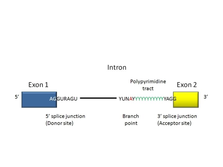

Components of the spliceosome • Small RNAs, found only in the nucleus, play an essential role in splicing. These “small nuclear RNAs—sn. RNAs, interact with small nuclear proteins to form small nuclear ribonucleoprotiens (sn. RNPs or snurps). • The sn. RNAs are rich in U and have been named “U 1, U 2, U 3, U 4, U 5, and U 6”

• Different “U” sn. RNAs bind to different parts of the intron (by complementary base-pairing) and facilitate interactions of sn. RNAs and protein components of the spliceosome. The branchpoint A becomes joined to the donor site, forming a loop. • Since the loop forms with nucleotides in the middle of the intron the 3’ end of the intron extends like a tail.

• Eventually there is a cleavage of the 3’ acceptor site which simultaneously releases the loop of intron and joins the nucleotides on either side of the intron. • The released intron resembles a lariat. • STUDY this in detail! Know the approximate order and specificity of each sn. RNA’s role in splicing.

Figure 13. 14

• Watch animation. • http: //www. dnalc. org/view/16938 -3 DAnimation-of-RNA-Splicing. html

• What are self-splicing introns? • What organisms/organelles/RNAs contain them? • Who discovered them and why was it truly remarkable? • What is alternative splicing? Show/sketch an example

Video has no sound—good practice to identify specific factors in transcription and splicing? http: //www. youtube. com/watch? v=p 2 u. C 5 j 0 h. K 0 A • Alternative splicing, again, partially explains how, what appears to be a rather low number of protein coding genes in the human genome, may encode many more proteins than we first estimated. Explain!

How important is splicing? • Mistakes in splicing have been linked to diseases. Usually we look for mutations only in exons---why? • How might a nucleotide change in an intron cause a protein to be altered?

• Great paper on splicing defects. Read headings and summarize. Look at table 1. Study 1 or 2 different examples of splicingdefect diseases, in detail. • https: //www. ncbi. nlm. nih. gov/pmc/articles/P MC 6060985/pdf/13353_2018_Article_444. pdf

One final post-transcriptional alteration • RNA editing – In the late 1980 s, it was discovered that a few eukaryotic RNAs had slightly different nucleotide sequence than the DNA from which they were derived.

• The changes seemed to be both: – Substitutions---changing one nucleotide to another. AND --Insertions or deletions of RNA nucleotides This was thought to be impossible, since the central DOGMA states that genes are the recipe for proteins. m. RNA was thought to be an exact RNA copy of a gene transcribed exactly from the DNA.

• RNA editing was first studied in a protozoan, parasite, Trypanosoma, which causes a disease called African Sleeping Sickness. • Special RNAs called a “guide RNA”/g. RNAs seems to direct the editing by binding to complementary RNA and directing editing enzymes to insert short stretches of RNA.

• We will discuss an example of RNA editing in a mammalian gene/RNA for Apolipoprotein B. • This protein is routinely measured in medical testing for its link to heart/vascular health and disease. It is considered a Low denistity lipoprotein (LDL). It is sometime called the “bad cholesterol” component. People with blocked arteries may have high LDL levels.

Great summary of cholesterol! • http: //www. nhlbi. nih. gov/healthtopics/hbc

• It appears that the Apo-B m. RNA is altered at a specific C nucleotide which gets changed to U. • This editing only occurs in small intestine cells and results in a shorter version of the protein and which changes its function of fat absorption in these cells.

Importa intestine

• Speculate on how RNA editing was discovered? Think about Apo. B as an example. • Read more on your own. Google!

Gene expression…Part 2 • Translation! – Despite the important roles we’ve discussed for RNA, we can’t underestimate the importance of proteins in cells. – Proteins are translated from m. RNA which is transcribed from genes in DNA.

Figure 13. 1

• Again, a number of scientists established the mechanism for translation, using a combination of biochemical, and molecular biology approaches. Details of the mechanism of translation were established during the 1960 s and involved many scientists.

The knowns… • Biochemists had studied both nucleic acids and proteins and had established: – DNA is made of linear sequence of 4 different d. NTPs ( A, T, C, G) – Proteins are more complex, in that they are built using 20 different amino acids.

• Despite many attempts to link DNA directly to protein synthesis, several scientists, including Francois Jacob, and Jacques Monod, hypothesized that an intermediate molecule was necessary to produce a protein from a DNA code.

• We’ve learned already that the intermediate was m. RNA transcribed from the DNA (gene) sequence. – Read again how scientists discovered m. RNA using bacteria and bacteriophages. (13. 9) – How did scientists show sequence similarity before we were able to sequence DNA?

Once it was established that m. RNA held the actual code for protein synthesis, lots of experiments were done to show the code worked. • m. RNA, like DNA is made with 4 different building blocks NTPs (U, C, G, A) • Sydney Brenner proposed, based on the numbers of building blocks in both types of molecules, a 3 -letter RNA code, would be sufficient to encode 20 amino acids.

Sidney Brenner was in the James Watson video you watched.

• The “extra” codes suggested a possibility for redundancy, that one amino acid might have more than one 3 -letter designation. • Later, the code was show to be degenerate. The actual nature of the degeneracy, helped establish details of the translation machinery.

• One of the first pieces of evidence supporting a triplet code used bacteriophages (go figure? ) and a method for creating insertion or deletions of nucleotides in the genes of the phage. • It was noticed that inserting or deleting one nucleotide usually wiped out the normal activity of a gene—it created a non-functional mutant.

• The same occurred if 2 nucleotides were inserted or deleted. – But if 3 or multiples of 3 nucleotides were inserted or deleted, the mutations were often reversed, and at least in some cases, a wild-type phenotype was restored. – This was consisted with the 3 -letter code proposed by Brenner. Adding or subtracting 3 nucleotides has only limited effect on how a m. RNA is translated.

Figure 14. 2 a

Figure 14. 2 b

• The term frame-shift has been used to describe mutations which alter the way in which a triplet code is read in translation.
- Slides: 38