Molecular Biology Biol 480 Lecture 18 March 1

Molecular Biology Biol 480 Lecture 18 March 1 st , 2019

Announcements/Assignments Handed out presentation assignment last week and asked you to read. We will discuss today. No lecture March 8 th. This is the ND Academy of Science meeting in Grand Forks. Will have exam 2 graded and left outside my office —Thursday by 3.

Where we? How proteins bind to DNA. So far we have discussed quite a few DNA binding proteins. We have named several eukaryotic transcription factors… Eukaryotic DNA-binding motifs (common protein shape patters in different proteins that allow binding to DNA).

• We will study more of these DNA binding proteins soon. They have cool names that describe a shape that allows them to bind DNA. – Helix-loop-helix – Helix-turn-helix – Leucine zipper – Zinc finger

Transcription Factor Myo. D is a H-L-H protein which binds genes encoding proteins needed for muscle development.
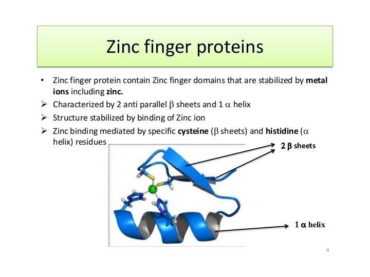

Figure 18. 12

The estrogen receptor protein binds DNA with a Zn finger motif
 that interact with each")
Leucine zipper—transcription Factors usually consist of 2 identical proteins (dimer) that interact with each other and DNA at the same time.

Figure 18. 13

AP-1 is actually 2 different proteins that bind each other through interactions between lots of leucines in both proteins. A second part of each protein interacts with DNA.
 modifications")
Where we… • Eukaryotic m. RNA (transcript) modifications

RNA processing • 5’ modification---addition of a modified guanosine tri-phosphate. • 3’ modification---cleavage of 3’ end addition of a poly A sequence. • Internal modification---removal of RNA known as introns and splicing together adjacent RNA known as exons. (RNA splicing)

Table 14. 8

• Most genes of eukaryotes have introns. The average number is ~8 introns/gene. • Some exceptions…Histone genes have no introns! • …We will return to splicing shortly…
 • http: //www. ncbi. nlm. nih. gov/books/NBK 9846")
Additional resources at NCBI (NCBI bookshelf) • http: //www. ncbi. nlm. nih. gov/books/NBK 9846 / • READ!

• Back to RNA splicing. Since introns are unique to eukaryotic genes and since much of the early work in transcription and translation was done in prokaryotes, the existence of introns was not even discovered until the 1970 s

• In 1977, three people, Susan Berget, Phil Sharp, and Richard Roberts noticed that a virus, in a family called adenovirus, made longer RNA molecules that were eventually shortened. • Using hybridization techniques and electron microscopy, they compared the longer and shorter RNAs and the RNAs to the DNA that encoded them.

• They noted that specific portions of the DNA sequence were missing in the RNA. – Sharp referred to this as “split genes”. The genes were shown to be interrupted with non-amino acid coding, intervening sequences. – These were later termed introns.
")
(from the cytoplasm)

Splicing mechanism • Although there are different types of introns based on the exact mechanism by which they are removed, we will focus first on introns that require a large protein/RNA complex for removal. – The complex is called the spliceosome.

• Not surprising---the removal of introns also relies on conserved nucleotide sequence. – Most m. RNA introns have at their 5’ end, GU (splice donor sequence), and at their 3’end, AG (splice acceptor sequence) – Somewhere in the intron sequence, closer to the 3’ end is a conserved A which will be utilized in the removal. The A nucleotide is known as the branch point. – Other parts of introns are less conserved, but may have some consensus.
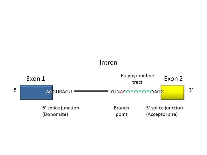

• In a sense, this is a loose consensus. DNA sequence can be analyzed to find introns in larger gene sequence.

Components of the spliceosome • Small RNAs, found only in the nucleus, play an essential role in splicing. These “small nuclear RNAs—sn. RNAs, interact with small nuclear proteins to form small nuclear ribonucleoprotiens (sn. RNPs or snurps). • The sn. RNAs are rich in U and have been named “U 1, U 2, U 3, U 4, U 5, and U 6”

• Different “U” sn. RNAs bind to different parts of the intron (by complementary base-pairing) and facilitate interactions of sn. RNAs and protein components of the spliceosome. The branchpoint A becomes joined to the donor site, forming a loop. • Since the loop forms with nucleotides in the middle of the intron the 3’ end of the intron extends like a tail.

• Eventually there is a cleavage of the 3’ acceptor site which simultaneously releases the loop of intron and joins the nucleotides on either side of the intron. • The released intron resembles a lariat. • STUDY this in detail!

Figure 13. 14

• Watch animation. • http: //www. dnalc. org/view/16938 -3 DAnimation-of-RNA-Splicing. html

• Interestingly, some introns, such as those found in r. RNA in eukaryotes undergo splicing without any protein enzymes. All the enzymatic activity comes from the RNA (intron) itself. • As such, these introns, known as Group 1 introns, undergo self-splicing.

Figure 14. 13

• Thomas Cech made this discovery using a protozoan called Tetrahymena. Self-splicing r. RNAs have been seen in other protozoans as well. • He was awarded a Nobel prize in 1989.

• Up until that point in time, no one had ever attributed catalytic/enzymatic activity to RNA. • Dogma said, “all enzymes are proteins”. • At this point, a new type of RNA-only enzyme was discovered. Catalytic RNA became known as ribozymes. • Now we know lots of examples of enzymatic activity of RNA.

• A similar process goes on in the m. RNA and t. RNA transcribed from DNA in mitochondria and chloroplasts! Amazing! The discover of catalytic RNA supports the notion that RNA was likely the first type of biomolecule in living cells. Why? Based on shapes that RNA can assume, is this reasonable?

• Recognizing RNA as a more versatile molecule, also helps us understand that the non-protein coding DNA in our cells is likely not really “junk DNA”. • Explain!

Alternative splicing • Some m. RNAs can be spliced in more than one way. Different exons are spliced together, creating different coding sequences for a protein.

Making more than one protein from a gene

http: //www. youtube. com/watch? v=p 2 u. C 5 j 0 h. K 0 A • Alternative splicing, again, partially explains how, what appears to be a rather low number of protein coding genes in the human genome, may encode many more proteins than we first estimated. Explain!

How important is splicing? • Mistakes in splicing have been linked to diseases. Usually we look for mutations only in exons---why? • How might a nucleotide change in an intron cause a protein to be altered?

Spinal Muscular Dystrophy • http: //www. dnalc. org/view/16941 -2 DAnimation-of-Alternative-RNA-Splicing. html

One final post-transcriptional alteration • RNA editing – In the late 1980 s, it was discovered that a few eukaryotic RNAs had slightly different nucleotide sequence than the DNA from which they were derived. – OH NO! Another exception to DOGMA!

• The changes seemed to be both: – Substitutions---changing one nucleotide to another. AND --Insertions or deletions of RNA nucleotides

• RNA editing was first studied in a protozoan, parasite, Trypanosoma, which causes a disease called African Sleeping Sickness. • Special RNAs called a “guide RNA”/g. RNAs seems to direct the editing by binding to complementary RNA and directing editing enzymes to insert short stretches of RNA.

• We will discuss and example of RNA editing in a mammalian gene/RNA for Apolipoprotein.
- Slides: 44