Mohammed ElKhateeb Chromosomal Disorders MGL4 March 11 th

Mohammed El-Khateeb Chromosomal Disorders MGL-4 March 11 th 2014 台大農藝系 遺傳學 601 20000 Chapter 1 slide 1

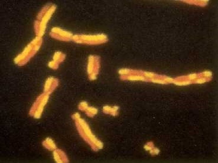
G-Banded Metaphase Chromosomes A D B C E F G
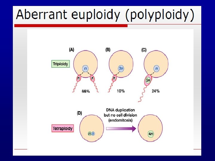
 Polyploidy")
Types of chromosome abnormalities n Numerical § § n Aneuploidy (monosomy, trisomy, tetrasomy) Polyploidy (triploidy, tetraploidy) Structural § § § § Translocations Inversions Insertions Deletions Rings Duplication Isochromosomes

Aneuploidy n n n Almost all been found in oocytes and early embryos, trisomies and monosomies Most lethal (miscarriage) Do not see in pregnancy or live born Exceptions sex chromosomes and Down Some aneuploidy is age related
 Tetraploidy (92, XXYY) Aneuploidy")
Numerical Abnormalities n Polyploidy § § n Triploidy (69, XXY) Tetraploidy (92, XXYY) Aneuploidy § § § Monosomy (45, X : Turner Syndrome) Trisomy (47, XY, +21 : Down Syndrome) Tetrasomy (48, XXXX)
 Diploid=2 n (in Normal somatic")
n n n Haploid = n (in normal gametes) Diploid=2 n (in Normal somatic cell) Euploid = An exact or multiple of n or of the monoploid number. A human with abnormal, but integral multiple of the monoploid number, (69 chromosomes) would also be considered as euploid e. g. ( 2 n, 3 n, 4 n etc)

POLYPLOID n n n Many organisms have more than two sets of homologous chromosomes and are called polyploid A chromosome number that is a multiple of haploid number of 23 other than the diploid number eg. 69 True polyploidy rarely occurs in humans, although it occurs in some tissues (especially in the liver).
")
TRIPLOIDY (all chromosomes threefold)

MEIOTIC NON DISJUNCTION n n May involve autosomes or sex chromosomes In females incidence increases with age 35 yrs or more. Meiosis I: Two members of homologous chromosomes fails to separate and both members of a pair move into one cell. Meiosis II: When sister chromatids fail to separate.

MITOTIC NONDISJUNCTION Mosaicism: n Some cells have abnormal chromosomal number and others have normal n Occurs in the earliest cell divisions n Affected individuals exhibit characteristics of a particular syndrome for e. g. down syndrome in 1% cases

Aneuploidy Caused by • Non-disjunction § Failure of homologous chromosomes to separate in anaphase I § Failure of sister chromatids to separate at meiosis II • Anaphase lag § Chromosomal loss via micronucleus formation caused by delayed movement of chromosome /chromatid during anaphase n results in daughter cell deficient of that chromosome or chromatid

Meiotic Nondisjunction Generates Aneuploidies abnormal gametes Zygotic Ploidy

Distribution of non-disjunction Meiosis II Mitosis Maternal 21, 15, 16 18 15, 18, 21, 8 Paternal - 18, 21

Genetic diversity Crossing over in MI 1. § swap pieces of DNA between maternal and paternal homologous chromosomes Independent assortment at end of MI 2. § paternally and maternally derived homologues assort randomly

Aneuploidy § Autosomal monosomy is rarely observed in spontaneously aborted fetuses or in live births. § Most autosomal trisomies are also lethal with one notable exception. Ø Trisomy 13 Ø Trisomy 18 Ø Trisomy 21 Patau syndrome Edward syndrome Down’s syndrome
 n")
Trisomy 21 (1 in 800 live births; incidence greater if mat. age >35) n n n Hypotonia Short neck with loose skin at nape Flat nasal bridge Brushfield spots around edge of iris Epicanthal folds Short, broad hands with single transverse palmar crease Congenital heart disease Mental retardation Increased risk for leukemia Alzheimer-like dementia Signs of hypothyroidism

Down Syndrome Wide “sandal” gap Simi

NON-DISJUNCTION FEATURES OF CHROMOSOME 21 n n n Occurs in all populations approximately at the same rate There is a significant loss of trisomy 21 pregnancy 1/150 The risk of having pregnancy affected with DS is increased with maternal age • <25 y/o 1/1600 • 30 to 34 y/o 1/800 • > 40 y/o 1/80

Changes in the Genome… n Trisomy: The zygote has three copies of a chromosome.
 Unbalanced translocation-4% b/w 21")
Recurrence Risks: Trisomy 21 Causes: Meiotic nondisjunction -95% (trisomy 21) Unbalanced translocation-4% b/w 21 and 13, 14, 15 Mosaicism due to mitotic non dysjunction-1% 46, i(21 q) recurrence risk is low (if not a carrier) t(14; 21) 15% if female carrier <1% if male carrier

MITOTIC NONDISJUNCTION Mosaicism: n Some cells have abnormal chromosomal number and others have normal n Occurs in the earliest cell divisions n Affected individuals exhibit characteristics of a particular syndrome for e. g. down syndrome in 1% cases

Chromosomal involvement and translocation in familial Down syndrome. The photograph shows the relevant chromosomes from a trisomy 21 offspring produced by a translocation carrier parent.
 §")
Trisomy 13, Patau syndrome Clinical Features (1 in 15 - 25, 000 births) § § § Microcephaly and mental retardation, scalp defect, microphthalmia, often blind Cleft lip/palate, polydactyly, rocker-botton feet, abnormal ears, apneic spells and myotonic seizures, cardiac dextroposition and VSD, extensive visceral defects CNS malformations presence of a single forebrain hemisphere or lobe Half of such individuals die within the first month-- the remainder by 1 year

§ Cleft palate, § Aseptal defect, inguinal hernia § Postaxial polydactyly of the left hand. Polydactyly, particularly of all extremities, strongly suggests trisomy 13.

Trisomy 13, Patau syndrome Karyotype Ø Ø Ø Trisomy 13 47, XX, +13 R. Translocation 46, XX, der(13; 14)+13) Mosaicism 47, XX, +13/46, XX Incidence >75% 20% 5%
 • • Prominent")
Clinical Features: Trisomy 18 Clinical feature (1 in 7, 500 births) • • Prominent occiput Short palpebral fissures Micrognathia Low-set, malformed ears Profound Mental Retardation Rocker-Bottom Feet Short Sternum Small Pelvis n n n Clenched Hands Renal anomalies Cleft Lip/Palate Hypoplastic thumbs Dorsiflexed halus (hammer toe) Renal Anomalies
, +18 Survival – 50%")
Trisomy 18 – Edwards syndrome Karyotype: 47, XX (or XY), +18 Survival – 50% within 1 month, 50% within less than 1 year
 Karyotype n n n Trisomy 18 Translocation Mosaicism 47, XX,")
Edwards Syndrome (Trisomy 18) Karyotype n n n Trisomy 18 Translocation Mosaicism 47, XX, +18 46, XX, +18, t(? ; 18 q) 47, XX, +18/46, XX Incidence 90% rare 10%

Trisomy 8, Mosaicism § 1/20, 000 births § Long face, high prominent forehead, wide upturned nose, thick everted lower lip, microretrognathia, low-set ears, cleft palate § Osteoarticular anomalies common § Moderate MR

MONOSOMIES n n One representative of a chromosome is present Nondisjunction: n One cell ends up with one copy (monosomic) and the other has 3 (trisomic) Anaphase lag: n chromosome fails to move into the new daughter cell All complete autosomal monosomies- lethal n Can survive in mosaic forms

Sex Chromosomes Abnormalities

Sex Chromosome Aneuploidy n n n Sex Chromosome Aneuploidy is more common than Autosome Aneuploidy! In order to survive and develop embryo needs at least one X chromosome. In healthy females, one X chromosome will be inactivated in each somatic cell. The inactivated X chromosome is referred to as a “Barr body”. An individual who has an intact Ychromosome will be male, regardless of the number of X chromosomes he possesses. In the absence of an intact Y chromosome an individual will be female.

X-Chromosomal Disorders n n n Imbalances of X-chromosomes are better tolerated than those of autosomes Increased number of X-chromosomes in either males or females lead to mental retardation Lyonization – Mary Lyon during 16 th day of embryonic life one Xchromosome in females is randomly inactivated n inactivation persists in all subsequent cells n

A woman with the chromosome constitution 47, XXX should have 2 Barr bodies in each cell. n XXY individuals are male, but have a Barr body. n XO individuals are female but have no Barr bodies. n

Nondisjunction of X chromosome

Sex Chromosome Aneuploidy Situation Normal Female Nondisjunction Oocyte Sperm Consequence X Y 46, XY normal male X X 46, XX normal female XX Y 47, XXY Klinefelter syndrome XX X 47, XXX triplo-X Y 45, Y nonviable X 45, X Turner syndrome Male Nondisjunction (meiosis I) X X XX 47, XXX triplo-X Male nondisjunction (meiosis II) X YY 47, XYY Jacobs syndrome X 45, X Turner syndrome

Turner syndrome Monosomy X or X 0 § § § 1 in every 5000 births varied degree of effects webbed neck short stature sterile KARYOTYPING: • Classical 45 X • 46, X, i(Xq) • 46 XXq • 46 XXp • 46, X r(X) • 45, X/46, XX

Turner Syndrome n n n Results from complete or partial monosomy of the X-chromosome in females Most common sex chromosome abnormality in females, incidence 1 in 1000 live births Classical cytogenetics 45, X (57%) n Structural abnormalities of X-chromosomes (14%) n Mosaics (29%) n

Mosaicism in Turner Syndrome n n 99% of conceptuses with 45, X are nonviable FISH and PCR studies show much higher incidence of mosaics than conventional studies Some authorities believe that there are no truly nonmosiac Turner syndrome patients Patients with a high proportion of 45, X cells are more severely affected
 n Deletion")
TURNER SYNDROME KARYOTYPING: n Classical 45 X n Isochromosome 46, X, i(Xq) n Deletion 46 XXqn Deletion 46 XXpn Ring 46, X r(X) n Mosaic e. g. 45, X/46, XX

47, XXY - Klinefelter syndrome Clinical Features • Tall, thin, long legs, hypogonadism, underdeveloped secondary sex characteristics, gynecomastiaexcessive development of the male mammary glands. usually infertile • Verbal comprehension and ability slightly lower than average • Increased risk of learning difficulties, esp. in reading KARYTYPING: Ø 60% are of maternal nondisjunction and maternal age increases the possibilities Ø 47 XXY Ø Mosaics XY / XXY Ø XXXY, XXYY, XXXXY. Severely mental retarded 1/1000 male births

Klinefelter Syndrome n n n A male hypogonadism that occurs when there are two or more X-chromosomes and one or more Y-chromosomes Incidence is 1 in 500 male births Usually (82% of cases) 47, XXY n n maternal (60%) or paternal (40%) nondisjunction during meiotic divisions 15% are mosaics, usually 46, XY/47, XXY

Clinical Features n Testicular abnormality does not develop before puberty seminiferous tubules are atrophic resulting in reduced spermatogenesis, infertility, small firm testes, and increased FSH n testosterone levels are reduced n impotence and increased LH n lack of secondary male sexual characteristics n n n Mental retardation is unusual but IQ may be below normal Mosaics are less severely affected

Clinical Features 47, XYY Not obviously abnormal No marked physical or behavioral phenotype Tall, fertile, may have severe acne during adolescence Incidence 1/1000 Increased risk of educational or behavioral problems IQ scores about 10 pts below average n n n Attention deficits Hyperactivity Impulsiveness

47, XYY X Y Origin of the error that leads to XYY karyotype must be paternal nondisjunction at meiosis II producing YY sperm. Fertilization of this sperm by an egg will lead to Turner syndrome YY

Sex Chromosome Tetrasomy Males: 48, XXXY Clinical Features § Reduction in intellectual functioning – IQ § § between 20 -80 Coarse facial appearance, gonadal hypoplasia common As a rule: additional X chromosomes cause a more abnormal phenotype-more defective sexual development and mental impairment.

Sex Chromosome Tetrasomy Males: 48, XXXY Clinical Features n n n Reduction in intellectual functioning – IQ between 20 -80 Coarse facial appearance, gonadal hypoplasia common As a rule: additional X chromosomes cause a more abnormal phenotype-more defective sexual development and mental impairment 1/6/2022 MSK/UJ/GL 2 48

Sex Chromosome Pentasomy 49, XYYYY Clinical Features • Tall stature • Aberrant behavior (impulsivity, low frustration tolerance) • Low-normal or subnormal intelligence • Developmental delay • Testicular abnormalities • Some craniofacial dysmorphisms (more common for 49, XYYYY) 49
, Y (red), 18 (blue),")
48, XXXY: X (green), Y (red), 18 (blue),

INSTABILITY OF CHROMOSOMES

Chromosome breaks n Once chromosome broken by some means n Unstable situation as telomeres not at end n Usually join up to other piece

Defects in DNA Repair or Replication All are associated with a high frequency of chromosome and gene (base pair) mutations; most are also associated with a predisposition to cancer, particularly leukemias n Xeroderma pigmentosum § § § caused by mutations in genes involved in nucleotide excision repair associated with a >1000 -fold increase of sunlightinduced skin cancer and with other types of cancer such as melanoma

Main Features Ataxia Telangiectasia • Cerebellar ataxia • Immune defects • Telangiectases of the • Ataxia telangiectasia caused by gene that detects DNA damage increased risk of X-ray associated with increased breast cancer in carriers • • • conjunctivae Predisposition to tumors (lymphoma, leukemia) Extreme radiation sensitivity Autosomal recessive Several gene loci
 § § § § Extreme intrauterine and postnatal")
Main features of Bloom syndrome (BS) § § § § Extreme intrauterine and postnatal growth retardation Chromosomal instability Predisposition to leukemias, lymphomas, and other tumors Immune defects Sunlight-induced erythema of the face Hypo- and hyper-pigmented skin areas Autosomal recessive Gene locus on chromosome 15
 • • caused by a gene involved in")
Main features of Fanconi Anemia (FA) • • caused by a gene involved in DNA repair increased risk of X-ray and sensitivity to sunlight • • • Growth retardation Skeletal defects (e. g. , radius and thumb) Bone marrow failure Skeletal and kidney malformation Localized pigment changes Autosomal recessive Several gene loci.

Defects in DNA Repair or Replication n Cockayne syndrome § § n caused by a defect in transcription-linked DNA repair sensitivity to sunlight Werner’s syndrome § § caused by mutations in a DNA helicase gene premature aging

Structural Chromosomal Abnormalities
 Polyploidy")
Types of chromosome abnormalities n Numerical § § n Aneuploidy (monosomy, trisomy, tetrasomy) Polyploidy (triploidy, tetraploidy) Structural § § § § Translocations Inversions Insertions Deletions Rings Duplication Isochromosomes

Chromosome Abnormalities Numerical Autosomal Disorders number of affected individuals AGE OF ONSET OF GENETIC DISORDERS chromosomal multifactorial single-gene (Mendelian) birth puberty adult

The Karyotype: an international description Total number of chromosomes, Sex chromosome constitution, 46, XY 47, XX, +21 47, XXX 69, XXY Anomalies/variants. Trisomy 21 (Down syndrome) Triple X syndrome Triploidy 45, XX, der(13; 14)(p 11; q 11) 46, XY, t(2; 4)(p 12; q 12) Robertsonian translocation Reciprocal translocation 46, XX, del(5)(p 25) 46, XX, dup(2)(p 13 p 22) 46, XY, inv(11)(p 15 q 14) 46, XY, fra(X)(q 27. 3) 46, XY/47, XXY Deletion tip of chromosome 5 Duplication of part of short arm Chr 2 Pericentric inversion chromosome 11 Fragile X syndrome Mosaicism normal/Klinefelter syndrome

Prenatal diagnosis cordocentesis amniocentesis Preimplantation genetic diagnosis Chorion villi sampling

Spontaneous Abortion Products 15% of first trimester pregnancies are lost , X +16 45 Other autosomal trisomy § 50% abnormal dy i o pl Tri r Othe 46, N § 50% normal

Chromosomal Findings in Early Miscarriages 40% apparently normal 60% abnormal: • Trisomy (47 chromosomes – one extra) 30% • 45, X (45 chromosomes – one missing) 10% • Triploidy (69 chromosomes – three sets) 10% • Tetraploidy (92 chromosomes – four sets) 5% • Other chromosome anomalies (e. g. structural anomalies) 5%
")
Most frequent numerical anomalies in liveborn Autosomes Down syndrome (trisomy 21: 47, XX, +21) Edwards syndrome (trisomy 18: 47, XX, +18) Patau syndrome (trisomy 13: 47, XX+13) Sex chromosomes Turner syndrome 45, X Klinefelter syndrome 47, XXY All chromosomes Triploidy (69 chromosomes)

Chromosome abnormalities and maternal age

Chromosome abnormalities seen in adults referred for: n Infertility 2. 5% n mostly sex chromosome aneuploidy rearrangements involving sex chromosomes Recurrent miscarriage 6% balanced chromosome rearrangements e. g. translocations and inversions However, up to 50% of first trimester loss is due to foetal chromosome abnormality – mostly de novo
- Slides: 68