
Mohammed ElKhateeb Chromosomal Disorders MGL 5 June 29
Mohammed El-Khateeb Chromosomal Disorders MGL - 5 June 29 th 2014 台大農藝系 遺傳學 601 20000 Chapter 1 slide 1
Human chromosome disorders On rare occasions, a chromosome’s structure changes; such changes are usually harmful or lethal, rarely neutral or beneficial • High frequency in humans § most embryos are spontaneously aborted § alterations are too disastrous § developmental problems result from biochemical imbalance • imbalance in regulatory molecules? • Certain conditions are tolerated § upset the balance less = survivable § characteristic set of symptoms = syndrome
Important Issues Pertinent To Structural Rearrangements § ARE THE INDIVIDUAL'S CHILDREN AT RISK? § A balanced rearrangement that does not cause a genetic disorder in the individual can still pose a risk for the individual's offspring § The chromosomes cannot line up evenly during meiosis § This may result in the egg or sperm having an unbalanced genetic complement, such as: • missing material, • extra material, • often a combination of both
Important Issues Pertinent To Structural Rearrangements IS THE REARRANGEMENT BALANCED OR UNBALANCED? Balanced = § § No DNA was lost when the chromosomes broke The individual has all his/her genes Rarely causes a genetic disorder Will only cause a genetic disorder if one of the breakpoints interrupts a gene - only 2 -4% of your DNA is protein-coding sequence Unbalanced = § DNA was lost when the chromosomes broke § The individual is missing one or more of his/her genes § Often causes a genetic disorder § Severity of effect is often proportional to the amount of DNA/genes lost
Types of chromosome abnormalities • Numerical § Aneuploidy (monosomy, trisomy, tetrasomy) § Polyploidy (triploidy, tetraploidy) • Structural § § § § Translocations Inversions Insertions Deletions Rings Duplication Isochromosomes
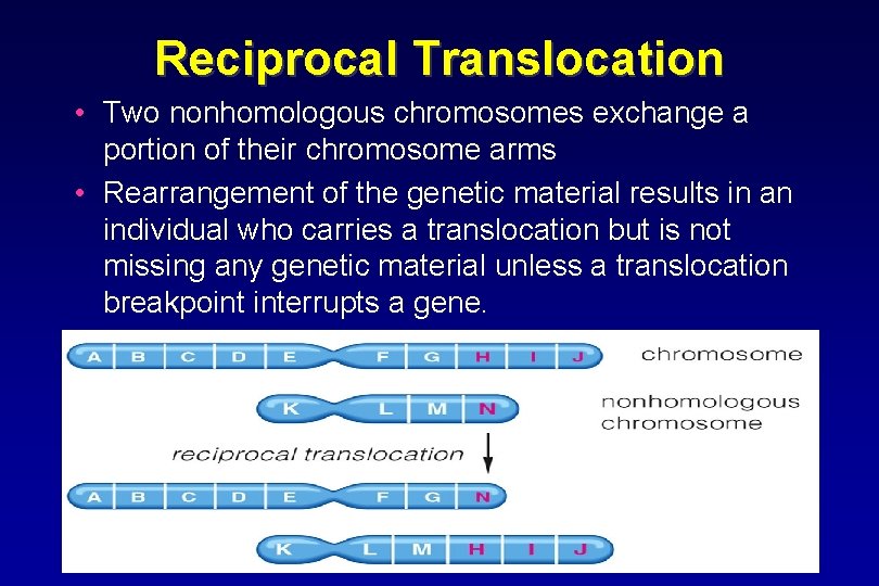
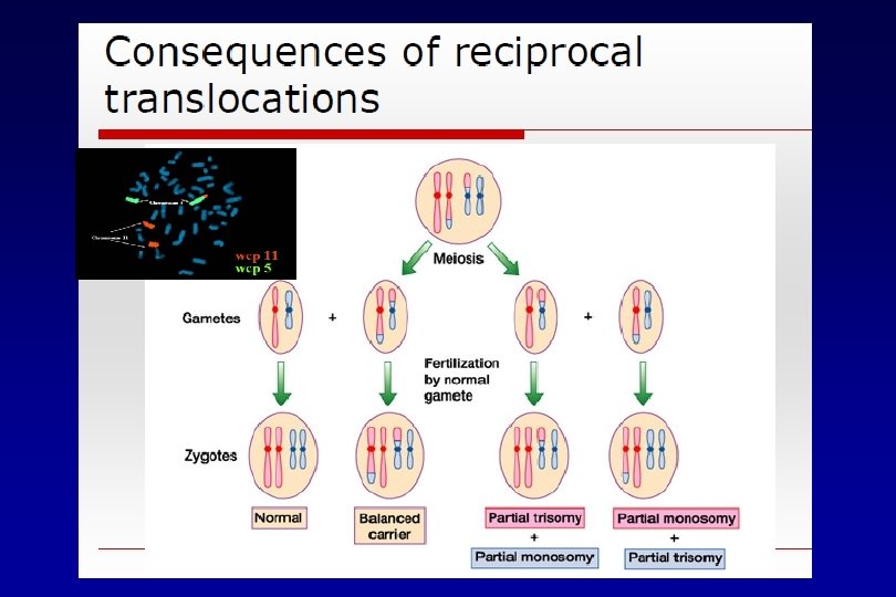
Reciprocal Translocation • Two nonhomologous chromosomes exchange a portion of their chromosome arms • Rearrangement of the genetic material results in an individual who carries a translocation but is not missing any genetic material unless a translocation breakpoint interrupts a gene.
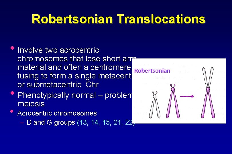
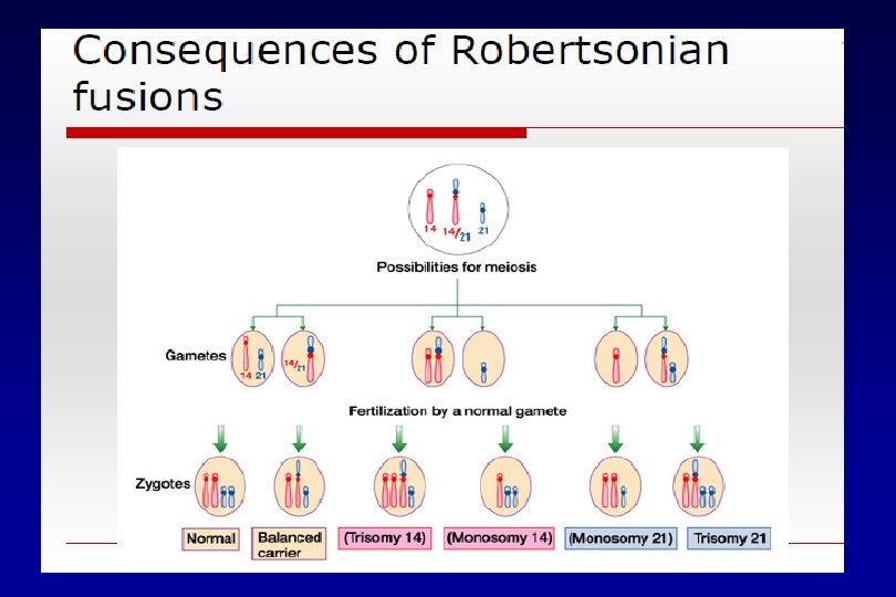
Robertsonian Translocations • Involve two acrocentric • chromosomes that lose short arm material and often a centromere, fusing to form a single metacentric or submetacentric Chr Phenotypically normal – problems at meiosis • Acrocentric chromosomes – D and G groups (13, 14, 15, 21, 22)
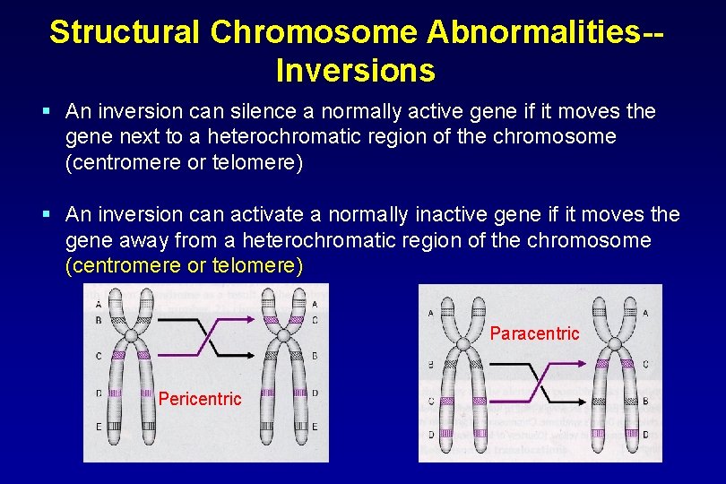
Structural Chromosome Abnormalities-Inversions § An inversion can silence a normally active gene if it moves the gene next to a heterochromatic region of the chromosome (centromere or telomere) § An inversion can activate a normally inactive gene if it moves the gene away from a heterochromatic region of the chromosome (centromere or telomere) Paracentric Pericentric
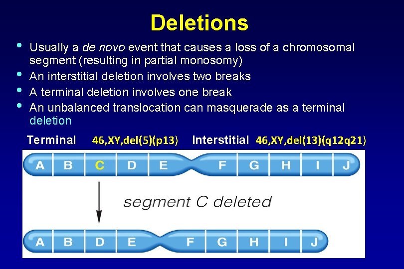
• • Deletions Usually a de novo event that causes a loss of a chromosomal segment (resulting in partial monosomy) An interstitial deletion involves two breaks A terminal deletion involves one break An unbalanced translocation can masquerade as a terminal deletion Terminal 46, XY, del(5)(p 13) Interstitial 46, XY, del(13)(q 12 q 21)
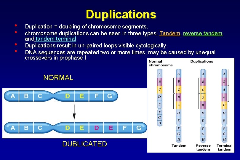
• • Duplications Duplication = doubling of chromosome segments. chromosome duplications can be seen in three types; Tandem, reverse tandem, and tandem terminal Duplications result in un-paired loops visible cytologically. DNA sequences are repeated two or more times; may be caused by unequal crossovers in prophase I NORMAL DUBLICATED
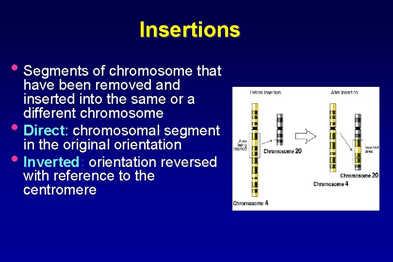
Insertions • Segments of chromosome that • • have been removed and inserted into the same or a different chromosome Direct: chromosomal segment in the original orientation Inverted: orientation reversed with reference to the centromere
Duplications And Deletions Affect The Phenotype § If a duplication produces one or more extra copies of a gene, the ratio of that gene’s protein to the proteins it interacts with is altered § A deletion can delete the dominant allele of a gene, allowing the remaining recessive allele to control the phenotype - pseudodominance § The phenotypic consequences of a deletion depend on whether the gene(s) in the deletion make their protein(s) in overabundance, or in just enough quantity to fill the body’s needs § If the protein is made in just enough quantity, the deletion will affect the phenotype - haploinsufficiency
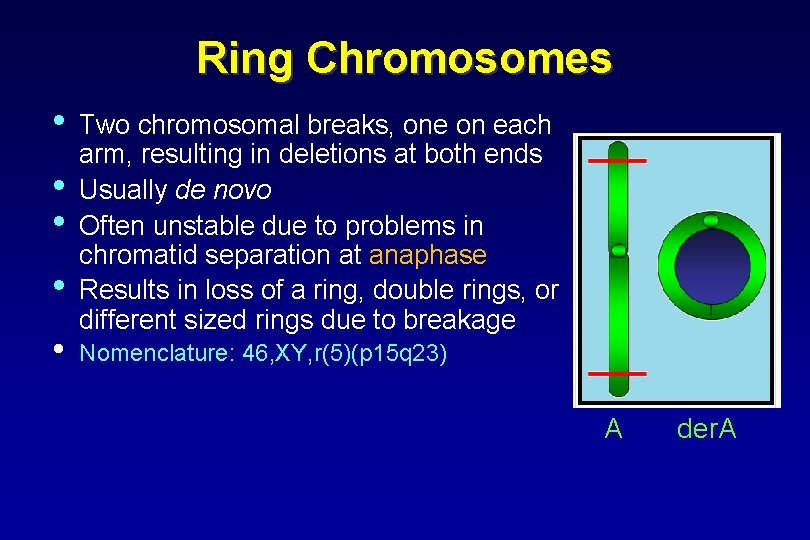
Ring Chromosomes • • • Two chromosomal breaks, one on each arm, resulting in deletions at both ends Usually de novo Often unstable due to problems in chromatid separation at anaphase Results in loss of a ring, double rings, or different sized rings due to breakage Nomenclature: 46, XY, r(5)(p 15 q 23) A der. A
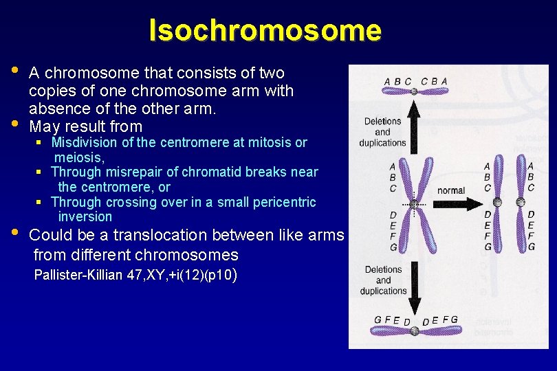
Isochromosome • • • A chromosome that consists of two copies of one chromosome arm with absence of the other arm. May result from § Misdivision of the centromere at mitosis or meiosis, § Through misrepair of chromatid breaks near the centromere, or § Through crossing over in a small pericentric inversion Could be a translocation between like arms from different chromosomes Pallister-Killian 47, XY, +i(12)(p 10)
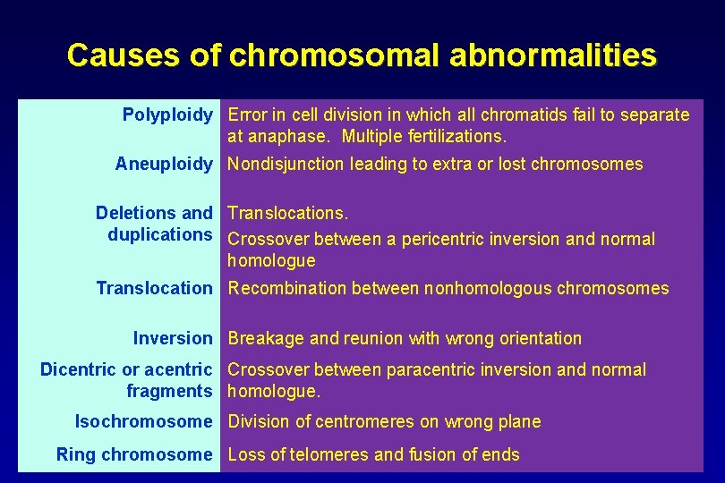
Causes of chromosomal abnormalities Polyploidy Error in cell division in which all chromatids fail to separate at anaphase. Multiple fertilizations. Aneuploidy Nondisjunction leading to extra or lost chromosomes Deletions and Translocations. duplications Crossover between a pericentric inversion and normal homologue Translocation Recombination between nonhomologous chromosomes Inversion Breakage and reunion with wrong orientation Dicentric or acentric Crossover between paracentric inversion and normal fragments homologue. Isochromosome Division of centromeres on wrong plane Ring chromosome Loss of telomeres and fusion of ends
CHROMOSOMAL DELETIONS Large Dilitions Micro Dilitions
Large Deletions • Cri du Chat (Cat-cry) Syndrome • Wolf-Hirschorn Syndrome • Di. George Syndrome (DGS)
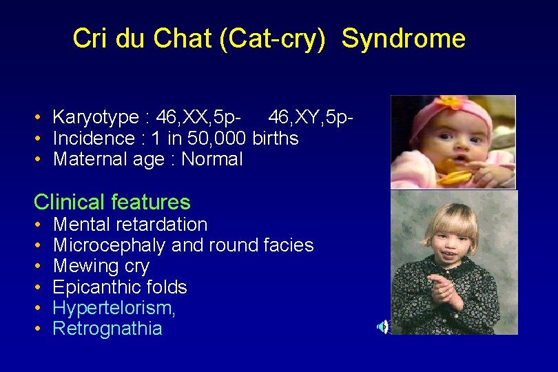
Cri du Chat (Cat-cry) Syndrome • Karyotype : 46, XX, 5 p- 46, XY, 5 p • Incidence : 1 in 50, 000 births • Maternal age : Normal Clinical features • • • Mental retardation Microcephaly and round facies Mewing cry Epicanthic folds Hypertelorism, Retrognathia
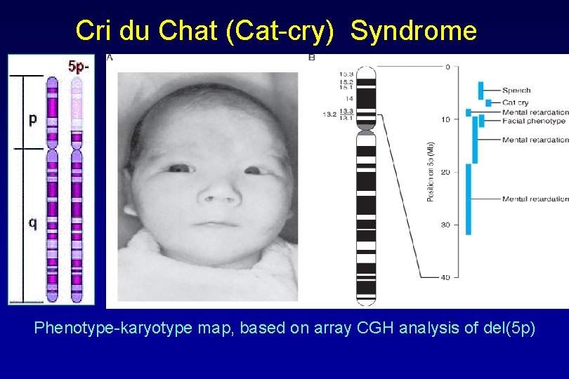
Cri du Chat (Cat-cry) Syndrome Phenotype-karyotype map, based on array CGH analysis of del(5 p)
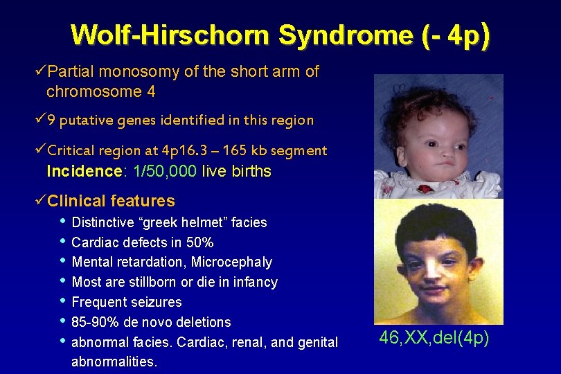
Wolf-Hirschorn Syndrome (- 4 p) üPartial monosomy of the short arm of chromosome 4 ü 9 putative genes identified in this region üCritical region at 4 p 16. 3 – 165 kb segment Incidence: 1/50, 000 live births üClinical features: • Distinctive “greek helmet” facies • Cardiac defects in 50% • Mental retardation, Microcephaly • Most are stillborn or die in infancy • Frequent seizures • 85 -90% de novo deletions • abnormal facies. Cardiac, renal, and genital abnormalities. 46, XX, del(4 p)
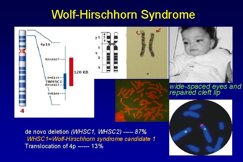
Wolf-Hirschhorn Syndrome wide-spaced eyes and repaired cleft lip de novo deletion (WHSC 1, WHSC 2) ----- 87% WHSC 1=Wolf-Hirschhorn syndrome candidate 1 Translocation of 4 p ------ 13%
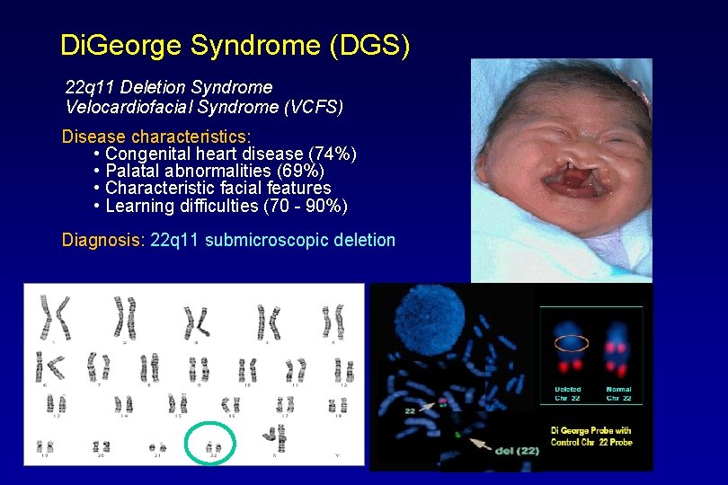
Di. George Syndrome (DGS) 22 q 11 Deletion Syndrome Velocardiofacial Syndrome (VCFS) Disease characteristics: • Congenital heart disease (74%) • Palatal abnormalities (69%) • Characteristic facial features • Learning difficulties (70 - 90%) Diagnosis: 22 q 11 submicroscopic deletion
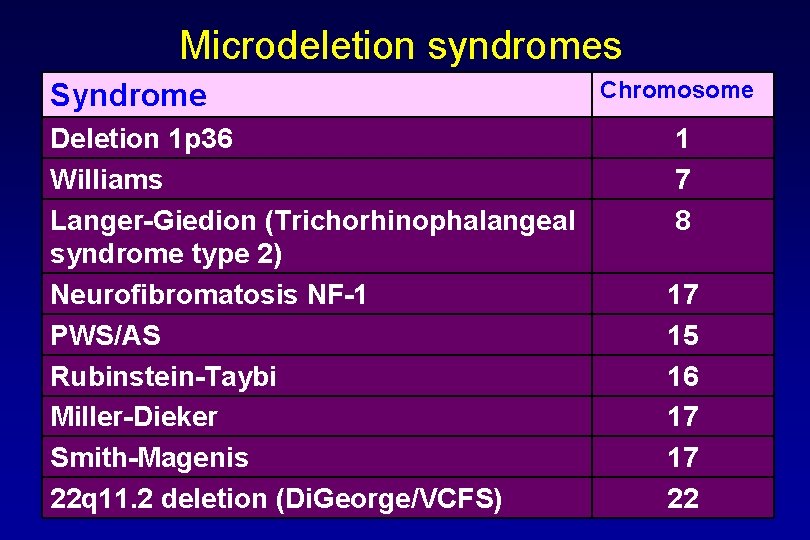
Microdeletion syndromes Syndrome Deletion 1 p 36 Williams Langer-Giedion (Trichorhinophalangeal syndrome type 2) Neurofibromatosis NF-1 PWS/AS Rubinstein-Taybi Miller-Dieker Smith-Magenis 22 q 11. 2 deletion (Di. George/VCFS) Chromosome 1 7 8 17 15 16 17 17 22
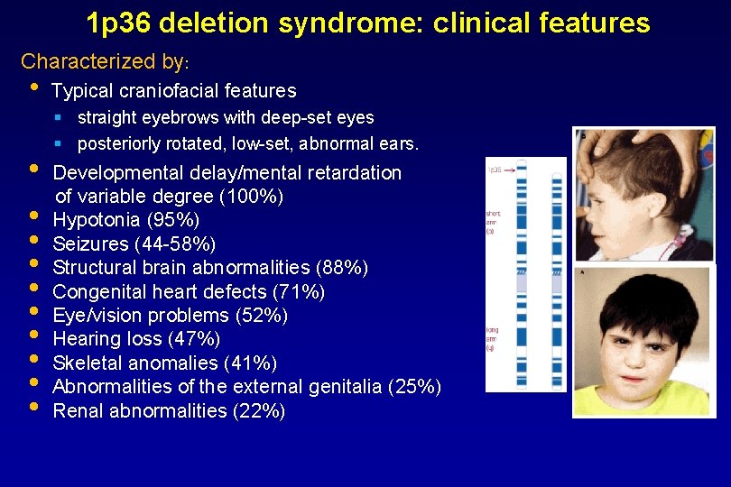
1 p 36 deletion syndrome: clinical features Characterized by: • • • Typical craniofacial features § straight eyebrows with deep-set eyes § posteriorly rotated, low-set, abnormal ears. Developmental delay/mental retardation of variable degree (100%) Hypotonia (95%) Seizures (44 -58%) Structural brain abnormalities (88%) Congenital heart defects (71%) Eye/vision problems (52%) Hearing loss (47%) Skeletal anomalies (41%) Abnormalities of the external genitalia (25%) Renal abnormalities (22%)
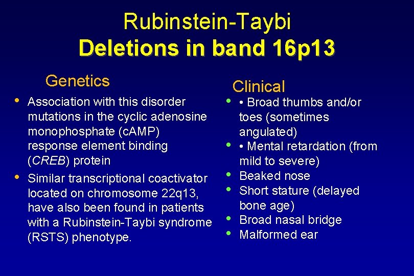
Rubinstein-Taybi Deletions in band 16 p 13 Genetics • • Association with this disorder mutations in the cyclic adenosine monophosphate (c. AMP) response element binding (CREB) protein Similar transcriptional coactivator located on chromosome 22 q 13, have also been found in patients with a Rubinstein-Taybi syndrome (RSTS) phenotype. • • • Clinical • Broad thumbs and/or toes (sometimes angulated) • Mental retardation (from mild to severe) Beaked nose Short stature (delayed bone age) Broad nasal bridge Malformed ear
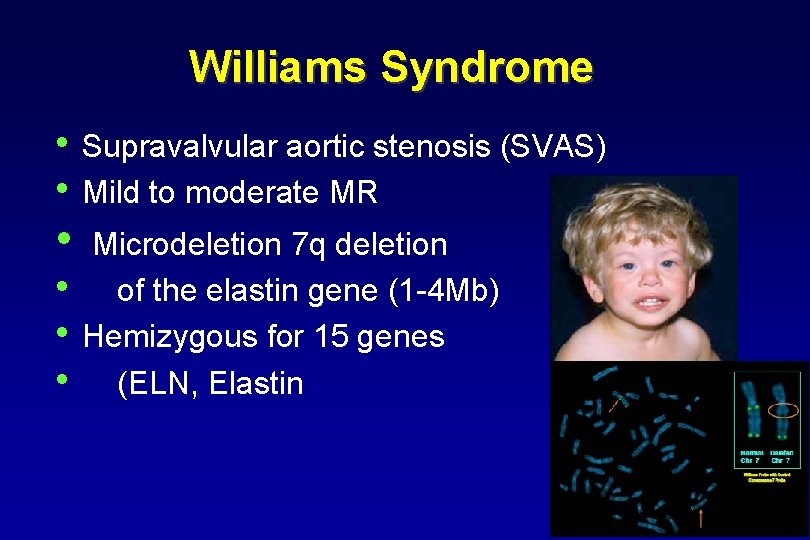
Williams Syndrome • Supravalvular aortic stenosis (SVAS) • Mild to moderate MR • • Microdeletion 7 q deletion of the elastin gene (1 -4 Mb) Hemizygous for 15 genes (ELN, Elastin
Langer-Giedion syndrome § intellectual deficit § redundant skin § multiple cartilaginous exostoses - affects mainly the extremities of the long bones § characteristic facies § cone-shaped phalangeal epiphyses. § Growth retardation, microcephaly, hypotonia and hearing problems have also been reported. § Prevalence is unknown § • Caused by a microdeletion in chromosome 8 q 23. 3 -q 24. 13 leading to the loss of at least two genes: TRPS 1 and EXT 1: encodes an endoplasmic reticulumresident type II transmembrane glycosyltransferase involved in the chain elongation step of heparan sulphate biosynthesis mutations in this gene cause the type I form of multiple exostoses TRPS 1: transcription factor that represses GATA-regulated genes plays a role in regulating growth of bone and cartilage loss of functional TRPS 1 protein contributes to short stature, cone-shaped ends of the long bones (epiphyses), and distinctive facial features in people with Langer. Giedion syndrome
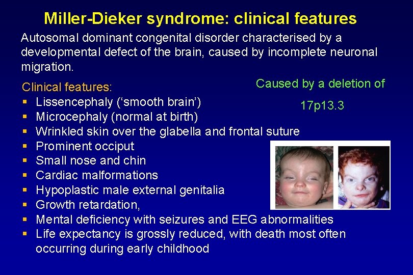
Miller-Dieker syndrome: clinical features Autosomal dominant congenital disorder characterised by a developmental defect of the brain, caused by incomplete neuronal migration. Caused by a deletion of Clinical features: § § § § § Lissencephaly (‘smooth brain’) 17 p 13. 3 Microcephaly (normal at birth) Wrinkled skin over the glabella and frontal suture Prominent occiput Small nose and chin Cardiac malformations Hypoplastic male external genitalia Growth retardation, Mental deficiency with seizures and EEG abnormalities Life expectancy is grossly reduced, with death most often occurring during early childhood
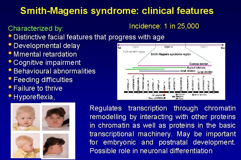
Smith-Magenis syndrome: clinical features Incidence: 1 in 25, 000 Characterized by: • Distinctive facial features that progress with age • Developmental delay • Mmental retardation • Cognitive impairment • Behavioural abnormalities • Feeding difficulties • Failure to thrive • Hyporeflexia, Regulates transcription through chromatin remodelling by interacting with other proteins in chromatin as well as proteins in the basic transcriptional machinery. May be important for embryonic and postnatal development. Possible role in neuronal differentiation
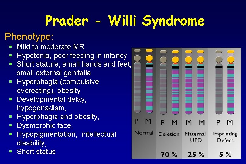
Prader - Willi Syndrome Phenotype: § Mild to moderate MR § Hypotonia, poor feeding in infancy § Short stature, small hands and feet, small external genitalia § Hyperphagia (compulsive overeating), obesity § Developmental delay, hypogonadism, § Hyperphagia and obesity, § Dysmorphic face, § Hypopigmentation, intellectual disability, § Short status
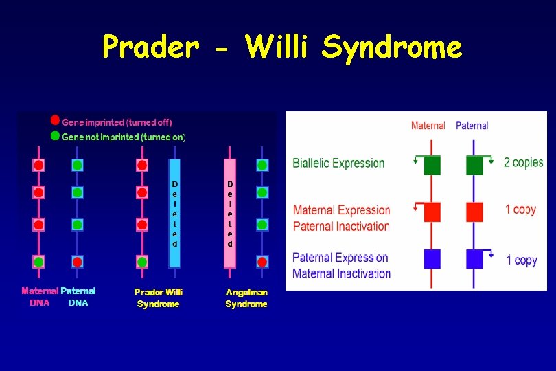
Prader - Willi Syndrome
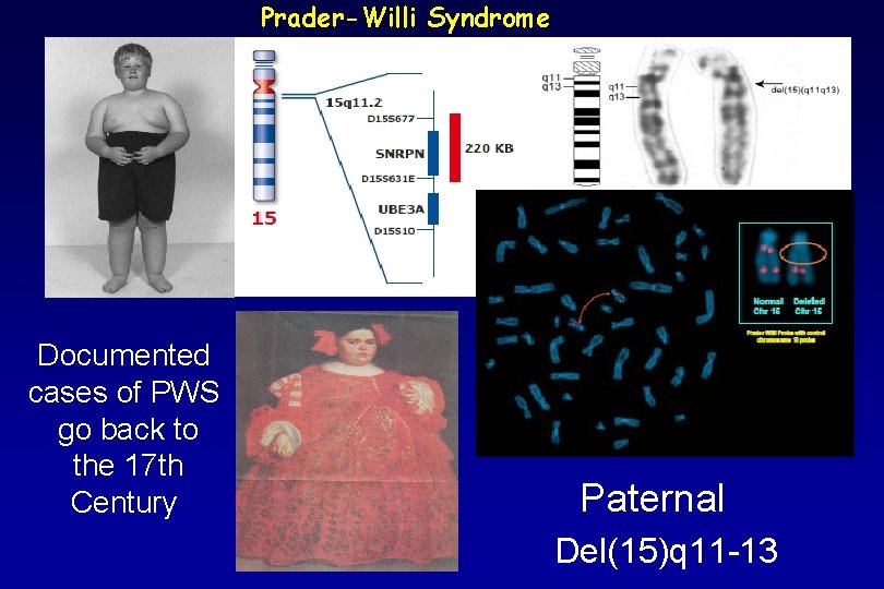
Prader-Willi Syndrome Documented cases of PWS go back to the 17 th Century Paternal Del(15)q 11 -13
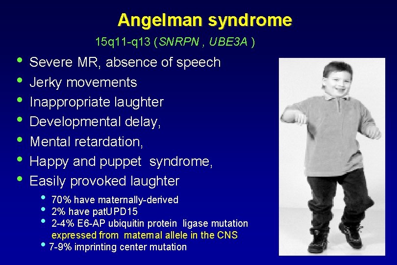
Angelman syndrome 15 q 11 -q 13 (SNRPN , UBE 3 A ) • • Severe MR, absence of speech Jerky movements Inappropriate laughter Developmental delay, Mental retardation, Happy and puppet syndrome, Easily provoked laughter • 70% have maternally-derived • 2% have pat. UPD 15 • 2 -4% E 6 -AP ubiquitin protein ligase mutation expressed from maternal allele in the CNS • 7 -9% imprinting center mutation
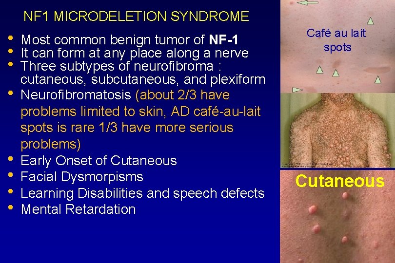
NF 1 MICRODELETION SYNDROME • • Most common benign tumor of NF-1 It can form at any place along a nerve Three subtypes of neurofibroma : cutaneous, subcutaneous, and plexiform Neurofibromatosis (about 2/3 have problems limited to skin, AD café-au-lait spots is rare 1/3 have more serious problems) Early Onset of Cutaneous Facial Dysmorpisms Learning Disabilities and speech defects Mental Retardation Café au lait spots Cutaneous
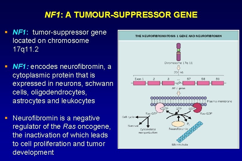
NF 1: A TUMOUR-SUPPRESSOR GENE § NF 1: tumor-suppressor gene located on chromosome 17 q 11. 2 § NF 1: encodes neurofibromin, a cytoplasmic protein that is expressed in neurons, schwann cells, oligodendrocytes, astrocytes and leukocytes § Neurofibromin is a negative regulator of the Ras oncogene, the inactivation of which leads to cell proliferation and tumor development
Neurofibromatosis 2 • Autosomal dominant, with 95% penetrance and linkage to 22 q 11 -q 13. • The gene has been isolated, and encodes a protein named merlin
DUPLICATION SYNDROMES • Beckwith-Wiedemann § Duplication - 11 p 15 (Paternal) • Duplication 17 p 11. 2 p 12 • Cat-Eye Syndrome § Duplication of 22 q • Velo-cardio-facial syndrome – features (VCF) Duplication – 22 q 11. 21 -q 11. 22 • PWS/AS Duplication – 15 q 11 -q 13
Marker Chromosomes • • • Chromosomes of unidentifiable origin (except now chromosomal origin can be identified using SKY, although specific bands cannot yet be identified) Occasionally occur as supernumerary chromosomes with or without phenotypic effect Parental chromosomes should be analyzed
OTHER ABNORMALITIES § Chromosome breaks • Once chromosome broken by some means • Unstable situation as telomeres not at end • Usually join up to other piece § Dicentric Chromosomes • Chromosomes with two centromeres § Double minutes • A minute is an acentric fragment smaller than the width of a chromatid. • Double minutes (dmin) are seen in tumor cells as double dots.
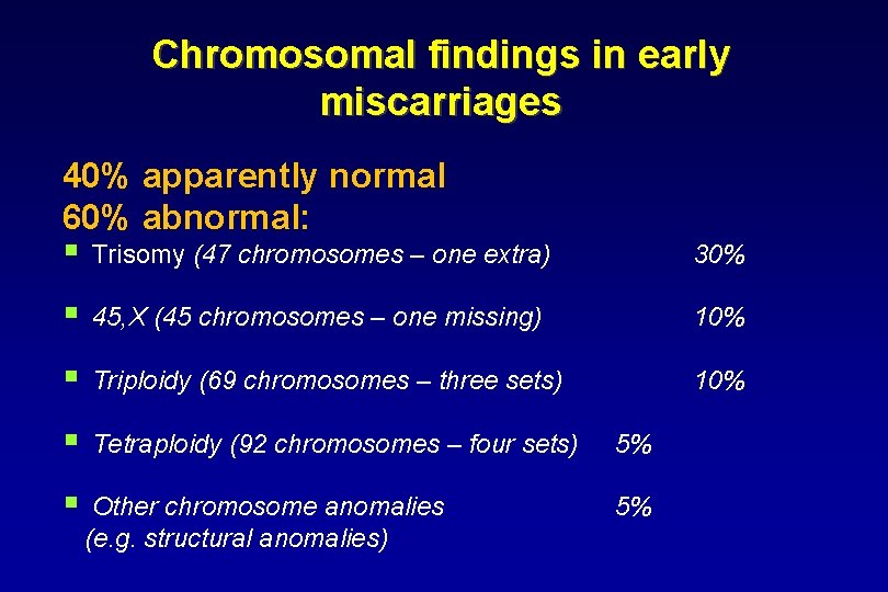
Chromosomal findings in early miscarriages 40% apparently normal 60% abnormal: § Trisomy (47 chromosomes – one extra) 30% § 45, X (45 chromosomes – one missing) 10% § Triploidy (69 chromosomes – three sets) 10% § Tetraploidy (92 chromosomes – four sets) 5% § Other chromosome anomalies (e. g. structural anomalies) 5%
Indications for postnatal chromosomal analysis • Suspicion to concrete chromosomal abnormality • • (concrete syndrome) Multiple congenital anomalies or developmental delay Mental retardation Gonadal dysgenesis Infertility Miscarriages Delivery of dead fetus or death of a newborn child Occurrence of certain malignancies
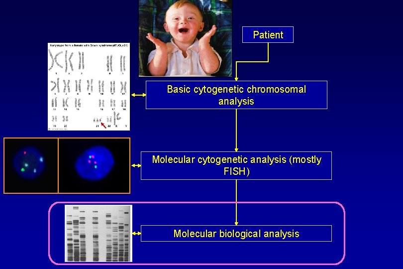
Patient Basic cytogenetic chromosomal analysis Molecular cytogenetic analysis (mostly FISH) Molecular biological analysis
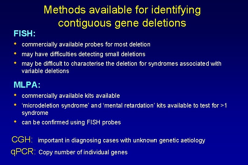
Methods available for identifying contiguous gene deletions FISH: • commercially available probes for most deletion • may have difficulties detecting small deletions • may be difficult to characterise the deletion for syndromes associated with variable deletions MLPA: • commercially available kits available • ‘microdeletion syndrome’ and ‘mental retardation’ kits available to test for >1 syndrome • can be confirmed using FISH probes CGH: important in diagnosing cases with unknown genetic aetiology q. PCR: Copy number of individual genes
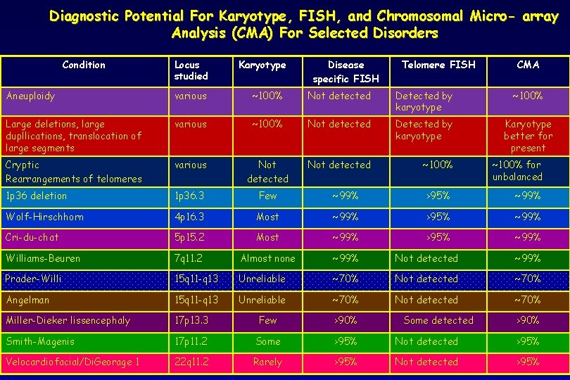
Diagnostic Potential For Karyotype, FISH, and Chromosomal Micro- array Analysis (CMA) For Selected Disorders Condition Locus studied Karyotype Disease specific FISH Telomere FISH CMA Aneuploidy various ~100% Not detected Detected by karyotype ~100% Large deletions, large dupllications, translocation of large segments various ~100% Not detected Detected by karyotype Karyotype better for present Cryptic Rearrangements of telomeres various Not detected ~100% 1 p 36 deletion 1 p 36. 3 Few ~99% >95% ~99% Wolf-Hirschhorn 4 p 16. 3 Most ~99% >95% ~99% Cri-du-chat 5 p 15. 2 Most ~99% >95% ~99% Williams-Beuren 7 q 11. 2 Almost none ~99% Not detected ~99% Prader-Willi 15 q 11 -q 13 Unreliable ~70% Not detected ~70% Angelman 15 q 11 -q 13 Unreliable ~70% Not detected ~70% Miller-Dieker lissencephaly 17 p 13. 3 Few >90% Smith-Magenis 17 p 11. 2 Some >95% Not detected >95% Velocardiofacial/Di. Georage 1 22 q 11. 2 Rarely >95% Not detected >95% Some detected ~100% for unbalanced >90%
- Slides: 46