
Module 3 Clinical Manifestations Introduction Intraoral cancers occur

- Slides: 37
Module 3 Clinical Manifestations
Introduction § Intraoral cancers occur most frequently on the: Tongue Floor of the mouth Soft palate and Oropharynx § Squamous cell carcinoma is the most common type, accounting for 90% of oral cancers.
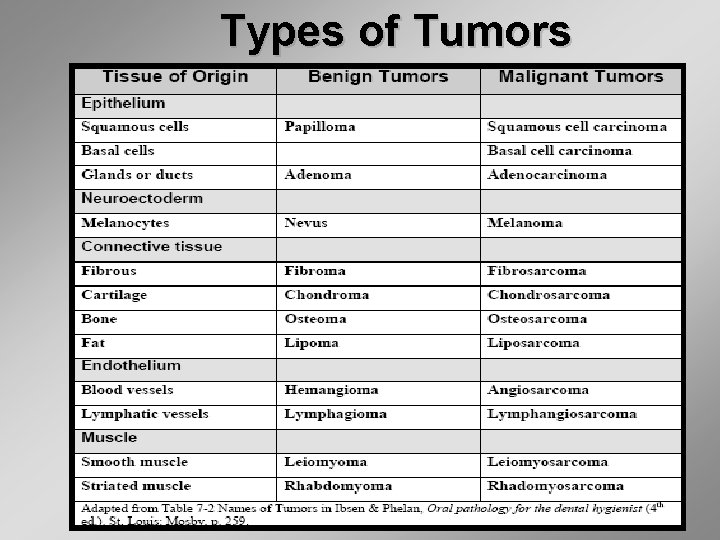
Types of Tumors
Squamous Cell Carcinoma § The most common presentations of intraoral squamous cell carcinoma are: – exophytic (mass forming) – endophytic (ulcerated) – leukoplakic (white patches) – erythroplakic (red patches) and – erythroleukoplakic (combined white and red patches)
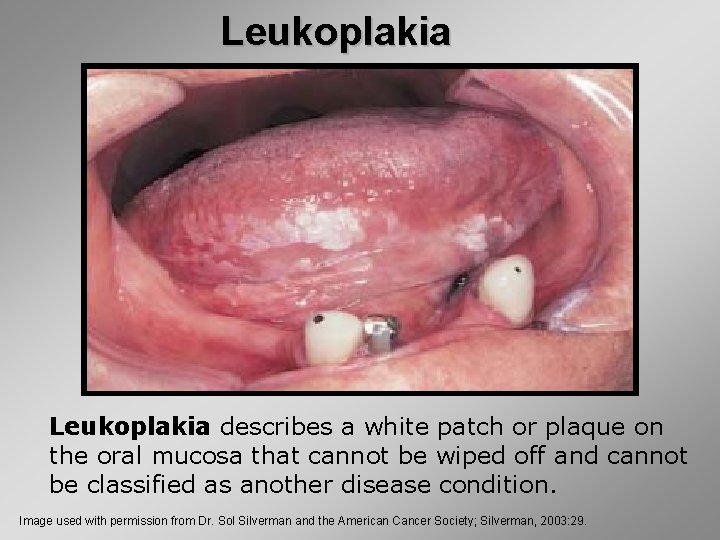
Leukoplakia describes a white patch or plaque on the oral mucosa that cannot be wiped off and cannot be classified as another disease condition. Image used with permission from Dr. Sol Silverman and the American Cancer Society; Silverman, 2003: 29.
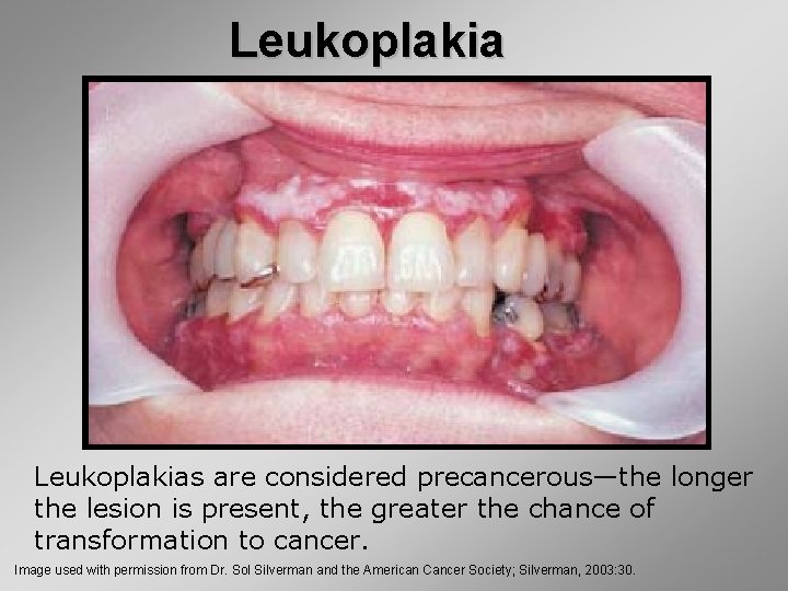
Leukoplakias are considered precancerous—the longer the lesion is present, the greater the chance of transformation to cancer. Image used with permission from Dr. Sol Silverman and the American Cancer Society; Silverman, 2003: 30.
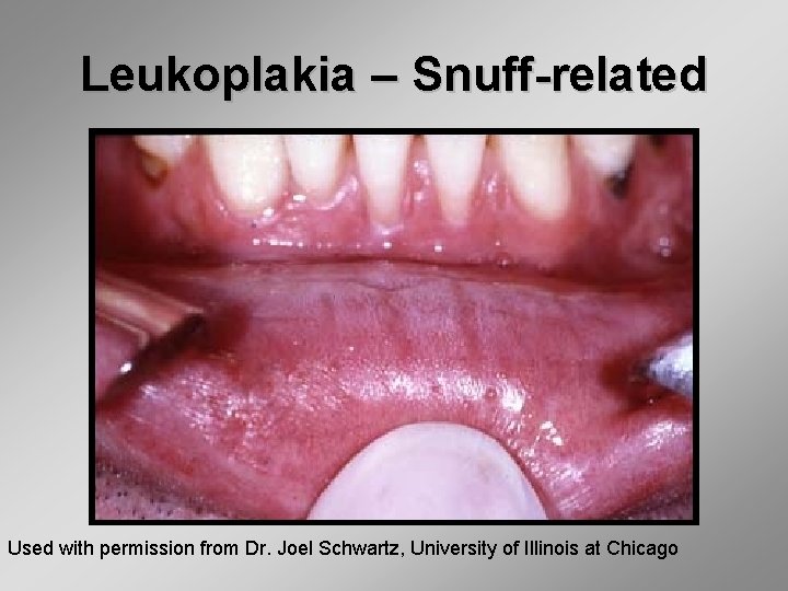
Leukoplakia – Snuff-related Used with permission from Dr. Joel Schwartz, University of Illinois at Chicago
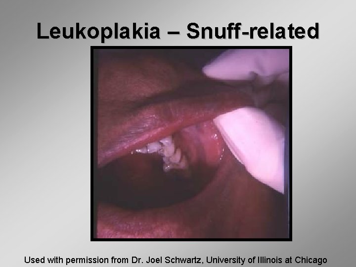
Leukoplakia – Snuff-related Used with permission from Dr. Joel Schwartz, University of Illinois at Chicago
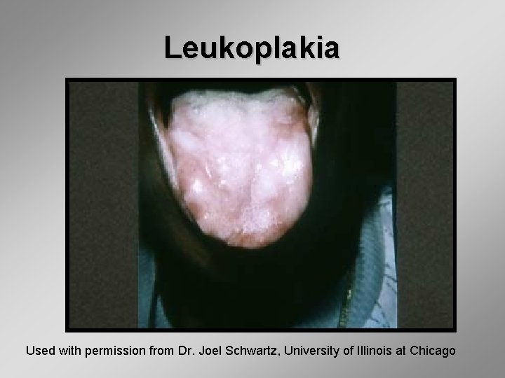
Leukoplakia Used with permission from Dr. Joel Schwartz, University of Illinois at Chicago
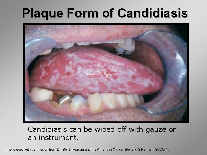
Plaque Form of Candidiasis can be wiped off with gauze or an instrument. Image used with permission from Dr. Sol Silverman and the American Cancer Society; Silverman, 2003: 41.
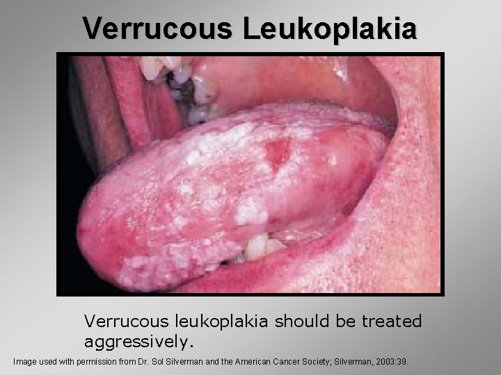
Verrucous Leukoplakia Verrucous leukoplakia should be treated aggressively. Image used with permission from Dr. Sol Silverman and the American Cancer Society; Silverman, 2003: 39.
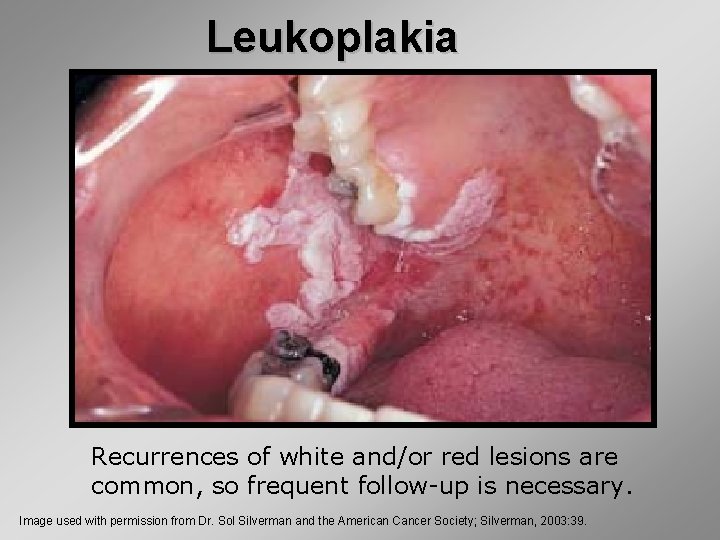
Leukoplakia Recurrences of white and/or red lesions are common, so frequent follow-up is necessary. Image used with permission from Dr. Sol Silverman and the American Cancer Society; Silverman, 2003: 39.
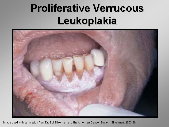
Proliferative Verrucous Leukoplakia Image used with permission from Dr. Sol Silverman and the American Cancer Society; Silverman, 2003: 39.
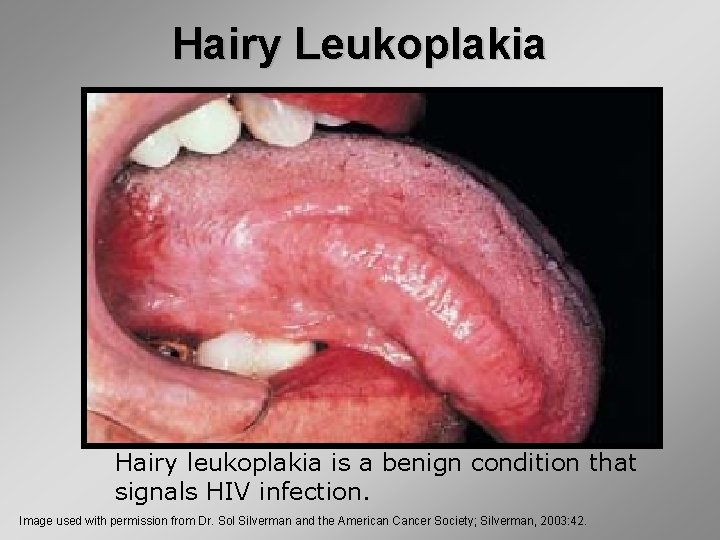
Hairy Leukoplakia Hairy leukoplakia is a benign condition that signals HIV infection. Image used with permission from Dr. Sol Silverman and the American Cancer Society; Silverman, 2003: 42.
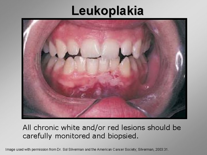
Leukoplakia All chronic white and/or red lesions should be carefully monitored and biopsied. Image used with permission from Dr. Sol Silverman and the American Cancer Society; Silverman, 2003: 31.
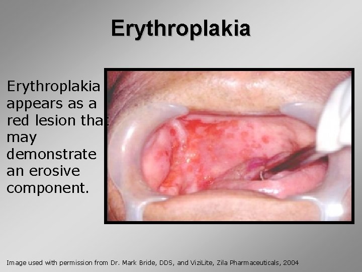
Erythroplakia appears as a red lesion that may demonstrate an erosive component. Image used with permission from Dr. Mark Bride, DDS, and Vizi. Lite, Zila Pharmaceuticals, 2004
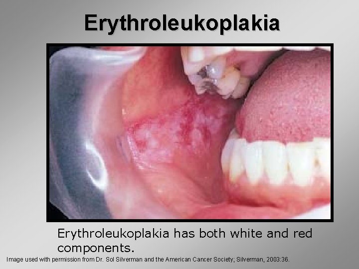
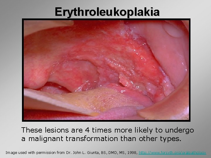
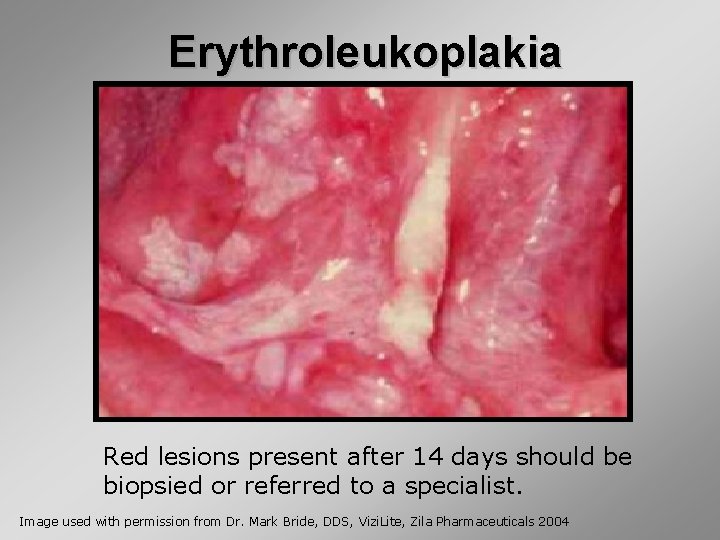
Erythroleukoplakia has both white and red components. Image used with permission from Dr. Sol Silverman and the American Cancer Society; Silverman, 2003: 36.
Erythroleukoplakia These lesions are 4 times more likely to undergo a malignant transformation than other types. Image used with permission from Dr. John L. Giunta, BS, DMD, MS, 1998, http: //www. forsyth. org/oralpathology
Erythroleukoplakia Red lesions present after 14 days should be biopsied or referred to a specialist. Image used with permission from Dr. Mark Bride, DDS, Vizi. Lite, Zila Pharmaceuticals 2004
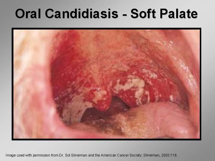
Oral Candidiasis - Soft Palate Image used with permission from Dr. Sol Silverman and the American Cancer Society; Silverman, 2003: 119.
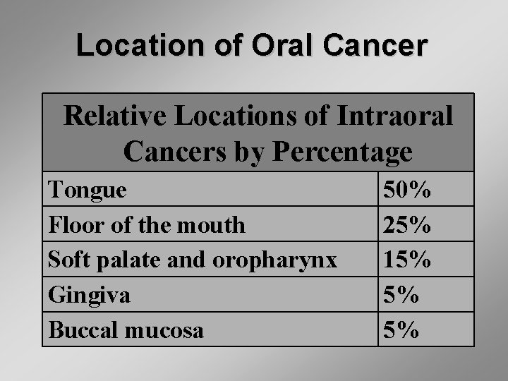
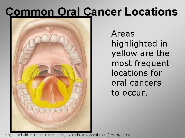
Location of Oral Cancer Relative Locations of Intraoral Cancers by Percentage Tongue Floor of the mouth Soft palate and oropharynx Gingiva Buccal mucosa 50% 25% 15% 5% 5%
Common Oral Cancer Locations Areas highlighted in yellow are the most frequent locations for oral cancers to occur. Image used with permission from Sapp, Eversole, & Wysocki (2004) Mosby, 190.
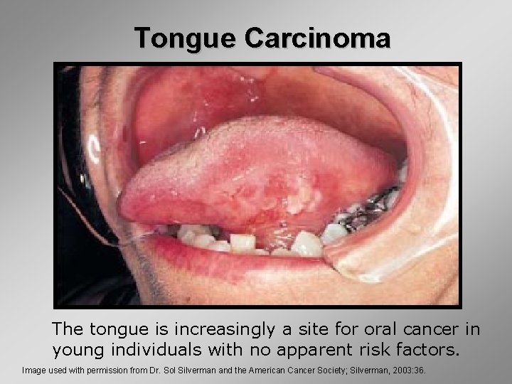
Tongue Carcinoma The tongue is increasingly a site for oral cancer in young individuals with no apparent risk factors. Image used with permission from Dr. Sol Silverman and the American Cancer Society; Silverman, 2003: 36.
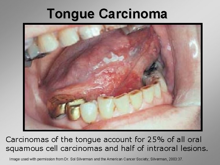
Tongue Carcinomas of the tongue account for 25% of all oral squamous cell carcinomas and half of intraoral lesions. Image used with permission from Dr. Sol Silverman and the American Cancer Society; Silverman, 2003: 37.
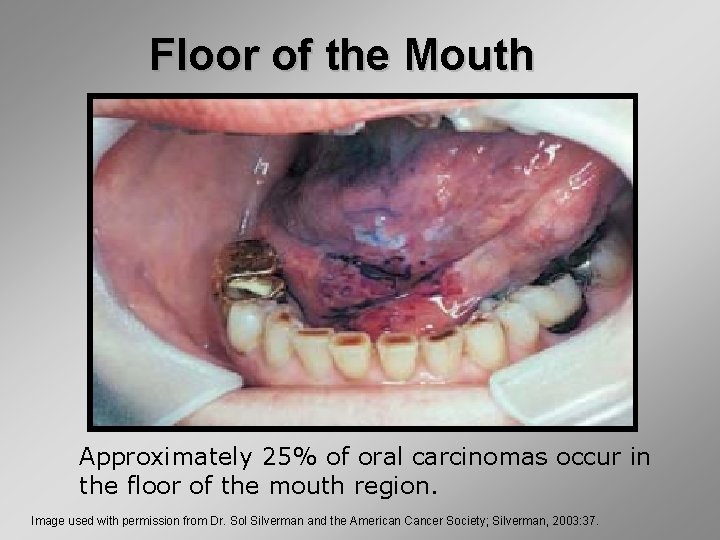
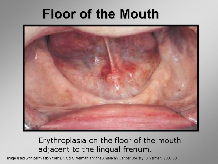
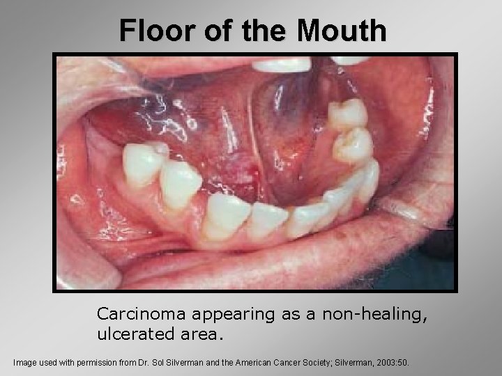
Floor of the Mouth Approximately 25% of oral carcinomas occur in the floor of the mouth region. Image used with permission from Dr. Sol Silverman and the American Cancer Society; Silverman, 2003: 37.
Floor of the Mouth Erythroplasia on the floor of the mouth adjacent to the lingual frenum. Image used with permission from Dr. Sol Silverman and the American Cancer Society; Silverman, 2003: 50.
Floor of the Mouth Carcinoma appearing as a non-healing, ulcerated area. Image used with permission from Dr. Sol Silverman and the American Cancer Society; Silverman, 2003: 50.
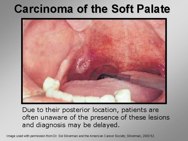
Carcinoma of the Soft Palate Due to their posterior location, patients are often unaware of the presence of these lesions and diagnosis may be delayed. Image used with permission from Dr. Sol Silverman and the American Cancer Society; Silverman, 2003: 52.
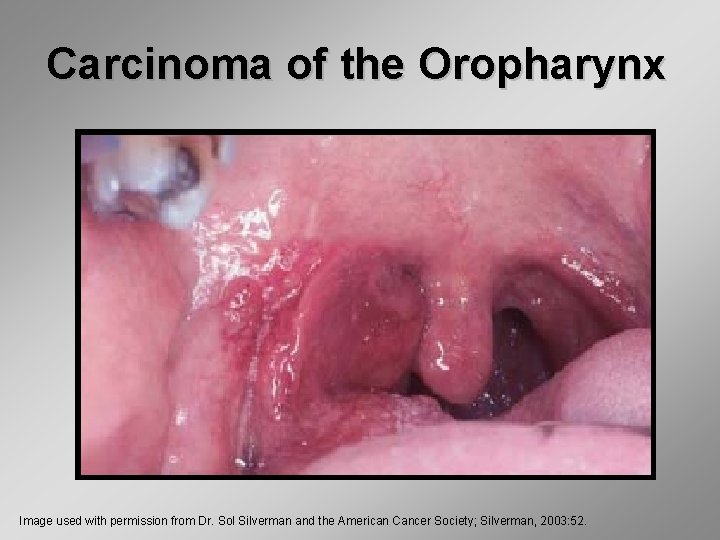
Carcinoma of the Oropharynx Image used with permission from Dr. Sol Silverman and the American Cancer Society; Silverman, 2003: 52.
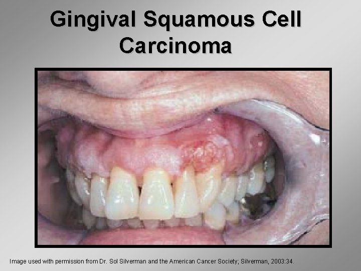
Gingival Squamous Cell Carcinoma Image used with permission from Dr. Sol Silverman and the American Cancer Society; Silverman, 2003: 34.
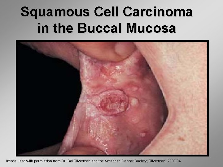
Squamous Cell Carcinoma in the Buccal Mucosa Image used with permission from Dr. Sol Silverman and the American Cancer Society; Silverman, 2003: 34.
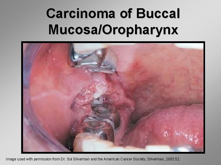
Carcinoma of Buccal Mucosa/Oropharynx Image used with permission from Dr. Sol Silverman and the American Cancer Society; Silverman, 2003: 52.
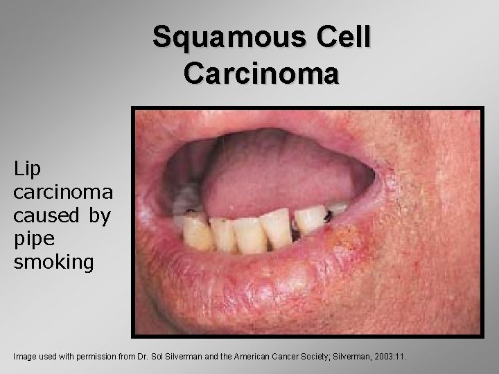
Squamous Cell Carcinoma Lip carcinoma caused by pipe smoking Image used with permission from Dr. Sol Silverman and the American Cancer Society; Silverman, 2003: 11.
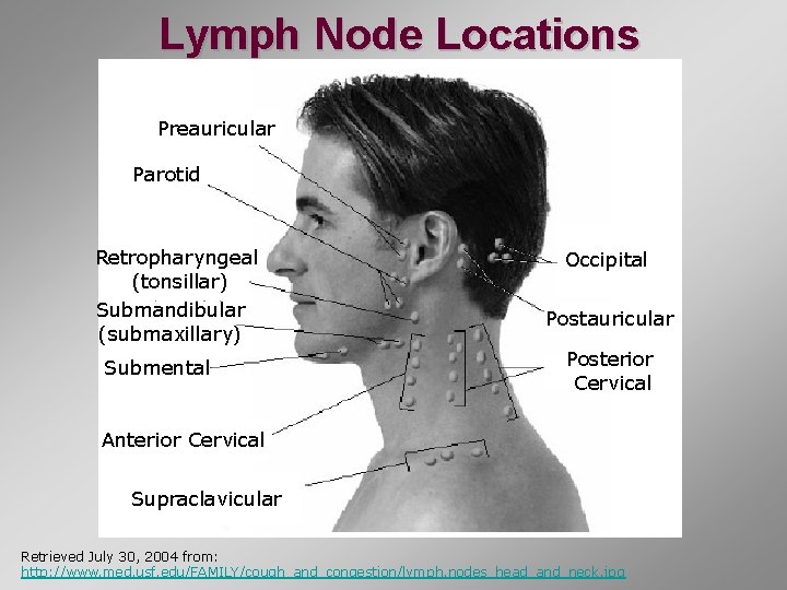
Lymph Node Locations Preauricular Parotid Retropharyngeal (tonsillar) Submandibular (submaxillary) Submental Occipital Postauricular Posterior Cervical Anterior Cervical Supraclavicular Retrieved July 30, 2004 from: http: //www. med. usf. edu/FAMILY/cough_and_congestion/lymph. nodes_head_and_neck. jpg
Signs and Symptoms § A sore or area in the mouth that does not heal after two weeks § Persistent pain in the mouth § Persistent lump or thickening in the cheek § Sore throat or feeling that something is caught in the throat § Difficulty chewing or swallowing § Difficulty moving the jaw or tongue § Voice changes
Signs and Symptoms § Numbness in the tongue or other area of the mouth § Swelling in the jaw that causes dentures to fit poorly or become uncomfortable § Loosening of teeth or pain around the teeth or jaw § Lump or mass in the neck § Weight loss (unexplained) § Persistent bad breath
Summary • Oral cancer may appear as erythroplakia, leukoplakia, or erythroleukoplakia. • Common locations include the tongue (50%), floor of the mouth (25%), soft palate/oropharynx (15%), and gingiva (5%) or buccal mucosa (5%) • Lesions that may mimic oral cancer include candidiasis, oral lichen planus or physical trauma.