Module 05 The Brain Module 05 The Brain

Module 05 The Brain

Module 05: The Brain Studying the Brain: Case Studies

Case Study • A research technique in which one person is studied in depth in the hope of revealing universal principles. • Phineas Gage’s Accident

Module 05: The Brain Studying the Brain: Scanning Techniques
 • A series of X-ray photographs taken from")
Computerized Axial Tomography (CT or CAT) • A series of X-ray photographs taken from different angles and combined by computer into a composite representation of a slice through the body. • Reveals the brain’s structure
 • A technique that sues magnetic fields and radio waves")
Magnetic Resonance Imaging (MRI) • A technique that sues magnetic fields and radio waves to produce computer-generated images that distinguish among types of soft tissue; • this allows us to see structures within the brain.
 • An amplified recording of the waves of electrical activity that sweep")
Electroencephalogram (EEG) • An amplified recording of the waves of electrical activity that sweep across the brain’s surface; • these waves, measured by electrodes placed on the scalp, are helpful in evaluating brain function.
 • A visual display of brain activity. •")
Positron Emission Tomography Scan (PET scan) • A visual display of brain activity. • Injection of a radioactive glucose • Reveals the brain’s functioning

Module 05: The Brain Lower-Level Brain Structures: The Brainstem

Brainstem • The oldest part and central core of the brain; • it begins where the spinal cord swells as it enters the skull and • is responsible for automatic survival functions.

Brainstem

Medulla • Located at the base of the brainstem, • it controls basic life-supporting functions like heartbeat and breathing. • Damage to this area can lead to death.

Medulla

Reticular Formation • A nerve network in the brainstem that plays an important role in controlling wakefulness and arousal. • Extending up and down the spinal cord into the brain • Controls an organism’s level of alertness • Damage to this area can cause a coma.

Module 05: The Brain Lower-Level Brain Structures: The Thalamus

Thalamus

Thalamus • The brain’s sensory switchboard, located on top of the brainstem; • it directs messages to the sensory receiving areas in the cortex.

Module 05: The Brain Lower-Level Brain Structures: The Cerebellum

Cerebellum

Cerebellum • The “little brain”, attached to the rear of the brainstem; • it helps coordinate voluntary movements and balance. • If damaged, the person could perform basic movements but would lose fine coordination skills.

Cerebellum

Module 05: The Brain Lower-Level Brain Structures: The Limbic System

Limbic System • it helps regulate functions such as memory, fear, aggression, hunger, and thirst, and

Hypothalamus • it helps regulates the body’s maintenance activities, such as eating, drinking, body temperature, and it linked to emotion. • Plays a role in emotions, pleasure, and sexual function

Hypothalamus

Hippocampus • it helps processing new memories for permanent storage.

Hippocampus

Amygdala • An almond shaped neural cluster in the limbic system that controls emotional responses such as fear and anger.

Amygdala

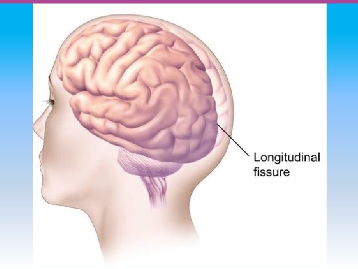
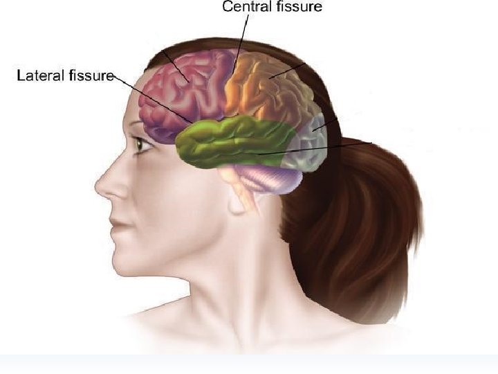
Module 05: The Brain The Cerebral Cortex

Cerebral Cortex • The intricate fabric of interconnected neurons that form the body’s ultimate control and information processing center. • Covers the brain’s lower level structures • Contains an estimated 30 billion nerve cells • Divided into four lobes

Corpus Callosum • The large band of neural fibers that connects the two brain hemispheres and • allows them to communicate with each other. • Is sometimes cut to prevent seizures

Corpus Callosum

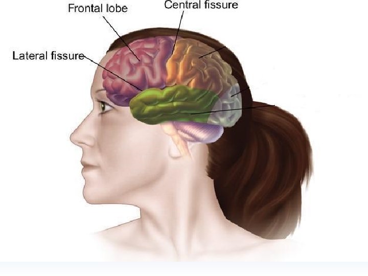
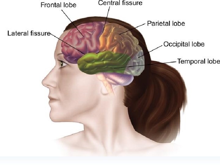
Frontal Lobes • The portion of the cerebral cortex lying just behind the forehead that is involved in planning and judgment; • it includes the motor cortex.

Parietal Lobes • The portion of the cerebral cortex lying on the top of the head and toward the rear; • it includes the somatosensory cortex and general association areas used in processing information. • Regions available for general processing, including mathematical reasoning • Designated as the association lobes • Behind the frontal lobes


Occipital Lobe • The portion of the cerebral cortex lying at the back of the head; • it includes the primary visual processing areas of the brain.


Temporal Lobes • The portion of the cerebral cortex lying roughly above the ears; • it includes the auditory (hearing) areas of the brain. • Where sound information is processed

Motor Cortex • A strip of brain tissue at the rear of the frontal lobes that controls voluntary movement. • Different parts of the cortex control different parts of the body. • The motor cortex in the left hemisphere controls the right side of the body and visa versa.

Somatosensory Cortex • The strip of brain tissue at the front of the parietal lobes that registers and processes body sensations. • Soma is Greek for “body. ”

Plasticity • The brain’s ability to change, especially during childhood, by reorganizing after damage or experience.

Module 05: The Brain Differences Between the Brain’s Two Hemispheres

Hemispheric Differences • “Left-brained” and “right-brained” debunked • Brain is divided into two hemispheres but works as a single entity. • Both sides continually communicate via the corpus callosum, except in those with split brains.

Module 05: The Brain Differences Between the Two Hemispheres: Language and Spatial Abilities

The Brain’s Left Hemisphere • For most people, language functions are in the left hemisphere. • For a small percentage of people, language functions are in the right hemisphere.

Broca’s Area • A brain area of the left frontal lobe that directs the muscle movements involve in speech. • If damaged the person can form the ideas but cannot express them as speech
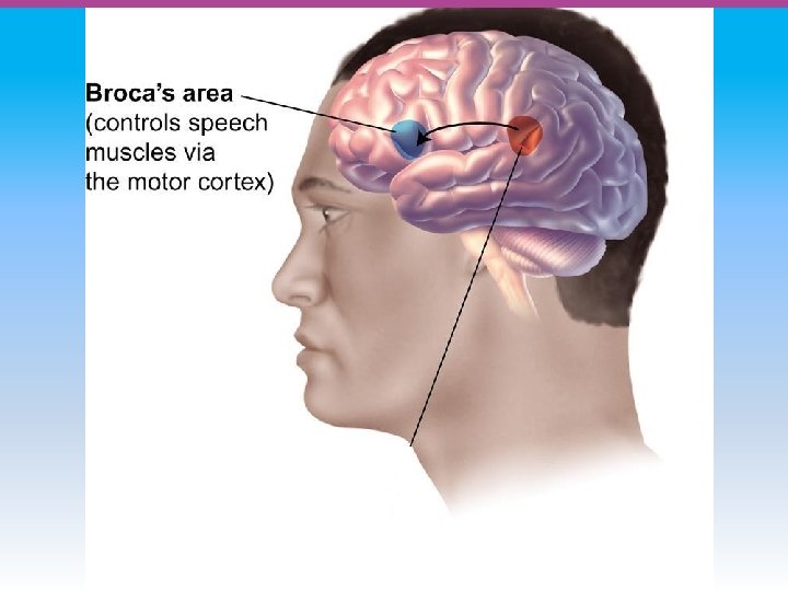

Wernicke’s Area • A brain area of the left temporal lobe involved in language comprehension and expression. • Our ability to understand what is said to us • Usually in the left temporal lobe


The Brain’s Right Hemisphere • Houses the brain’s spatial abilities • Our spatial ability allows us to perceive or organize things in a given space, judge distance, etc. • Helps in making connections between words

Split Brain Research

Split Brain Research
- Slides: 57