Mitosis Meiosis and Gametogenesis Prof Abdulameer AlNuaimi Email

Mitosis, Meiosis and Gametogenesis Prof. Abdulameer Al-Nuaimi E-mail: a. al-nuaimi@sheffield. ac. uk E. mail: abdulameerh@yahoo. com

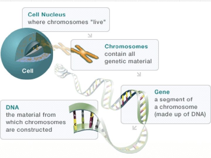
Chromosomes are structures located in nucleus of the cell. Each chromosome is made up of DNA tightly coiled many times around proteins called histones that support its structure. Chromosomes are long thin threads inside the nucleus, they are called chromatin until cell division occurs, then they become visible as rod-like chromosomes. Chromosomes are composed of genes. All the genes on chromosomes make up the cell’s genome, Genes contain genetic information vital for proper cell function. Genes control the physical characteristics of a species. All organisms of the same species contain the same number of chromosomes in their nuclei.

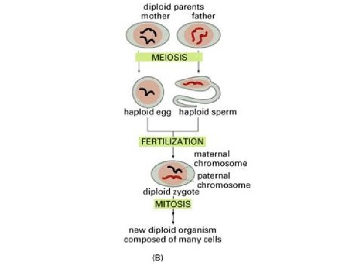
Diploid and Haploid CELLS Diploid cells contain 2 sets of chromosomes in their nuclei. The human species contain 46 chromosomes in their nuclei i. e. 23 sets of two copies of autosomal chromosomes, it is a diploid (2 n) number. Haploid cells contain 1 set of chromosomes in their nuclei. Human sex cells contain 23 chromosomes in their nuclei. This is the haploid (n) number. When fertilisation takes place the 23 chromosomes (n) from the father (called Paternal chromosomes) and the 23 chromosomes (n) from the mother (called the Maternal chromosomes) combine to form the diploid (2 n=46) number of chromosomes in the fertilised egg cell.

Chromatid Scheme of a replicated Chromosome. (two identical parts of a chromosome after S phase) Centromere (1) Chromatid: One of the two identical parts of the chromosome after S phase (synthesis phase). (2) Centromere: The point where the two chromatids touch, and where the microtubules attach. (3) Short arm (4) Long arm. In accordance with the display rules in Cytogenetics, the short arm is on top

Meiosis is the program used by sexually reproducing organisms to produce haploids from diploid precursors It is a specialized type of cell division that reduces the chromosome number by half, creating four haploid cells, In Meiosis DNA replication is followed by two rounds of cell division to produce four potential daughter cells, each with half the number of chromosomes as the original parent cell Phases of meiosis 1 -Premeiotic S-phase (synthesis phase): the DNA of each chromosome is replicated so that it consists of two identical sister chromatids, which remain held together through cohesion.

2 -Meiotic prophase: homologous chromosomes can swap different parts of their DNA by “crossing over” to create new genetic combinations. In this stage, DNA is cut and then repaired, which allow homologous chromosomes to exchange some of their genetic information 3 -Meiosis I: Each pair of homologous chromosomes segregate away from other pair 4 -Meiosis II: the cohesion between sister chromatids is released and they segregate from one another
 meiotic prophase (2) (3) Meiosis I (4)Meiosis II")
Meiosis p premeiotic S-phase (1) meiotic prophase (2) (3) Meiosis I (4)Meiosis II
 and female")
Gametogenesis • Gametogenesis is a process of formation and development of male(sperm) and female (ovum)gametes. • During this process a reduction of the number of chromosomes takes place from diploid number (46) of primary germ cells to the haploid number (23) in the mature germ cell. Copyright © 2006/07 The University of Sharjah slide 10

Male Gametogenesis • The large rounded primary male germ cells, lose most of their cytoplasm and develop head, tail and neck to assist in the movement of the sperms. • The rounded female germ cells become larger as a result of increase in the a mount of their cytoplasm and called ovum. Copyright © 2006/07 The University of Sharjah Mature sperm Mature oocyte slide 11

Introduction Mature Male Genital Organ www. google. co. uk/search?

Seminiferous tubules get canalised at puberty Cross section in the Testis

Testis Cross section in the seminiferous tubule Testis and Sperm www. google. co. uk/search?

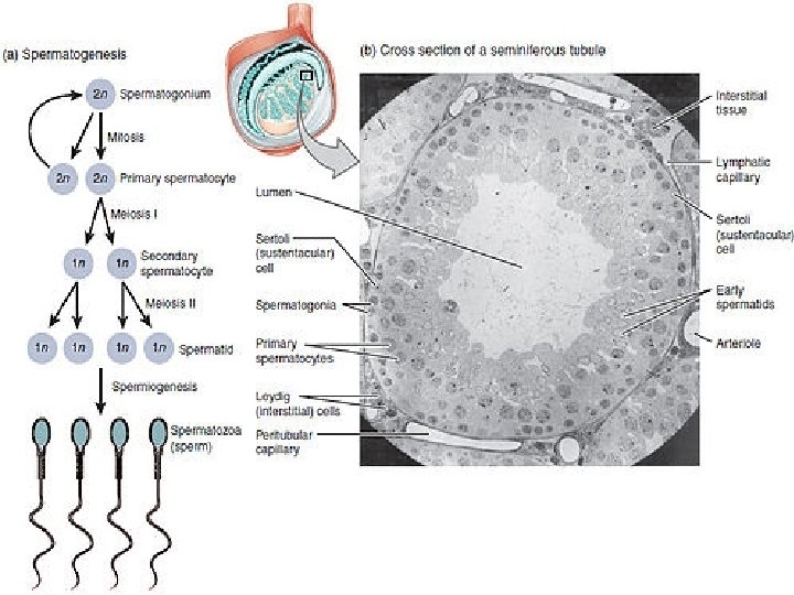
Spermatogenesis is the process in which spermatozoa are produced from spermatogonial stem cells by way of mitosis and meiosis The initial cells in this pathway are called spermatogonia, it divides mitotically into two primary spermatocytes. The primary spermatocyte divides meiotically (Meiosis I) into two secondary spermatocytes; each secondary spermatocyte divides into two spermatids by Meiosis II. Spermatids develop into mature spermatozoa, also known as sperm cells. Thus, the primary spermatocyte gives rise to two secondary spermatocytes. Secondary spermatocytes by their subdivision produce four spermatozoa

Seminiferous Tubule in the Testis
 Mature Female Genital Organ www. google. co. uk/search?")
Introduction (Uterine tube) Mature Female Genital Organ www. google. co. uk/search?

During Embryonic life ovaries appear as a pair of longitudinal Genital ridges on each side of the midline. Primordial (primitive) Germ Cells originate in the Epiblast migrate to the genital ridges, invade them and producing the ovaries If Germ cells fail to reach the Ridge, the gonads do not develop Inside these ovaries, clusters of cells begin to surround each Germ cell, Oogonium (oocyte) with a layer of epithelial cells called Follicular cells Producing ovarian follicles. Between 16 and 20 weeks of pregnancy, the ovaries of a female fetus contain 6 to 7 million oocytes. Most of the oocytes gradually degenerate, leaving about 1 to 2 million present at birth.

At puberty, only about 300, 000 are present and only a small percentage of oocytes mature into eggs (ova). The many thousand of oocytes that do not mature degenerate. Degeneration progresses more rapidly in the 10 to 15 years before menopause. All oocytes are gone by menopause. Only about 400 ova are released during a women's reproductive life, usually one during each menstrual cycle. Until released, an ovum remains dormant in its follicle. Thus, the ovum is one of the longest lived cells in the body
")
Migration of Primordial germ cells to the Gonadal ridge Allantois (Langman’s Medical Embryology)
")
primitive Mesonephric duct = Follicular cells + Germ cell Ovary (Langman’s Medical Embryology)

Oogenesis Oogonium divides mitotically giving rise to Primary Oocyte this develops into primary follicle, the functional unit of the ovary. Primary Oocyte (diploid)-(Meiosis I) First Polar Body (Discarded afterward) + Secondary oocyte (haploid) Secondary oocyte—(Meiosis II)—> Second Polar Body (Discarded afterward) + Ovum (haploid) Oogonium Mitosis

Female Gametogenesis • The large rounded primary male germ cells, lose most of their cytoplasm and develop head, tail and neck to assist in the movement of the sperms. • The rounded female germ cells become larger as a result of increase in the a mount of their cytoplasm and called ovum. Copyright © 2006/07 The University of Sharjah Mature sperm Mature oocyte slide 24

Mitosis Meiosis II

Fertilization • The fertilization occurs when sperm contacts an ovum to form a zygote in the uterine tube. • The zygote, which contains 46 chromosomes, rapidly divide by mitosis to form mass of cells as 2, 4, 8, 16, 32…. • This mass of cells continue dividing in order to give the different tissues then organs of the human body. Copyright © 2006/07 The University of Sharjah slide 26

Sun Set Malaysia 16 Feb. 2015 Thank You
- Slides: 27