Mitosis Cell Growth There are two reasons cells


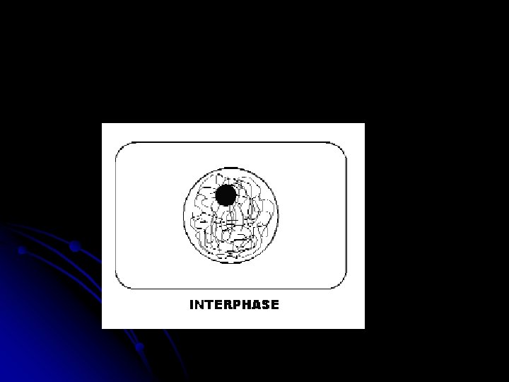
 Interphase is divided")


- Slides: 36
Mitosis
Cell Growth There are two reasons cells divide rather than continue to grow extra large: l 1. The larger the cell grows, the more demands the cell places on its DNA l 2. The larger the cell, the more trouble it has moving nutrients and waste across the membrane. l
As a result: l l Before a cell grows too large it undergoes cell division. This mean it forms two new daughter cells. First it must copy all its DNA. Each daughter cell gets one complete set of genetic information. The division causes the two daughter cells to have an increased surface area to volume ration, allowing an efficient exchange of nutrients and waste across the cell membrane.
What would happen if the cell divided without copying its genetic information? Would each cell have a complete set of genetic instructions? No. That is why the cell must make a copy of the genetic information before dividing. Each daughter cell gets a complete copy.
Chromosomes The genetic information that each daughter cell gets is carried by chromosomes. l Chromosomes are made of DNA which carries the cells coded genetic information and proteins. l Each organism has a specific number of chromosomes. l
l Humans have 46, fruit flies have 8, and carrots have 18 chromosomes.
Chromosomes They are not visible inside the cell except during cell division. l They condense into compact structures and are now called chromatin. l The DNA has already been copied or replicated at this point l The condensed Chromosome consists of two identical sister chromatids. l
l Chromosome: genetic information in the nucleus that is passed from one generation to the next. Made of DNA and proteins. l Chromatin: condensed DNA in the nucleus that is visible. DNA is coiled around proteins. l Chromatid: one of two sister parts of the duplicated chromosome.
l Chromosome chromatin chromatid
Each pair of chromatids is attached by a centromere, usually towards the middle. l Human body cells contain 46 chromosomes, each chromosome is made of 2 chromatids. l
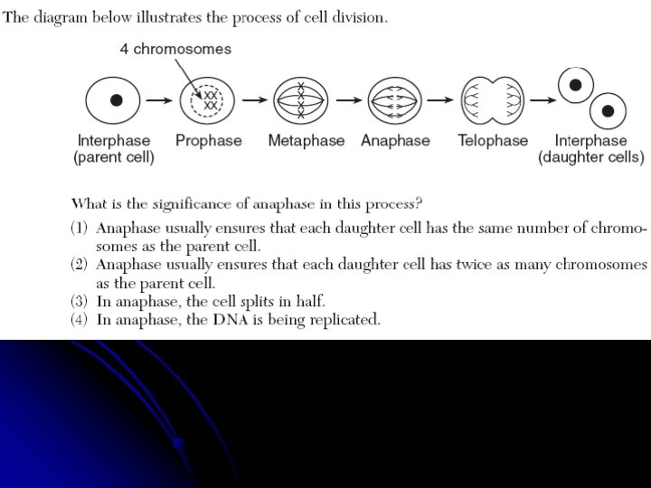
The Cell Cycle The cell cycle is a series of events that the cell goes through as they grow and divide. l During the cycle, a cell grows, prepares for division and divides to form two daughter cells. l Each daughter cell then begins the cycle again. l
The cell cycle consists of four phases. 1. 2. 3. 4. M Phase G 1 Phase S Phase G 2 Phase
1. M Phase: known as mitosis. l What happens: division of the nucleus and cytokinesis l Four stages of mitosis are: l 1. Prophase l 2. Anaphase l 3. Metaphase l 4. Telophase l
l 2. G 1 Phase: “Gap 1” : cell does most of its growing. Cell increases in size Makes proteins and new organelles. 3. S Phase: What happens: copying of the chromosomes making a duplicate set of DNA. 4. G 2 Phase: “Gap” intense growth and activity. Shortest of these 3 phases. After G 2 phase, the M phase begins. l G 1, G 2 and S Phase make up Interphase. l l l l l
Section 10 -2 Concept Map Cell Cycle includes M phase (Mitosis) Interphase is divided into G 1 phase Go to Section: S phase is divided into G 2 phase Prophase Metaphase Anaphase Telophase
1. Prophase First and longest step of mitosis l Chromatin condense into chromosomes. l Centrioles separate and move to opposite sides of the nucleus. l Spindle begins to form l Nuclear membrane breaks down. l
Section 10 -2 Figure 10– 5 Mitosis and Cytokinesis Spindle forming Centrioles Nuclear envelope Chromatin Interphase Centromere Chromosomes (paired chromatids) Prophase Cytokinesis Go to Section: Spindle Centriole Telophase Nuclear envelope reforming Centriole Individual chromosomes Anaphase Metaphase
2. metaphase Second stage of mitosis l Chromosomes line up at the center of the cell l Each chromosome is connected to a spindle fiber. l
Section 10 -2 Figure 10– 5 Mitosis and Cytokinesis Spindle forming Centrioles Nuclear envelope Chromatin Interphase Centromere Chromosomes (paired chromatids) Prophase Cytokinesis Go to Section: Spindle Centriole Telophase Nuclear envelope reforming Centriole Individual chromosomes Anaphase Metaphase
3. Anaphase Third stage of mitosis. l Sister chromatids separate into individual chromosomes and move apart. l They become individual chromosomes. l Anaphase ends when the chromosomes stop moving near the end of the poles. l
Section 10 -2 Figure 10– 5 Mitosis and Cytokinesis Spindle forming Centrioles Nuclear envelope Chromatin Interphase Centromere Chromosomes (paired chromatids) Prophase Cytokinesis Go to Section: Spindle Centriole Telophase Nuclear envelope reforming Centriole Individual chromosomes Anaphase Metaphase
4. Telophase Fourth and final stage of mitosis. l Chromosomes de-condense and become less visible. l Nuclear envelope reforms l Spindle breaks apart. l Cell division is not yet complete though. l
Section 10 -2 Figure 10– 5 Mitosis and Cytokinesis Spindle forming Centrioles Nuclear envelope Chromatin Interphase Centromere Chromosomes (paired chromatids) Prophase Cytokinesis Go to Section: Spindle Centriole Telophase Nuclear envelope reforming Centriole Individual chromosomes Anaphase Metaphase
Cytokinesis The M-phase is cytokinesis. l Cytokinesis is the division of the cyotplasm. l The plasma membrane moves inward until the cytoplasm is divided into two equal parts. l Each part has its own nucleus and organelles. l Usually occurs during telophase. l
Section 10 -2 Figure 10– 5 Mitosis and Cytokinesis Spindle forming Centrioles Nuclear envelope Chromatin Interphase Centromere Chromosomes (paired chromatids) Prophase Cytokinesis Go to Section: Spindle Centriole Telophase Nuclear envelope reforming Centriole Individual chromosomes Anaphase Metaphase
Figure 10– 4 The Cell Cycle Section 10 -2 G 1 phase M phase S phase G 2 phase Go to Section:
Cell Regulators Even cells have a limited life span. l Proteins in the cell when its life is over and mitosis stops. l Cancer cells ignore these protein signals. l Result: masses of cells that can damage the surrounding tissue. l Cancer cells can break loose from the tumor and infect other areas such as the lungs. l
Good animation l http: //highered. mcgrawhill. com/olc/dl/120073/bio 14. swf
Can you find all the phases? Interphase, prophase, metaphase, anaphase, telophase
l What would most likely result if mitosis was not accompanied by cytoplasmic division? 1. two cells, each with one nucleus 2. two cells, each without a nucleus 3. one cell with two identical nuclei 4. one cell without a nucleus
During which phase does the chromosomes line up along the center of the cell? l Prophase l Metaphase l Interphase l Anaphase l Telophase l
l During mitosis, a double-stranded chromosome is attached to a spindle fiber at the 1. centriole 2. centromere 3. centrosome 4. cell plate
During which phase does the cell begin to divide? l Prophase l Metaphase l Interphase l Anaphase l Telophase l