Mitosis Cell Division How do cells divide and


 • Chromosomes appear as")


 attach to the")

 separate and")


 Beads (centromeres) Macaroni (centrioles)")


- Slides: 31
Mitosis: Cell Division
How do cells divide and reproduce?
Cell Division 1. Mitosis: division of nucleus 2. Cytokinesis: division of cytoplasm
Cell division occurs in a series of stages, or phases. • Interphase • Prophase • Metaphase • Anaphase • Telophase • Cytokinesis
Interphase occurs before mitosis begins G 1 S G 2 Interphase
Interphase occurs before mitosis begins Chromosomes are copied (# doubles) • Chromosomes appear as threadlike coils (chromatin) at the start, but each chromosome and its copy (sister chromosome) change to sister chromatids at end of this phase • Nucleus CELL MEMBRANE Cytoplasm
Interphase Animal Cell Plant Cell Photographs from: http: //www. bioweb. uncc. edu/biol 1110/Stages. htm
Prophase 1 st step in Mitosis • • • Mitosis begins (cell begins to divide) Centrioles appear and begin to move to opposite ends (or poles) of the cell. Spindle fibers form between the poles. Sister chromatids Centrioles Spindle fibers
Prophase Animal Cell Plant Cell Spindle fibers Centrioles Photographs from: http: //www. bioweb. uncc. edu/biol 1110/Stages. htm
Metaphase 2 nd step in Mitosis Chromatids (or pairs of chromosomes) attach to the spindle fibers. • Chromatids line up in the middle of the dividing cell. • Centrioles Spindle fibers
Metaphase Animal Cell Plant Cell Photographs from: http: //www. bioweb. uncc. edu/biol 1110/Stages. htm
Anaphase 3 rd step in Mitosis • Chromatids (or pairs of chromosomes) separate and begin to move to opposite ends of the cell. Centrioles Spindle fibers
Anaphase Animal Cell Plant Cell Photographs from: http: //www. bioweb. uncc. edu/biol 1110/Stages. htm
Telophase 4 th step in Mitosis Two new nuclei form. • Chromosomes appear as chromatin (threads rather than rods). • Mitosis ends. • Nuclei Chromatin Nuclei
Telophase Animal Cell Plant Cell Photographs from: http: //www. bioweb. uncc. edu/biol 1110/Stages. htm
Cytokinesis occurs after mitosis • Cell membrane moves inward to create two daughter cells – each with its own nucleus with identical chromosomes.
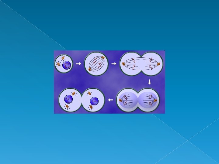
Animal Mitosis – Review Interphase Metaphase Telophase Prophase Anaphase Interphase
Plant Mitosis – Review Interphase Metaphase Telophase Prophase Anaphase Interphase
REMEMBER! Interphase Prophase Metaphase Anaphase Telophase Cytokinesis IPMATC I Pray More At The Church
MITOSIS Mitosis animation
CYTOKINESIS Animal cells – contractile ring forms around the midpoint and pinches the membrane inwards
CYTOKINESIS Plant cells – cell plate forms in the middle and creates new cell wall
Cell Cycle 23
- Cell Division The Cell Cycle 24 24
Cell Cycle Class Project Assignment: Create a comic strip of the cell cycle. 8 groups: G 1 S Interphase G 2 Prophase Metaphase Anaphase M Telophase Cytokinesis
Cell Cycle Class Project Materials: Paper Markers Glue Yarn (chromosomes) Beads (centromeres) Macaroni (centrioles) Thread (spindle fibers)
Cell Cycle Class Project Make sure you consult with other groups so that cell shape/size is consistent and chromosome colors are the same. Process: 1. Draw the shape of your cell(s) on the paper. 2. Sketch the structures inside your cell(s) – what do the insides look like during this stage? 3. Glue the appropriate materials on top of the sketch to represent the cell structures. 4. Label everything! 5. At the end, we will connect all the stages in order to form a complete comic strip of the cell cycle. «
Observing Mitosis Lab
Observing Mitosis Lab Whitefish embryo Onion root tip
Observing Mitosis Lab Use the photos below as a guide to identifying the stages of mitosis.