Mitosis and Meiosis Chapter 8 0 8 24
")


 1. Duplicating chromosomes")


 https: //www. youtube. com/ watch? v=C 6 hn 3")


 https: //www. youtube. com/watch? v=QVCjd. Nx.")

")
 and")

 involves meiosis")


 https: //www. youtube. com/watch? v=zr. Kdz 93 Wl.")


- Slides: 53
Mitosis and Meiosis (Chapter 8. 0 -8. 24)
Learning Objectives for Mitosis • • Develop a clear understanding of the role of mitosis Explain what chromosomes are and where they are found Explain how chromosomes are affected by mitosis. Understand that all cells arise from pre-existing cells Explain binary fission in prokaryotic cells Explain sister chromatids and centromeres Provide an explanation of the eukaryotic cell cycle Provide a simple diagram and explanation for each of the stages of mitosis (interphase, prophase, metaphase, anaphase, telophase, and cytokinesis. Use the following terms to explain the events of each stage (chromatin, chromosomes, nuclear envelope, centrosome position, kinetochore, mitotic spindle, daughter chromosomes, cleavage furrow)
Learning Objectives for Mitosis • Explain the role of growth factors in the cell-cycle control system. Explain how an improperly functioning cell cycle control system could lead to cancer. • Provide an explanation of how cytokinesis differs between plant and animal cells • Explain how anchorage, cell density (density dependent inhibition), and chemical growth factors affect cell division
Learning Objectives for Meiosis • • Explain how chromosomes are matched in homologous pairs Explain the difference between somatic and sex cells. Explain autosomes Understand why gametes have a single set of chromosomes Explain haploid, diploid, zygote Explain how gametes acquire a single set of chromosomes Provide a brief explanation and diagram of the events that occur in each stage of meiosis. Compare and contrast meiosis and mitosis (provide three major differences) • Given the haploid number of an organism, be able to calculate the number of • chromosomal combinations that meiosis can package into gametes (2 n) Example: A fictional creature has a haploid number of n=5. How many chromosomal combinations are possible?
Learning Objectives for Meiosis • • Explain how crossing over causes genetic variability Explain the cause of Down’s Syndrome in terms of chromosomes Explain 4 types of chromosome alterations (deletion, duplication, inversion, and translocation) that can cause birth defects and cancer What is a karyotype and how it is useful? Learn how to interpret and analyze karyotypes. Explain nondisjunction Explain the role of sex chromosomes in sex determination of humans
Cell Division • Cells can only arise from pre-existing cells • Cell division is the basis of reproduction for all organisms • The creation of genetically identical offspring from one parent is called asexual reproduction • Sexual reproduction requires fertilization of an egg by a sperm
Binary Fission • Prokaryotes reproduce by binary fission (dividing in half) 1. Duplicating chromosomes move towards opposite ends of the cell 2. The cell elongates 3. The plasma membrane growns inward and more cell wall forms, dividing the parent cell into two daughter cells.
Chromosomes • Found in the nucleus of eukaryotic cells • Chromatin duplicate and condense into chromosomes before cell division • Each chromosome contains two identical copies, called sister chromatids joined by a centromere
Function of Mitosis • Ensures correct distribution of chromosomes during cell division • Responsible for Asexual reproduction of singlecelled organisms and growth and maintenance of multi-cellular organisms
The Cell Cycle • The cell cycle proceeds in an ordered sequence of events • Consists of a growing stage called interphase and the cell division stage (mitotic phase)
The Cell Cycle
Interphase • Period of cell growth where molecules and organelles are synthesized • In late interphase, the cell doubles its contents and chromosomes are duplicated. • Centrosomes with two centriole pairs are present
Prophase • In the nucleus, chromatin become tightly coiled to form chromosomes • In the cytoplasm, spindle fibers grow out from the centrosomes to form the mitotic spindle
Prometaphase • Nuclear envelope disappears • Spindle fibers attach to the kinetochore of the chromosomes • Spindle fibers move the chromosomes toward the centre of the cell
Metaphase • Mitotic spindle forms poles at either ends of the cell • Chromosomes align on the metaphase plate with sister chromatids facing the opposite poles
Anaphase • Two centromeres of each chromosome come apart, separating the sister chromatids • The poles move farther apart, elongating the cell • The chromosomes reach the two poles of the cell
Telophase • The cell continues to elongate • Daughter nuclei appear at the poles • Nuclear envelope forms around the chromosomes • Spindle fibers disappear and chromatin fibers uncoil
Cytokinesis • Division of the cell cytoplasm occurs • Two daughter cells completely separate • In animal cells, a cleavage furrow forms and the cell pinches into two • In plant cells, a cell plate forms to separate the two daughter cells
Video: mitosis (6: 10 min) https: //www. youtube. com/ watch? v=C 6 hn 3 s. A 0 ip 0
Factors on Cell Division • Body cells secrete protein growth factors to stimulate other cells to divide • Density dependent inhibition stops crowded cells from dividing when they come in contact with each other • Most animal cells must be in contact with a surface to divide, the is called anchorage dependence
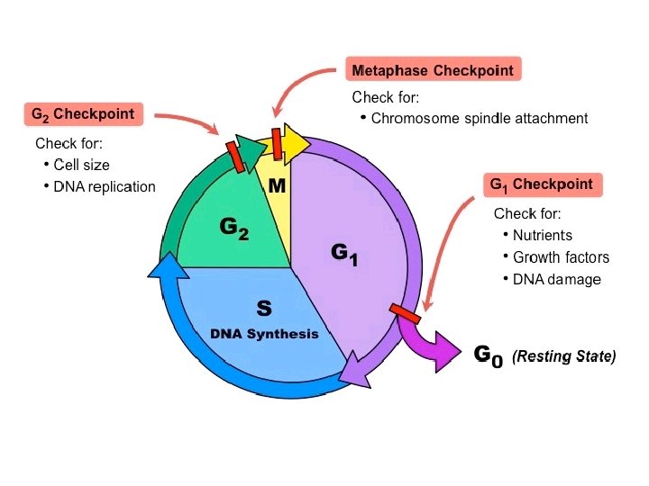
Cell Cycle Controls • The cell cycle control system consists of a set of molecules that triggers and coordinates key events in the cell cycle • Critical points (checkpoints) in the cell cycle have a default stop state unless specific go-ahead signals override the “brakes” at the checkpoints
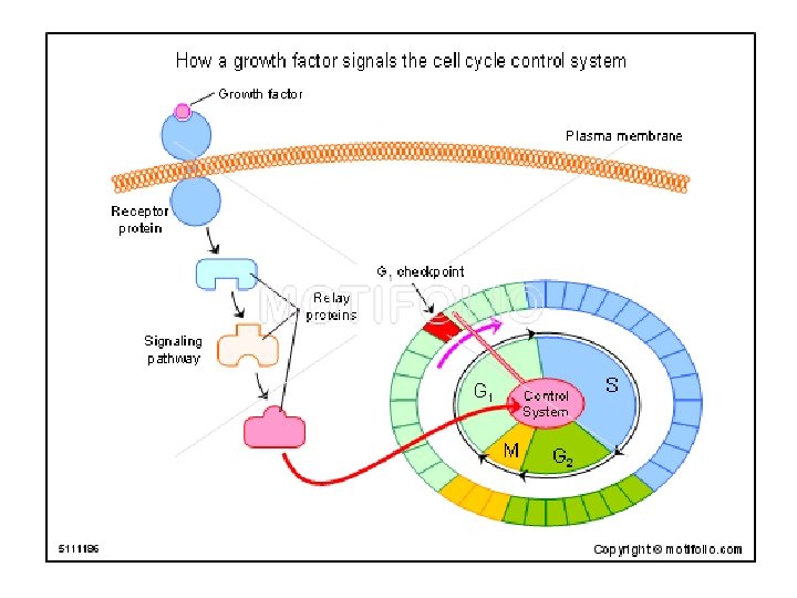
How do growth factors affect cell cycle controls? • Binding of a growth factor on receptor proteins on the plasma membrane results in a signal transduction pathway • The pathway leads to a signal override of the checkpoint (ie. cell division at checkpoint G 1)
Cancer and the Cell Cycle • Cancer is a disease of the cell cycle • A cell mutation leads to transformation into a cancer cell which evades cell death of abnormal cells (apoptosis) • Cancer cells do not respond to the normal signals that regulate the cell cycle and divide excessively, forming a tumour • Benign tumours remain in the original site • Malignant tumours spread into neighbouring tissue (metastasis)
Video: The cell cycle and cancer (9: 18) https: //www. youtube. com/watch? v=QVCjd. Nx. Jre. E
Homologous Chromosomes • Homologous chromosomes are matching pairs of chromosomes that are similar in length and centromere position • They carry genes that control the same genetic traits in the same locus (position in the chromosome) • However, homologous chromosomes have different versions of the same gene
Somatic cells • Somatic cells are typical body cells (eg. skin cells, intestinal cells) • They contain pairs of homologous chromosomes (23+23 = 46 in humans) • Cells that contain pairs of homologous chromosomes are called diploid (2 N) cells
Sex Cells • Sex cells are called gametes (ie. egg and sperm cells) and are haploid (1 N) • They contain only one set of chromosomes (eg. 23 chromosomes in humans) • There is only one member of each homologous pair
Sex cells • Gametes are involved in sexual reproduction by fusing during fertilization • The product of fertilization is called a zygote and will have two sets of chromosomes • In the zygote, one set of chromosomes comes from each parent
Function of Meiosis • The production of egg and sperm cells (gametes) involves meiosis • Results in the formation of genetically different daughter cells with ½ of the chromosomes Video; Meiosis (6: 45 min) https: //www. youtube. c om/watch? v=c 5 h. A 0 W Cv 1 lg
Meiosis • Interphase duplicates each of the chromosomes • Meiosis I segregates the two chromosomes of the homologous pair • Meiosis II separates the sister chromatids
Meiosis I • Homologous chromosomes separate
Prophase I - Formation of tetrads - exchange of DNA portions between homologous chromosomes occurs at chiasma
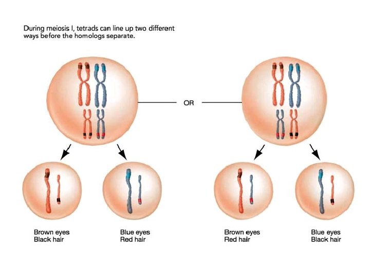
Metaphase I - random alignment of homologous chromosomes at equator
Anaphase I - separation of homologous pairs result in reduction of chromosome number
Telophase I and Cytokinesis I • Formation of 2 genetically different cells
Meiosis II • Starts with haploid cells • DNA is not duplicated • 4 haploid daughter cells, called gametes, are formed
Prophase II - spindle fibers form and attach to kinetochores of sister chromatids
Metaphase II and Anaphase II • Metaphase II - Chromatids line up at the metaphase plate • Anaphase II – chromatids separate
Telophase and Cytokinesis II Telophase II - Chromosomes arrive at poles and nuclear envelope surrounds chromosomes Cytokinesis II - Formation of 4 haploid gametes
Comparing Mitosis and Meiosis
Video: mitosis v meisosis (6: 34) https: //www. youtube. com/watch? v=zr. Kdz 93 Wl. Vk
Meiosis and Genetic Variability In addition to random fusing of sperm and egg during fertilization, the process of meiosis also increases genetic variability through 1. Random segregation of tetrads 2. Independent Assortment of homologous chromosomes during Metaphase 3. Crossing Over of gene segments between chromatids
Random Segregation and Indepedent Assortment • The way homologous chromosomes line up at the metaphase plate is not dependent on other pairs, this leads to genetic variety in the gametes • The total number of chromosomal combinations that meiosis can package into gametes is 2 n • For a haploid number of 2, 22 = 4 • For humans, n = 23, 223 = 8 million!
Crossing Over • Crossing over is the exchange of corresponding segments between non-sister chromatids of homologous chromosomes • Crossing over produces new combinations of genes due to genetic recombination
Chromosomal Alterations • Deletion - when a chromosomal fragment is lost • Duplication – when chromosomal fragments are joins a sister chromatid or homologous chromosome • Inversion – when chromosomal fragments reattach to original chromosome in reverse order • Translocation - when chromosomal fragments attach to non-homologous chromosomes (e. g Chronic Myolegenous Leukemia) • Note: chromosomal changes to somatic cells are not inherited
Types of Chromosomal Alterations
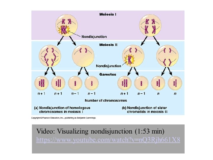
Non-disjunction • Chromosome pair fails to separate • Results in abnormal chromosome number • Examples: Down Syndrome (extra chromosome 21) Klinefelter Syndrome (XXY)
Karyotyping • A karyotype is an ordered display of an individual’s chromosomes arranged in pairs • Karyotypes provide a photographic inventory in order to detect chromosomal abnormalities
Sex Determination • Chromosomes not directly involved in determining sex are called autosomes • Mammalian cells usually operate with only one functioning X chromosome • The presence of the Y chromosome is crucial in determining sex • Eg. XX, XO – “female” • Eg. XY, XXXY – “male”