MITOCHONDRIA STRUCTURE Diagrame of the mitochondria structure Mitochondria


6 + H 2 O O 2")


 that can be")


, are donated")


- Slides: 31
MITOCHONDRIA STRUCTURE
Diagrame of the mitochondria structure
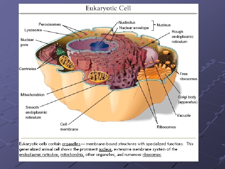
Mitochondria contain two membranes, separated by space. Inside the space enclosed by inner membrane is the matrix. These appears moderatly dense and one may find strands of DNA, Ribosome, or small granula in the matrix. The above diagrame shows the diagram of the mitochondrial membranes and the enclosed compartement.
Contd Mitochondria is the Power of house of the cell. How are mitochondria organized to be power house. The food we eat is oxidized to produce high energy electrons that converted to store energy. This energy is stored in high energy phosphat bond in a molecule called Adenosine Triphosphate (ATP). ATP is converted from Adenosine Diphosphat by adding the phophat group with high energy bond. Various reaction in the cells can be either use energy ( where by the ATP is converted back to ADP( releasing the high energy bond).
REAKSI PEMBENTUKAN ENERGI C 6 (H 2 O)6 + H 2 O O 2 + 4 H+ + 4 e NADP+ + 2 H+ + 2 e Energi + ADP + H 3 PO 4 NADPH + H+ + Energi ATP
Why are mitochondria important The food we eat must first be converted to basic chemicals that the cell can use. Some of the best energy supplaying foods contain sugar or carbohydrates. The sugars are broken down by enzymes that split them into simplest form sugar which called glucose. Then glucose enters the cell by special molecules in the membrane called “ Glucose transporter”.
Contd Once inside the cell, glucose is broken down to make ATP in two pathways. The first pathways requires no oxygen and is called “ anaerobic metabolism” this pathway is called glycolysis and it occur in the cytoplasm, outside of mitochondria. During glycolysis, glucose is broken down into pyruvate.
Contd Each reaction is designed to produce some hydogen ions (electron) that can be used to make energy packet ( ATP ). However, only 4 ATP molecules can be made by one molecule of glucose run through this pathway. That is why mitochondria and oxygen are so important. We need to continou the breakdown with the Krebs cycle inside the mitochondria in orde to enough ATP to run all the cell function.
Figure 2. Mitochondria as power house of the cell
Figure : Glycolysis diagrame
Figure : Anaerob and aerob meabolism
Figure 2. Information Pyruvat is carried into the mitochondria and it converted into Acetyl Coa which enter the Krebs cycle. This first reaction produce carbon dioxide, because it involves the removal of one carbon from pyruv, atc. How does the Krebs Cycles work. The whole idea behind respiration in the mitochondria is to use the Krebs ( also called the Citric acid Cycle ) to get many electron ( in the form of hydrogen ions ), are then used to drive pumps that produce ATP. The energy carried by ATP is than used for all kind of cellular function, like movement, transport, entry and exit products, devision, etc.
Contd First, you need pyruvate, which is made by glycolysis from glucose. Next you need some carrier molecule for the electrons. There are two types of these : one called Nicotinamide Adenin Dinucleotide ( NAD+ ), and the other is called Flavin Adenin Dinucleotid ( FAD+ ), The third molecule, of course is oxygen. Pyruvat is a 3 carbon molecule. After inter the mitochondria, it is broken down to a 2 carbon molecule by special enzyme. This release
contd molecule are called acetyl Coa and it enters the Krebs Cycle by joining to 4 carbon molecule called Citric acid ( 2 carbon + 4 carbon = 6 carbon). That is where the citric acid cycle got it name. ( from the first reaction, that make citric acid). Citric acid is then broken down, and modified in a stepwise fashion ( See text for details), and as the happens, hidrogen ions and carbon molecules are released. The carbon molecules are used to make more carbon dioxide and the hydrogen ions are picked up by NAD and FAD.
Contd Eventually the process produces the 4 carbon oxalo acetat again. The reason, the process called cycle, is because its ends up always where it started, with oxalo acetat available to combine with more acetyl Coa.
Oxydative Phosphorilation First some basic difinition. When you take hydrogen ion or electron away from molecule, you “ oxydaze” that molecule. When you give hydrogen ion or electron to a molecule, you “ reduce” that molecule. So, oxidative phosphorilation ( very simply) mean, the process that couples the removal of hydrogen ion from molecule and giving phosphat molecule to another molecule. How does this apply to mitochondria?
Contd As the Krebs cycle runs, hydrogen ion ( or electron ), are donated to the two carrier molecules in the 4 of the steps. They are picked up by either NAD or FAD, and this molecules became NADH and FADH ( becauce they now are carrying a hydrogen ion). The following diagrame shows what heppens next ( Figure 3).
Figure 3.
More information Figure 3 The electron are carried chemically to the respiratory or electron transport chain found in the mitochondria crestae ( see diagram above and bellow). The NADH and FADH essentially serve as Ferry in the lateral plane of the membrane diffusing from one complex to the next. At each site is the hydrogen (or proton) pump which transfers hydrogen from one side of the membrane the other. This creates a gradient a cross the inner membrane with a higher concentration of hydrogen ion in the intercrestae space ( The space between inner and outer membranes). ( Figure 4). The diagram shows the individual complexes in electron transport chain.
The following diagram shows the individual complex in the electron transport chain. The elecron are carried from complex to complex by ubiquinon and cytochrome C. In the third pump in the series catalyzes the transfer of electron to oxygen to make water. This semiosmotic pumping creates. An electrochemical proton gradient a cross the membrane which is used to drive the “Energy Producing Machine”. The ATP Synthase this molecule is found in small elementary particle that project from crestae. See figure 5 ).
Figure 4.
More information figure 5 As started above, this process requires oxygen, which is called “ aerobic metabolism” The ATP Synthase uses energy of the hydrogen ion ( also called proton) gradient to form ATP from ADP and Phosphat. It also produces water from hydrogen and oxygen. Thus, each compartement of the mitochondria is specialized for one phase of this reaction. How oxidation is coupled to phophorilation To Review : NAD and FAD remove the electron that are donated during some of the steps of the Krebs or citric acid cycle.
Figure 5
Contd They carried the electron to one electron transport Pump and donate them to the pump. So NAD and FAD are “ Oxidized” because they loss the hydrogen ion to the pump. The pump then transport the hidrogens ion to space between two membranes, where they accumalate in high enough concentration fuel to the ATP pumps. With sufficient fuel, they “ Phosphorylate” the ADP : That is how “ oxidation” is coupled to phosphorilation”. The hydrogen that get pumped back into the matrix by ATP pump than combine with oxygen to make water. And that is very important because, without oxygen, they will accumulate and the concentration gradient needed to turn the ATP pump will not allow the pump work.
So, Why do we need mitochondria The whole idea behind process to get as much ATP out of glucose (or other food product) as posible. If we have no oxygen, we get only 4 molecule ATP s for energy packet each glucose molecule (in glycolysis). However, if we have oxygen, then we get to run Krebs cycles to product many more hydrogen ion, can run those ATP pumps, from the Krebs cycle we get 24 - 28 ATP molecules out of one molecule of glucose converted to pyruvate. So you can see how much more energy we can get out of a molecule of glucose, if mitochondria are working, and if we have oxygen.
Importance of the crestae Not only do they contain and organize the electron transport chain and the ATP pump, they also servte to separate the matrix from the space that will contain the hydogen ion, allowing the gradient needed to drive the pump. As shown in the above diagam, the molecules in the electron transport chain are found as cluster organized in the crestae. These membrane sheves maybe more numerous in mitochondria that are active in production ATP ( Gambar 6 ).
More information of figure 6 Mitochondria can be separated and the inner and outer membrane can be dissociated. This will result in a fraction containing only the inner membrane and matrix. These have been called : Mitoplast”. They are functional and have helped us learn more about the compartementation of mitochondria.
Figure 6 Cyochrome C lying just outside the inner membrane
Figure 7 Cytochrome is on the inner membrane