
Missing Cirrhosis on CT Scan History 33 year
- Slides: 13
Missing Cirrhosis on CT Scan
History 33 year old male referred for cirrhosis and liver masses Abnormal LFT’s since 1992 PMH of DM, HTN, dyslipidemia and migraine No alcohol and quit smoking Home meds – pioglitazone, metformin, ursodiol, olmesartan, liothyronine, insulin
Exam Vitals stable, BMI of 39 Alert and oriented No icterus Enlarged liver and spleen No ascites or edema
Laboratory Data Hemoglobin 15. 1 WBC 6. 1 Platelets 134 INR 1. 0 AST 41, ALT 50 Alk phosphatase 83 T. Bilirubin 0. 6 Albumin – 4. 5 INR – 1. 0 MELD score of 6 ANA, AMA M 2, ASMA, ceruloplasmin, Cu, ferritin and hepatitis panel – negative Alpha 1 -AT level normal and phenotype MM
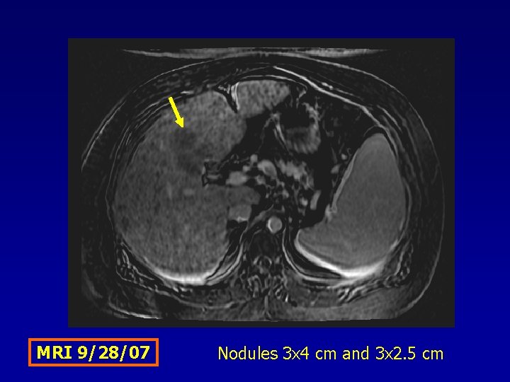
MRI 9/28/07 Nodules 3 x 4 cm and 3 x 2. 5 cm
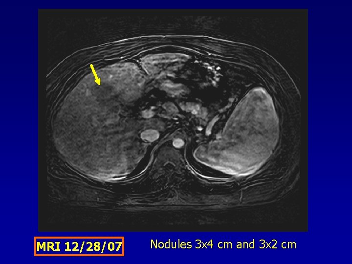
MRI 12/28/07 Nodules 3 x 4 cm and 3 x 2 cm
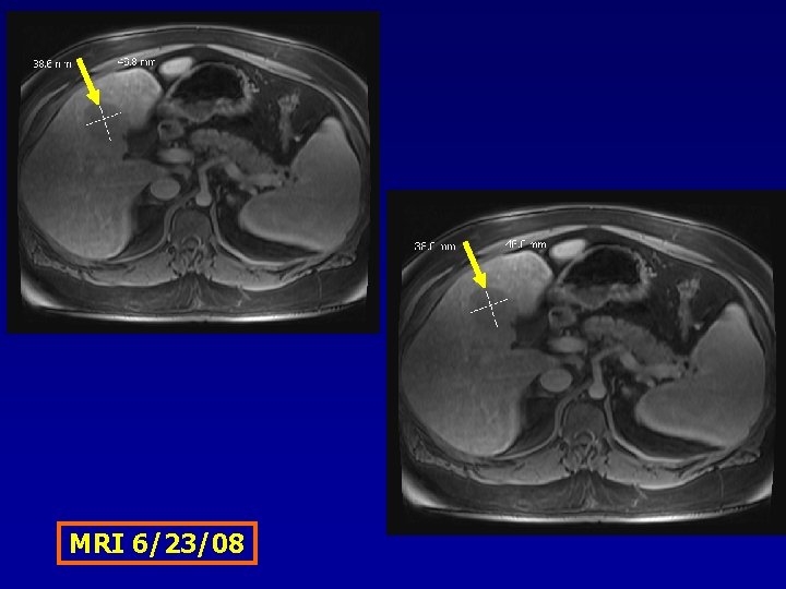
MRI 6/23/08
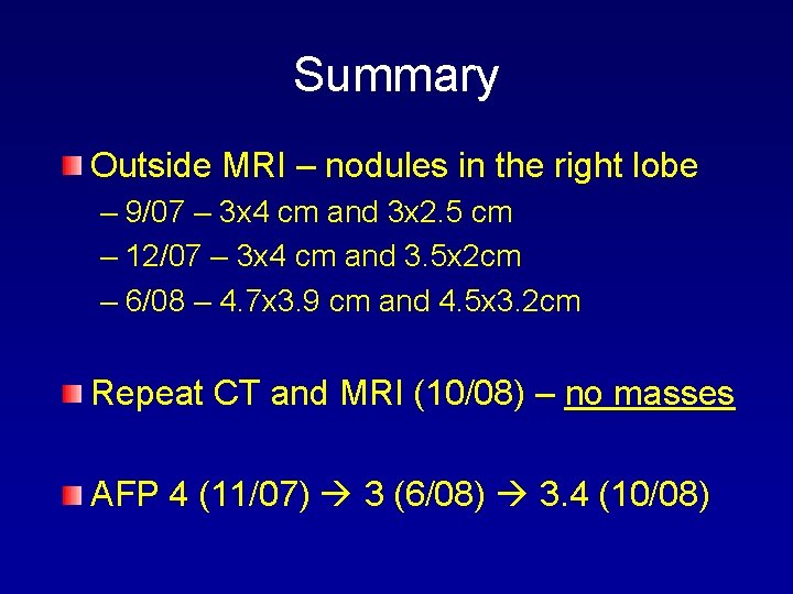
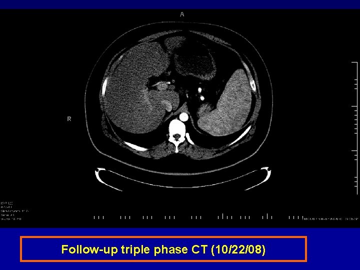
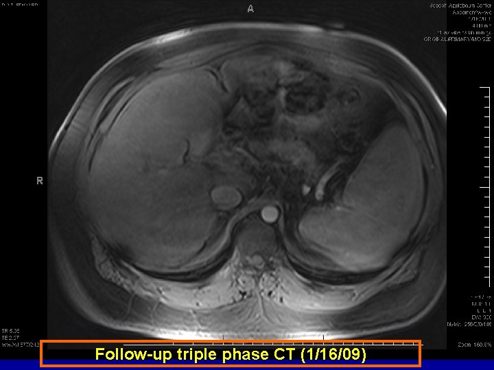
Summary Outside MRI – nodules in the right lobe – 9/07 – 3 x 4 cm and 3 x 2. 5 cm – 12/07 – 3 x 4 cm and 3. 5 x 2 cm – 6/08 – 4. 7 x 3. 9 cm and 4. 5 x 3. 2 cm Repeat CT and MRI (10/08) – no masses AFP 4 (11/07) 3 (6/08) 3. 4 (10/08)
Follow-up triple phase CT (10/22/08)
Follow-up triple phase CT (1/16/09)
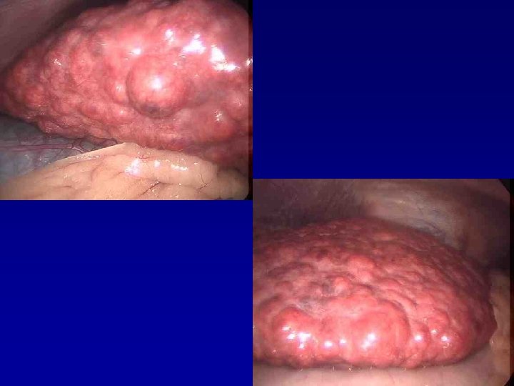
Work Up NO varices on EGD Etiology is NASH related cirrhosis Diagnostic laparoscopy
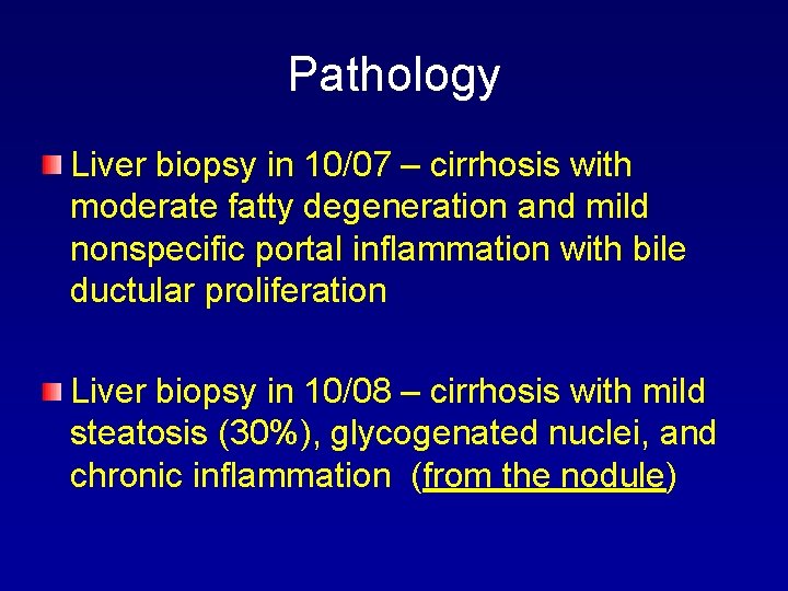
Pathology Liver biopsy in 10/07 – cirrhosis with moderate fatty degeneration and mild nonspecific portal inflammation with bile ductular proliferation Liver biopsy in 10/08 – cirrhosis with mild steatosis (30%), glycogenated nuclei, and chronic inflammation (from the nodule)