Microscopy Chapter 6 Objectives To be able to

Microscopy Chapter 6

Objectives • To be able to describe the light path through a simple lens • To be able to define a compound microscope and describe the light path through it • To be able to name the parts of a compound microscope • To be able to describe how a comparison microscope is constructed

Objectives • To be able to describe how a stereo microscope is constructed • To be able to define plane polarized light • To be able to describe how a polarized light microscope works • To be able to describe how a scanning electron microscope works • To be able to define and describe energy dispersive x-ray analysis

Introduction • The instruments you will encounter most often in a forensics lab is the microscope • Most evidence is of the trace variety – There is a small amount of it so it must be conserved • Examination with a microscope does not destroy evidence • Sometimes the only instrument needed is a microscope

Types of Microscopes • The two major types of microscopes are compound and electron microscopes • Microscopes discussed in this chapter include: – Simple magnifier (magnifying glass) – Compound (basic, stereo, polarized light, comparison, microspectrophotometer) – Electron • Table 6. 1 lists common evidence types and the microscopes that are used

Lenses: How Objects are Magnified • The most simple type of all microscopes is the simple convex lens • Convex lenses bend (refract) light rays as they pass through the lens • Light rays form a virtual image • The shape of the lens determines the magnification • The thicker the lens is in the middle, the higher the magnification – However, thickness causes distortion
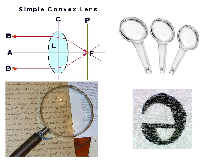

Lenses: How Objects are Magnified • The practical limit of a single lens is 50 x • Improvements can be made by using two convex lenses – The first lens magnifies the object, the second magnifies the virtual image • The total magnification is the product of the magnification of each lens – 10 x and 20 x = 200 x
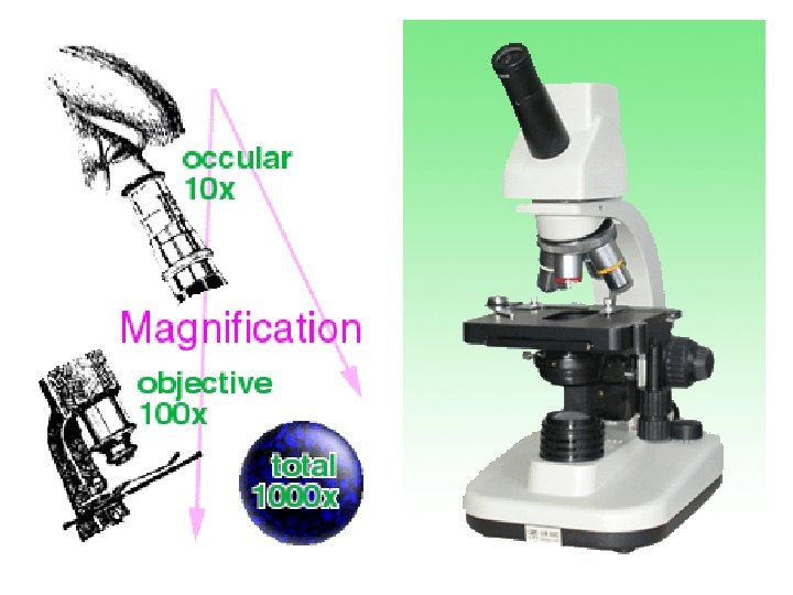

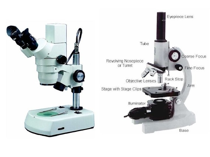
The Compound Microscope 1 • A microscope made from two convex lenses is called a compound microscope – Lens 1 = eyepiece, lens 2 = objective • The evidence sits on the stage and a light source shines through the object • The body tube is above the stage with the objective lenses mounted beneath it • Most microscopes are parfocal - once an object is in focus, lenses can be changed and the object will remain in focus

The Compound Microscope 2 • At the top of the body tube is the eyepiece (ocular lens) – Single lens = monocular – Two lenses (both eyes) = binocular – Three lenses (for photomicrographs) = trinocular • A course and fine focus are used to focus the object • The diaphragm is beneath the stage and controls the amount of light that reaches the object • Filters can limit the wavelengths of light that reach the object

The Compound Microscope 3 • Reflected light microscopes are used for opaque objects such as bullets – The light source is mounted above the stage • The most important characteristics of compound microscopes are: – Magnification, resolution, field of view, and depth of focus

The Compound Microscope 4 • Magnification: the product of the magnification of the ocular and objective lens (up to 1000 x) • Resolution: The ability of a lens to separate details of an object into distinct images rather than one blurred image • Field of view: How much of an object is visible at one time • Depth of focus: how far inside the object the image will be in focus

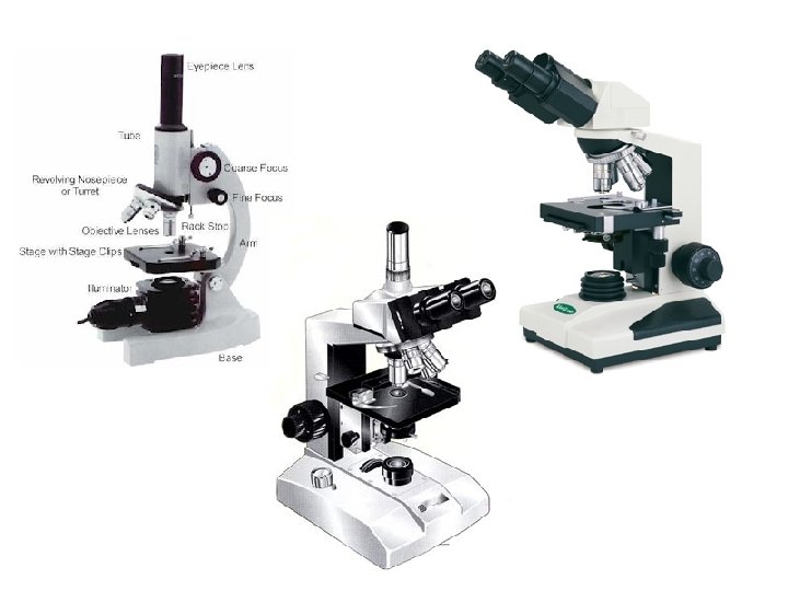
Microscopes Derived from CM’s • The compound microscope can be modified in a number of useful ways to accommodate special circumstances • Examples of modified compound microscopes include: – Comparison microscope – Stereo microscope – Polarized light microscope – Microspectrophotometer

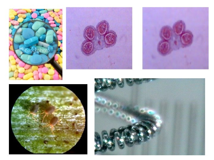
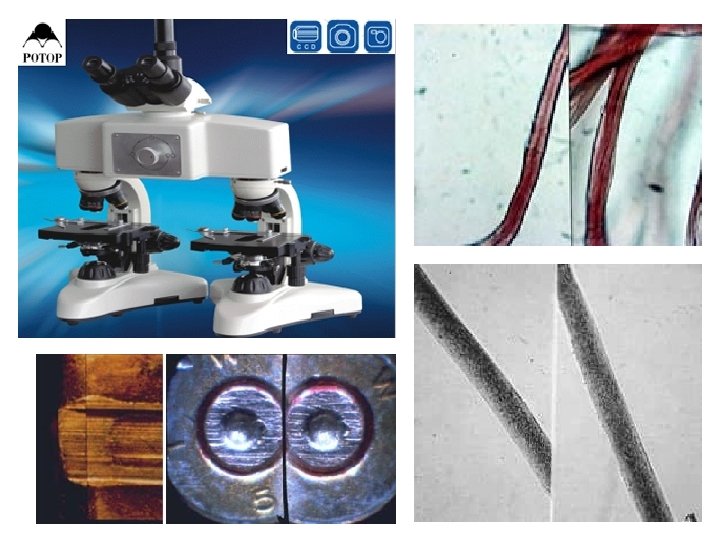
The Comparison Microscope • Evidence often needs to be microscopically compared – Fired bullets, hairs, fibers, etc. • The comparison microscope enables the examiner to view two objects, side-by-side, at the same time • The comparison microscope consists of two compound microscopes connected with a comparison bridge
, the")
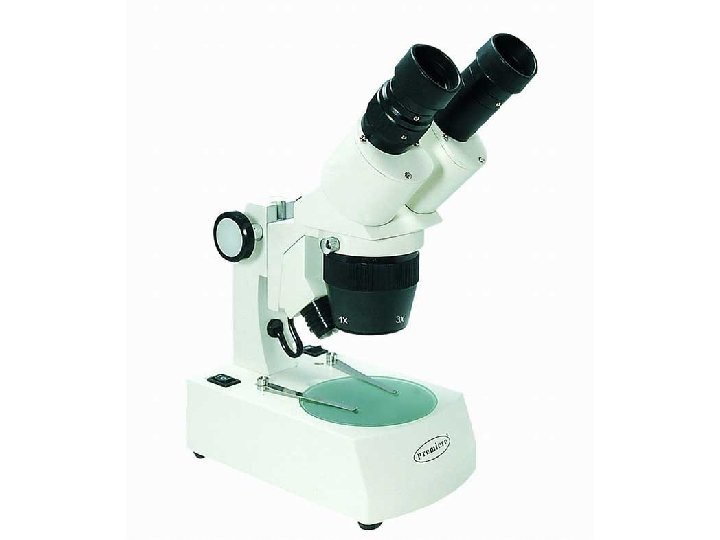
The Stereo Microscope • Stereo microscopes typically have low magnification (25 -50 x), the ability to manipulate the material, and the ability to see it in three dimensions • It is the most versatile and commonly used in forensics labs • It has a long working distance – Allows the examiner to get hands and tools under the lenses • Stereo microscopes consist of two monocular compound microscopes aligned with slightly different viewing angles to create a 3 D image

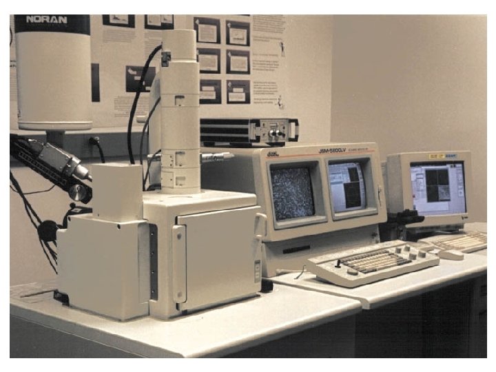
Scanning Electron Microscopy • Electrons microscopes use electrons instead of light to magnify an image • A scanning electron microscope (SEM) can magnify from 10 to 200, 000 times • A beam of electrons is aimed at the object and are backscattered • Backscattered electrons are captured, amplified, and aimed at a cathode ray tube (TV) to create an image
- Slides: 23