Microscopy Bright field microscope Ordinary microscope is called

Microscopy

Bright field microscope • Ordinary microscope is called a bright – field microscope, because it forms a dark image against a brighter background • objective lens forms an enlarged real image • eyepiece (ocular) lens further magnifies this primary image into the final virtual image • total magnification is multiplying the objective and eyepiece magnification together

Resolution • Resolution is the ability of a lens to separate or distinguish between small objects that are close together 0. 5 λ • d = ------n sin θ • Where λ is the wavelength of light used for illumination • n sin θ Is the numerical aperture (NA) • As d becomes smaller, the resolution increases
 is defined as")
• Numerical aperture = n sin θ • Theta (θ) is defined as ½ the angle of the cone of light entering an objective • The maximum theoretical resolving power of a microscope with an oil immersion objective, bluegreen light is approximately 0. 2 µ m

Light rays not refracted In oil

Properties of objective lenses Property Magnification Objective lenses Oil immersion Scanning Low High power 4 x 10 x 0. 10 0. 25 Approx. focal length 40 mm 16 mm 4 mm 0. 1 mm Approx. resolving power 2. 3 µ m 0. 9 µ m 0. 35 µ m 0. 18 µ m NA with light of 450 nm (blue light) Ideally a microscope should be Par focal 40 – 45 x 90 – 100 x 0. 55 – 0. 65 1. 25 – 1. 4 (0. 2 µ m)

Inverted microscope

Dark field Microscopy • Dark field microscope is used for examining live microorganisms which are invisible in the ordinary light microscope, that can not be stained • dark field condenser has a opaque disc that blocks light that would enter the objective directly • light that is reflected off the specimen reaches the objective lens • specimen appears bright against a black background

Dark Field Microscopy

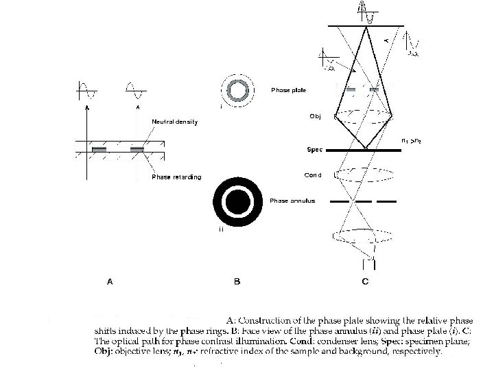
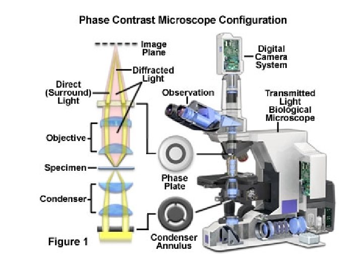
Phase – contrast microscopy • Can be used to study internal structures in living microorganisms • No need to fix or stain • Principle of phase-contrast microscopy is based on slight variations in refractive index • Light rays passing through the specimen are diffracted differently and travel in different pathways • These phase differences are seen through as different degrees of brightness

• A phase contrast microscope uses a special condenser that contains a ring-shaped annular diaphragm • The diaphragm allows a ring of light to pass through the condenser, focusing light on the specimen and a ring shaped phase plate in the objective lens • diffracted and undiffracted rays are then brought into phase with each other to produce the image

Phase contrast microscopy

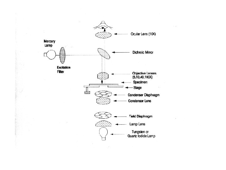
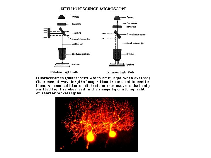
Fluorescence Microscope • Fluorescence microscope exposes a specimen to ultra violet light • Usually a mercury vapour arc lamp produces an intense beam • The light passes through an exciter filter • A dark field condenser provides a black background against which the fluorescent objects glow • Usually the specimens are stained with fluorescent dyes, called fluorochromes • The fluorescence microscope can be used for direct detection of microorganisms fluorescent antibody techniques direct microscope counts (DMC)

Electron Microscope • Electrons are used to illuminate the specimen for magnification • Resolving power increases by about a thousand fold, 0. 3 nm – 0. 5 nm • Transmission electron microscope (TEM) • Scanning electron microscope (SEM)

Transmission Electron Microscope • Electron gun generates a beam of electrons that is focused on the specimen by the condenser • Magnetic lenses (electromagnets) are used to focus the beam • Specimen must be kept under high vacuum • Focused by magnetic lenses to form an enlarged visible image of the specimen on a fluorescent screen • Denser region in the specimen scatters more electrons and appears darker in the image • Electron-transparent regions are brighter • Image can be captured on photographic film
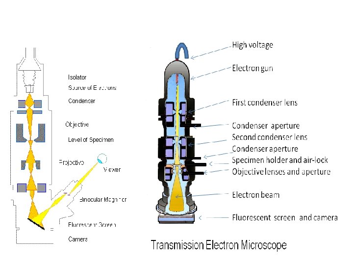

TEM

Scanning Electron Microscope • To study external surface features of microorganisms • SEM provides three dimensional views • electromagnetic lenses and are directed over the surface of the specimen • The primary electron beam knocks electrons out of the surface of the specimen • secondary electrons are transmitted to an electron collector, amplified, used to produce an image • SEM has a resolution 20 nm, 10000 x

SEM

Most advanced • Aluminum alloy sample seen through the lens of the Titan 80 -300 Cubed. (Credit: Image courtesy of Mc. Master University ) • Scanning Tunneling microscope • An advanced electron microscope • This microscope can easily identify atoms, measure their chemical state and even probe the electrons that bind them together
- Slides: 24