Microscopical features Paucicellular aspirate Small and large loosely


 Ø Sclerosing hemangioma Ø")


- Slides: 19
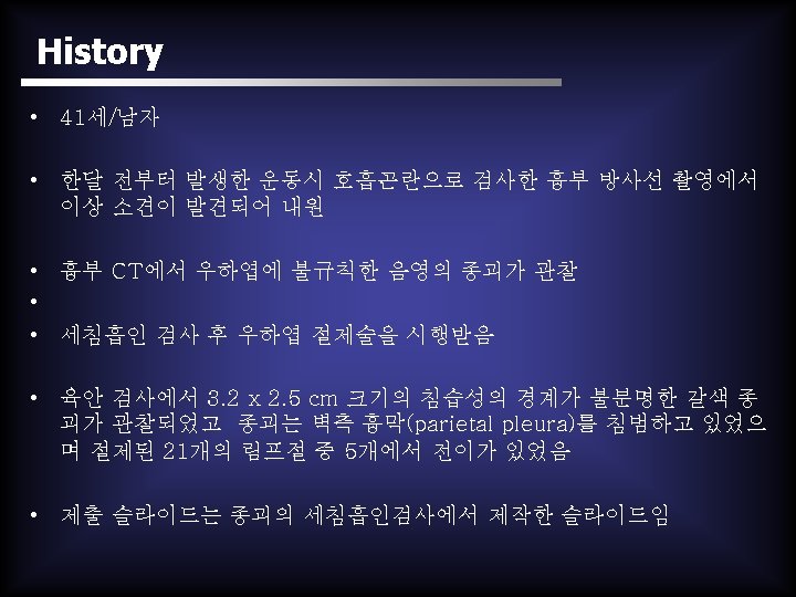
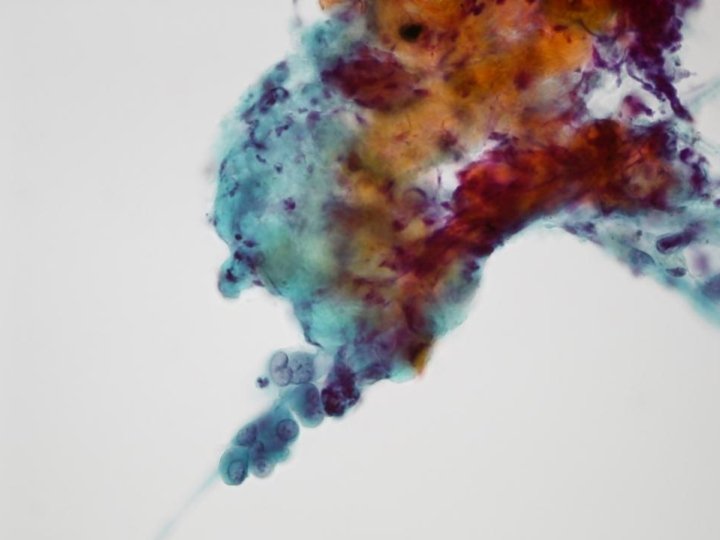
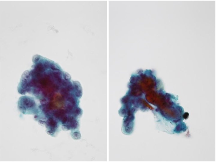
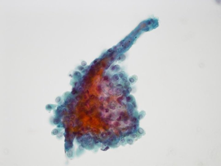
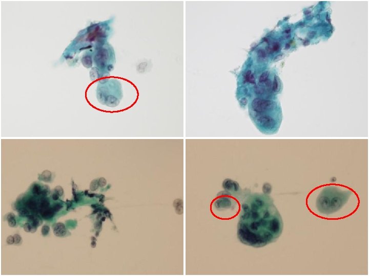
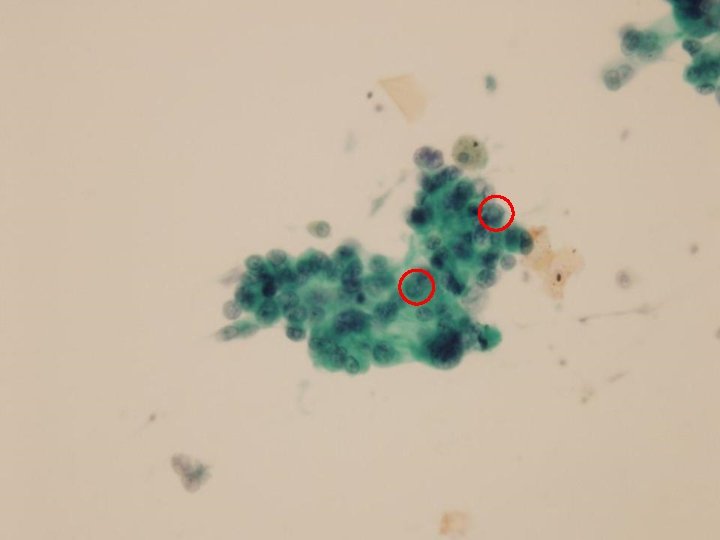
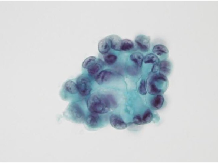
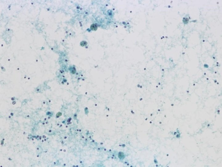
Microscopical features • Paucicellular aspirate • Small and large loosely cohesive cell groups of round to polygonal epithelioid cells • Focally, Papillary or acinar –like structures • Intermediate to large cells, with round to oval nuclei with frequent binucleation and multinucleation • Folded or wrinkled nuclear membrane • Abundant cytoplasm • Eccentrically placed nuclei (plasmacytoid) and granular chromatin with small nucleoli • Intracytoplasmic vacuoles and intranuclear pseudoinclusion • Connective tissue fragment • Lacks necrosis or mitotic figures • Foamy histiocytes and lymphocytes in background
Differential diagnosis Ø Well differentiated adenocarcinoma (Such as bronchioloalveolar carcinoma) Ø Sclerosing hemangioma Ø Mesothelioma, epithelioid type Ø Epithelioid hemangioendothelioma
Well differentiated adenocarcinoma • Peripheral, ill-defined nodule • Lymph node metastases • Non-mucinous BAC - Papillary formation - Small tissue fragments with undulated borders • Intranuclear cytoplasmic inclusion - commonly in BAC • Gray-white color • Uniform round nuclei with bland chromatin pattern and prominent nucleoli • Scant cytoplasm
Sclerosing hemangioma • Moderately cellular aspirate • Solitary and peripheral • Arranged singly, in sheet or papilla • Hyalinized stromal tissue fragments • Abundant eosinophilic cytoplasm • Intranuclear cytoplasmic inclusion • Mitotically inactive • Foamy, sometimes hemosiderinladen macrophages in the background
Sclerosing hemangioma • Marked female predilection • Well-circumscribed • Lymph node metastases: Rare • Dual cell population: Surface cells and round cells • Uniform round to oval nuclei, scanty cytoplasm • Hemorrhagic aspirate
Mesothelioma • 3 -dimentional papillary clusters • Central core of connective tissue • Eosinophilic cytoplasm and bland nuclei • Abundant faintly vacuolated cytoplasm – clear periphery and denser perinuclear area • Occupational disease (not all) • Old age (Largely, > 60 yrs) • Multiple small nodules with large pleural effusion • Increased cellularity • Mitotic activity
Epithelioid hemangioendothelioma • Usually, young women • Bilateral, multiple • Gray-white, non-encapsulated, discrete nodules • Lymph node meta: 10. 8% • Loosely cohesive sheets • Pseudopapillary or acinar structure • Round to polygonal epithelioid cells with abundant cytoplasm • Intracytoplasmic vacuolation and intranuclear pseudoinclusions • bi- or multinucleation • Small or inconspicuous nucleoli • Low mitotic activity • Excessive hyaline matrix
Epithelioid hemangioendothelioma