Microscopic Structure of Compact Bone Consists of multiple

Microscopic Structure of Compact Bone • Consists of multiple cylindrical structural units known as osteons or haversian systems. • Imagine these osteons as weight-bearing pillars that are arranged parallel to one another along the long axis of a compact bone. The diagram below represents a long bone shaft in cross-section. Each yellow circle represents an osteon. The blue represents additional matrix filling in the space between osteons. The white in the middle is the marrow cavity.

Osteons • Each osteon consists of a single central canal, known as a haversian canal, surrounded by concentric layers of calcified bone matrix. • Haversian canals allow the passage of blood vessels, lymphatic vessels, and nerve fibers. • Each of the concentric matrix “tubes” that surrounds a haversian canal is known as a lamella. • All the collagen fibers in a particular lamella run in a single direction, while collagen fibers in adjacent lamellae will run in the opposite direction. This allows bone to better withstand twisting forces.

Running perpendicular to the haversian canals are Volkmann’s canals. They connect the blood and nerve supply in the periosteum to those in the haversian canals and the medullary cavity.

Osteons • Lying in between intact osteons are incomplete lamellae called interstitial lamellae. These fill the gaps between osteons or are remnants of bone remodeling. • There also circumferential lamellae that extend around the circumference of the shaft. There are inner circumferential lamellae surrounding the endosteum and outer circumferential lamellae just inside the periosteum.

• Osteocytes occupy small cavities known as lacunae at the junctions of the lamellae. Hairlike canals called canaliculi connect the lacunae to each other and to the central canal. • Canaliculi allow the osteocytes to exchange nutrients, wastes, and chemical signals to each other via intercellular connections known as gap junctions.
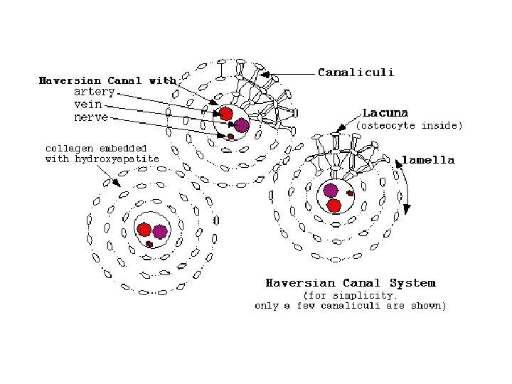
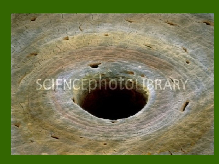

Here, we have a close up and a far away view of compact bone. You should be able to identify haversian canals, concentric lamellae, interstitial lamellae, lacunae, and canaliculi.

Microscopic Structure of Spongy Bone • Appears poorly organized compared to compact bone. • Lacks osteons. • Trabeculae align along positions of stress and exhibit extensive cross-bracing. • Trabeculae are a few cell layers thick and contain irregularly arranged lamellae and osteocytes interconnected by canaliculi. • No haversian or Volkmann’s canals are necessary.
- Slides: 9