Microscopic Anatomy Modifications for Absorption Small intestine is

Microscopic Anatomy Modifications for Absorption • Small intestine is highly adapted for nutrient absorption • Length provides a huge surface area and its wall has three structural modifications that amplify its absorptive surface enormously: – Plicae circulares – Villi – Microvilli • Most absorption occurs in the proximal part of the small intestine, so these specializations decrease in number toward its distal end

Microscopic Anatomy Plicae Circulares • Circular folds that are deep, permanent folds of the mucosa and submucosa (a) • These folds force chyme to spiral through the lumen, slowing its movement and allowing time for full nutrient absorption
 (b) • Give the")
Microscopic Anatomy Villi • Fingerlike projections of the mucosa (a) (b) • Give the mucosa a velvety texture • Epithelial cells of the villi are chiefly absorptive columnar cells • In the core of each villus is a dense capillary bed and a wide lymph capillary called a lacteal

Microscopic Anatomy Villi • Digested food is absorbed through the epithelial cells into both the capillary blood and the lacteal • A slit of smooth muscle (b) (muscularis mucosae) in the villus core allows it to alternately shorten and lengthen, pulsations that: – 1. Increase the contact between the villus and the contents of the intestinal lumen, making absorption more efficient – 2. Milk lymph along through the lacteals

Microscopic Anatomy Microvilli • Tiny projections of the plasma membrane of the absorptive cells of the mucosa (b) (c) • Gives the mucosal surface a fuzzy appearance called the brush border • The plasma membrane of the microvilli bear enzymes referred to as brush border enzymes, which complete the digestion of carbohydrates and proteins in the small intestine

SMALL INTESTINE MODIFICATIONS

VILLI

Histology of the Wall • The four tunics typical of the GI tract (mucosa, submucosa, muscularis externa, adventita) are also seen here, but the mucosa and submucosa are modified to reflect the intestine’s functions in the digestive pathway

Histology of the Wall • • Epithelium of the mucosa is largely simple columnar absorptive cells bound by tight junctions and richly endowed with microvilli Many secretory cells: – Goblet cells: mucus-secreting (d) – Enterogastrones: secretes • Secretin: – A hormone that stimulates sodium bicarbonate secretion by the pancreas and bile secretion by the liver – Decreases gastrointestinal peristalsis and motility • Cholecystokinin: – A hormone that stimulates contraction of the gallbladder and pancreatic secretions – Intraepithelial lymphocytes (T cells): represent an important immunological compartment • Generated locally (not in the Thymus)

Histology of the Wall • Between the villi, the mucosa is studded with pits that lead into tubular intestinal glands called intestinal crypts (crypts of Lieberkuhn): – The epithelial cells that line these crypts secrete intestinal juice: • A watery mixture containing mucus that serves as a carrier fluid for absorbing nutrients from chyme – Deep in the crypts are specialized secretory cells called Paneth cells: • Release lysozyme, an antibacterial enzyme

Histology of the Wall • • • Submucosa is typical areolar connective tissue – Contains both individual and aggregated lymphoid follicles (Peyer’s patches) – In the duodenal (Brunner’s) glands: (b) • Produce an alkaline (bicarbonate-rich) mucus that helps neutralize the acidic chyme moving in from the stomach Muscularis is typical and bilayered Adventitia: covered by visceral peritoneum (serosa)

SMALL INTESTINE MODIFICATIONS

Intestinal Juice: Composition and Control • Major stimulus is distension or irritation of the intestinal mucosa by hypertonic or acidic chyme • Slightly alkaline (7. 4 -7. 8) • Isotonic with blood plasma • Largely water but it also contains some mucus, which is secreted both by the duodenal glands and by goblet cells of the mucosa • Relatively enzyme-poor because intestinal enzymes are largely limited to the bound enzymes of the brush border

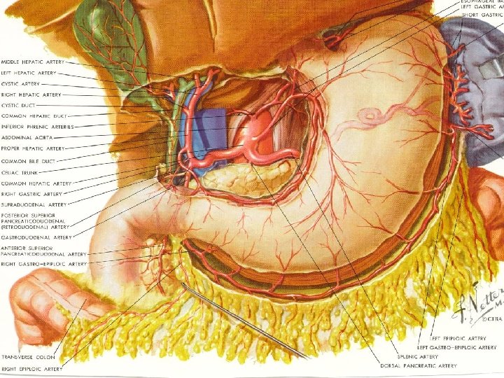
Associated Structures Liver and Gallbladder • Liver: one of the body’s most important organs, has many metabolic and regulatory roles – It’s digestive function is to produce bile for export to the duodenum: • Bile is a fat emulsifier; that is, it breaks up fats into tiny particles so that they are more accessible to digestive enzymes – Also processes nutrient-laden venous blood delivered to it from the digestive organs, this is a metabolic rather than a digestive role • Gallbladder is chiefly a storage organ for bile

LIVER • Largest gland in the body • Occupies most of the right hypochondriac and epigastric regions • Located under the diaphragm • Lies almost entirely within the rib cage, which provides some protection
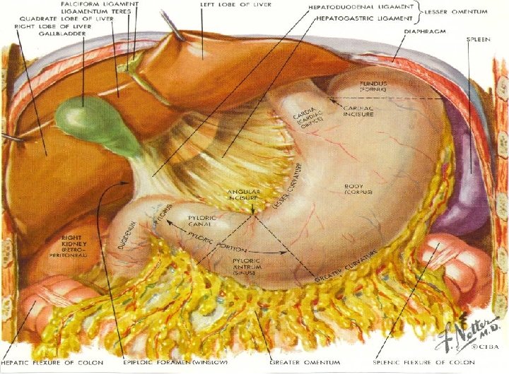

LIVER • Has four primary lobes • A mesentery, separates the right and left lobes anteriorly and suspends the liver from the diaphragm and anterior abdominal wall • Entire liver is enclosed by the visceral peritoneum

LIVER

LIVER • Bile leaves the liver through several bile ducts that ultimately fuse to form the large common hepatic duct, which travels downward toward the duodenum – Along its course, it fuses with the cystic duct draining the gallbladder to form the bile duct

LIVER • The liver is composed of liver lobules, each lobule is a roughly hexagonal (sixsided) structure consisting of plates of liver cells (hepatocytes), organized like bricks in a garden wall – The hepatocyte plates radiate outward from a central vein running in the longitudinal axis of the lobule

LIVER • The liver’s main function is to filter and process the nutrientrich blood delivered to it • At the end of the six corners of a lobule is a portal triad (three structures) – Branch of the hepatic artery: supplying oxygen-rich arterial blood to the liver – Branch of the hepatic portal vein: carrying venous blood laden with nutrients from the digestive viscera – Bile duct

LIVER • Between the hepatocyte plates are enlarged, leaky capillaries, the liver sinusoids – Blood from both the hepatic portal vein and the hepatic artery percolates from the triad regions through these sinusoids and empties into the central vein • From the central veins blood eventually enters the hepatic veins, which drain the liver, and empty into the inferior vena cava
(d)")
LIVER • Forming part of the sinusoid walls are starshaped hepatic macrophages (Kupffer cells)(d) – Remove debris such as bacteria and worn-out blood cells from the blood as it flows past

LIVER • The versatile hepatocytes have large amounts of both rough and smooth ER, Golgi apparatuses, peroxisomes, and mitochondria • Thus equipped, the hepatocytes not only produce bile but can also: – 1. Process the bloodborne nutrients in various ways • Store glucose as glycogen • Use amino acids to make plasma proteins – 2. Store fat-soluble vitamins – 3. Play important roles in detoxification, such as ridding the blood of ammonia by converting it to urea • Blood leaving the liver contains fewer nutrients and waste materials than the blood that entered it

LIVER

LIVER

HOMEOSTATIC IMBALANCE • Hepatitis: inflammation of the liver (most often due to viral infection – Hepatitis A virus (HVA): • Transmitted enterically (pertaining to the small intestine) – Sewage contaminated food, raw shellfish, water, feces-mouth route • • Infection is self-limiting 32% of hepatitis cases Benign form Frequently in day care centers – Hepatitis B virus (HVB): • • • Transmitted via blood, contaminated needles, sexual contact Linked to chronic hepatitis and liver cirrhosis 40% of hepatitis cases Elevated risk of cancer vaccine – Hepatitis C virus (HVC): • Transmitted via blood • Linked to chronic hepatitis and liver cirrhosis • Most important liver disease in the U. S. because it produces persistent or chronic liver infections
: • Mutated virus that needs HVB to")
HOMEOSTATIC IMBALANCE – Hepatitis D virus (HVD): • Mutated virus that needs HVB to be infectious – Hepatitis E virus (HVE): • Transmitted enterically (pertaining to the small intestine) – Sewage contaminated food, raw shellfish, water, feces-mouth route • Infection is self-limiting • Waterborne epidemics in developing countries – Major case of death in pregnant women – Hepatitis F virus (HVF): • Little is known

HOMEOSTATIC IMBALANCE • Nonviral causes of acute hepatitis include drug toxicity and wild mushroom poisoning • Cirrhosis: – Diffuse and progressive chronic inflammation of the liver that typically results from chronic alcoholism or severe chronic hepatitis • Alcohol-poisoned or damaged hepatocytes regenerate, but the liver’s connective (scar) tissue regenerates faster – As a result, the liver becomes fatty and fibrous and its activity is depressed – As the scar tissue shrinks, it obstructs blood flow throughout the hepatic portal system, causing portal hypertension

COMPOSITION of BILE • Bile is a yellow-green, alkaline solution containing bile salts, bile pigments (primarily bilirubin), cholesterol, neutral fats, phospholipids (lecithin and others), and a variety of electrolytes – Of these only bile salts and phospholipids aid the digestive process • Bile salts: – Primarily cholic acid and chenodeoxycholic acids » Cholesterol derivatives – Role is to emulsify fats—that is, to distribute them throughout the watery intestinal contents » As a result, large fat gobules entering the small intestine are physically separated into millions of small fatty droplets that provide large surface areas for the fatdigesting enzymes to work on – Facilitate fat and cholesterol absorption and help solubilize cholesterol – Although many substances secreted in bile leave the body in feces, bile salts are not among them » Instead, bile salts are conserved by means of a recycling mechanism, called the enterohepatic circulation, and returned to the liver by means of the hepatic portal blood after being reabsorbed by the ileum

COMPOSITION of BILE • Chief bile pigment is bilirubin, a waste product of the heme of hemoglobin formed during the breakdown of worn-out erythrocytes – Absorbed by the liver cells, excreted into bile, and metabolized in the small intestine by resident bacteria • One of the breakdown products, urobilinogen, gives feces a brown color – In the absence of bile, feces are gray -white in color and have fatty streaks (because essentially no fats are digested or absorbed) • Stimulus of bile secretion: – Bile salts – Secretin (released by the intestinal cells)

MECHANISMS OF BILE PRODUCTION

Gallbladder • Thin-walled green muscular sac • Size of a kiwi fruit • Stores and concentrates bile (absorbing some of its water and ions) that is not needed immediately for digestion • When its muscular wall contracts, bile is expelled into its duct, the cystic duct, and then flows into the bile duct

Regulation of Bile Release into the Small Intestine • When no digestion is occurring, the hepatopancreatic sphincter (guarding the entry of bile and pancreatic juice into the duodenum) is closed and the released bile backs up the cystic duct into the gallbladder, where it is stored until needed • Although the liver makes bile continuously , bile does not usually enter the small intestine until the gallbladder contracts • Major stimulus for gallbladder contraction is when stimulated by cholecystokinin (CCK), an intestinal hormone – Released into the blood when acidic, fatty chyme enters the duodenum – Stimulates the secretion of pancreatic juice – Relaxes the hepatopancreatic sphincter so that bile and pancreatic juice can enter the duodenum – Parasympathetic impulses delivered by the vagus nerves are a minor stimulus for gallbladder contraction

HOMEOSTATIC IMBALANCE • Bile is the major vehicle for cholesterol excretion from the body, and bile salts keep the cholesterol dissolved within bile – Too much cholesterol or too few bile salts leads to cholesterol crystallization, forming gallstones (biliary calculi) which obstruct the flow of bile from the gallbladder • When the gallbladder or its duct contracts, the sharp crystals cause agonizing pain that radiates to the right thoracic region • Treatment: – Ultrasound: pulverization (lithotripsy) – Lasers: vaporization – Surgically removal

PANCREAS • Is retroperitoneal and lies deep to the greater curvature of the stomach • Pancreatic juice (exocrine product) consists mainly of water and contains enzymes (delivered to the duodenum) that break down all categories of foodstuffs and electrolytes • Pancreatic duct generally fuses with the bile duct just as it enters the duodenum – A smaller accessory pancreatic duct empties directly into the duodenum just proximal to the main duct

PANCREAS • Acini: cluster of secretory cells surrounding ducts – Full of rough endoplasmic reticulum and exhibit deeply staining zymogen granules containing the digestive enzymes they manufacture – Scattered amidst the acini are the more lightly staining pancreatic islets (islets of Langerhans) • Release hormones: insulin and glucagon

STRUCTURE OF ACINAR TISSUE OF PANCREAS

Pancreatic Juice • Consists mainly of water, and contains enzymes and electrolytes (primarily bicarbonate ions—making it alkaline-p. H 8) • Acinar cells produce the enzyme-rich component of pancreatic juice • The high p. H (8) enables it to neutralize acid chyme entering the duodenum and provides the optimal environment for activity of intestinal and pancreatic enzymes
 are produced and released in inactive forms,")
Pancreatic Juice • Pancreatic proteases (protein-digesting enzymes) are produced and released in inactive forms, which are activated in the duodenum, where they do their work (This prevents the pancreas from selfdigestion) – Trypsinogen is activated to trypsin by enterokinase (an intestinal brush border enzyme) – Proteases (procarboxypeptidase and chymotrypsinogen) are activated to their active forms (carboxypeptidase and chymotrypsin) by trypsin – Amylase, lipases, and nucleases—are secreted in active form, but require that ions or bile be present in the intestinal lumen for optimal activity

Activation of pancreatic proteases in the small intestine

Regulation of Pancreatic Secretion • Regulated by: – Intestinal hormones: • Both act on the pancreas • Secretin released in response to the presence of HCl in the intestine targets the duct cells, prompting their release of watery bicarbonaterich pancreatic juice • Cholecystokinin (CCK) released in response to the entry of proteins and fats, stimulates the acini to release enzyme-rich pancreatic juice – Parasympathetic nervous system: • Vagal stimulation causes release of pancreatic juice primarily during the cephalic and gastric phases of gastric secretion

Regulation of pancreatic juice secretion by hormonal and neural factors

Digestive Processes Occurring in the Small Intestine • Food reaching the small intestine is far from being digested chemically – Carbohydrates and proteins are partially degraded, but virtually no fat digestion has occurred to this point • Food takes 3 to 6 hours to complete its digestive path through the small intestine, the site of virtually all nutrient absorption

Requirements for Optimal Intestinal Digestive Activity • Although the primary functions of the small intestine are digestion and absorption, intestinal juice provides little of what is needed to perform these functions: – Most substances required for chemical digestion within the small intestine are imported from the pancreas and the liver – Hence, anything that impairs liver or pancreatic function or delivery of their juices to the small intestine severely hinders our ability to digest food and absorb nutrients • Optimal digestive activity in the small intestine depends on a slow, measured delivery of chyme from the stomach (controlled by the pumping action of the stomach pylorus): – Why is this so? • Entering chyme is usually hypertonic – Thus, if large amounts of chyme were rushed into the small intestine, the osmotic water loss from the blood into the intestinal lumen would result in dangerously low blood volume – Additionally, the low p. H of entering chyme must be adjusted upward and the chyme must be well mixed with bile and pancreatic juice for digestion to continue

Motility of the Small Intestine • • Intestinal smooth muscle mixes chyme thoroughly with bile and pancreatic and intestinal juices, and moves food residues through the ileocecal valve into the large intestine (colon) In contrast to the peristaltic waves of the stomach, which both mix and propel food, segmentation is the most common motion of the small intestine – Chyme is moved backward and forward in the lumen a few centimeters at a time by alternating contraction and relaxation of rings of smooth muscle • Like the peristalsis of the stomach, segmentation is initiated by intrinsic pacemaker cells in the longitudinal smooth muscle layer (b)

Motility of the Small Intestine • Intensity of segmentation is altered by long and short reflexes (which parasympathetic activity enhances and sympathetic activity decreases) and hormones • True peristalsis occurs only after most nutrients have been absorbed: – At this point, segmenting movements wane, and peristaltic waves initiated in the duodenum begin to sweep slowly along the intestine – Each successive wave is initiated a bit more distally, and this pattern of peristaltic activity is called the migrating mobility complex

Motility of the Small Intestine • Most of the time, the ileocecal sphincter is constricted and closed • However, two mechanisms—one neural and the other hormonal—cause it to relax and allow food residues to enter the cecum when ileal mobility increases – Neural: enhanced activity of the stomach initiates the gastroileal reflex that enhances the force of segmentation – Hormonal: gastrin released by the stomach increases the motility of the ileum and relaxes the ileocecal sphincter • Once the chyme has passed through, it exerts backward pressure that closes the valve’s flaps, preventing regurgitation into the ileum


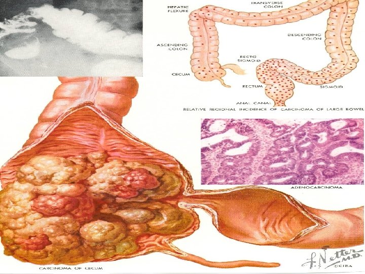

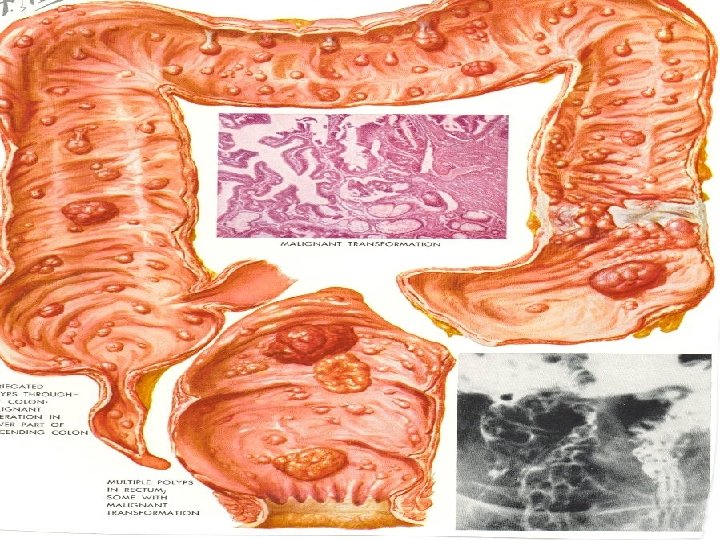
Large Intestine • • In terms of nutrient absorption, its major function is to absorb water from indigestible food residues (delivered to it in a fluid state) and eliminate them from the body as semisolid feces The large intestine exhibits three unique features: – Teniae coli: ribbons of the colon – Haustra: pocketlike sacs – Epiploic appendages: small fatfilled pouches of visceral peritoneum that hang from its surface • Significance is not known

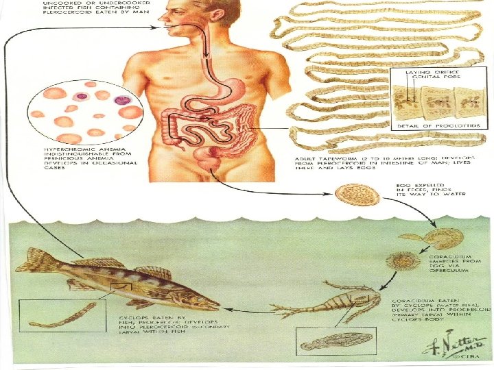
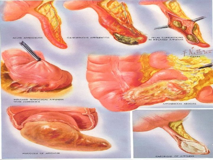
Large Intestine • Has the following subdivisions: cecum, appendix, colon, rectum, and anal canal • Saclike cecum: lies below the ileocecal valve in the right iliac fossa, is the first part of the colon – Attached to its posteromedial surface is the blind, wormlike vermiform appendix • Contains masses of lymphoid tissue, and as part of mucosaassociated lymphatic tissue (MALT), it plays an important role in body immunity

LARGE INTESTINE

HOMEOSTATIC IMBALANCE • Appendicitis: acute inflammation of the appendix – Results from a blockage (often by feces) that traps infectious bacteria in its lumen – Unable to empty its contents, the appendix swells, squeezing off venous drainage, which may lead to ischemia (deficiency of blood to an organ) and necrosis (death and decay) of the appendix – If the appendix ruptures, feces containing bacteria spray over the abdominal contents, causing peritonitis (inflammation of the serous membrane that lines the abdominal cavity and its viscera)

COLON • • Ascending colon Transverse colon Descending colon Sigmoid colon

ORGANS AND MESENTERIES OF ABDOMINAL REGION

ORGANS AND MESENTERIES OF ABDOMINAL REGION
 called rectal")
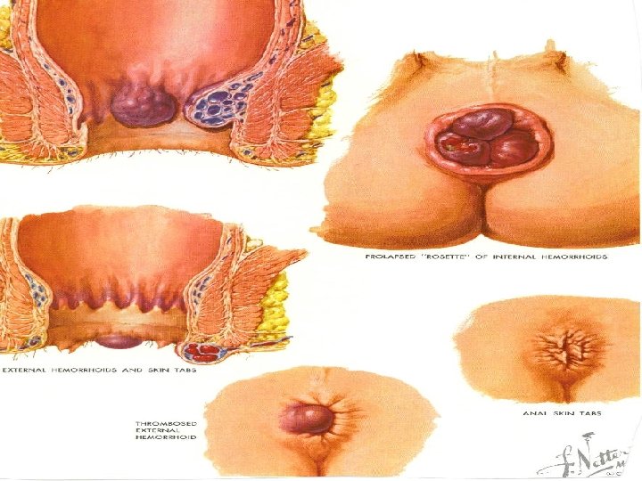
COLON • Rectum: – Has three lateral curves or bends (transverse folds) called rectal valves • Separate feces from flatus (gas), that is, they stop feces from being passed along with gas • Anal canal: has two sphincters (ordinarily closed except during defecation) – Involuntary internal anal sphincter composed of smooth muscle – Voluntary external anal sphincter composed of skeletal muscle

ANAL CANAL

Large Intestine – The mucosa of the large intestine is thick and has crypts with a large number of mucusproducing goblet cells • Mucus produced by goblet cells eases the passage of feces and protects the intestinal wall from irritating acids and gases released by resident bacteria in the colon

MUCOSA OF COLON

ANAL CANAL • Anal sinuses recesses between the anal columns, exude mucus when compressed by feces, which aids in emptying the anal canal • Hemorrhoidal veins: if inflamed—itchy varicosities results called hemorrhoids

Bacterial Flora • Although most bacteria entering the cecum from the small intestine are dead (having been killed by the action of lysozyme, defensins, HCl, and protein-digesting enzymes), some are still alive • Together with bacteria that enter the GI tract via the anus, these constitute the bacterial flora (microbial) of the colon: – Ferment some of the indigestible carbohydrates (cellulose and others), releasing irritating acids and a mixture of gases (dimethyl sulfide, H 2, N 2, CH 4, and CO 2) – Synthesize B complex vitamins and most of the vitamin K the liver requires to synthesize some of the clotting proteins

Digestive Processes Occurring in the Large Intestine • Except for a small amount of digestion of food residue by the enteric bacteria, no further food breakdown occurs in the large intestine • Although the colon harvest vitamins made by the bacterial flora and reclaims most of the remaining water and some of the electrolytes (particularly sodium and chloride), absorption (of nutrients) is not the major function of this organ

Digestive Processes Occurring in the Large Intestine • Colon is not essential for life – Ileostomy: • Removal of the colon and the terminal end of the ileum is attached to the abdominal wall – Food residues are eliminated into a sac – Ileoanal juncture: • Surgically linking the ileum to the anal canal

Motility of the Colon • The movement seen in the large intestine include: – Haustral contractions: slow segmenting movements that occur every 30 minutes • As a haustrum fills with food residue, the distension stimulates its muscle to contract, which propels the luminal contents into the next haustrum • These movements also mix the residue, which aids in water absorption – Mass movements: mass peristalsis • Slow-moving but powerful contractile waves that move over large areas of the colon (3 -4 times/day) • Force contents toward the rectum • Bulk, or fiber, in the diet increases the strength of colon contractions and softens the stool • • The primary concerns of the colon are propulsive activities that force the fecal material toward the anus and then eliminate it from the body (defecation) The semisolid product delivered to the rectum, feces, or the stool, contains undigested food residues, mucus, sloughed-off epithelial cells, millions of bacteria, and just enough water to allow its smooth passage

HOMEOSTATIC IMBALANCE • Diverticulosis: – Diet lacks bulk and the volume of residues in the colon is small – Colon narrows and its contractions become more powerful, increasing the pressure on its walls – Promotes formation of verticula, small herniations of the mucosa through the colon wall – Most common in the sigmoid colon • Diverticulitis: – Diverticula become inflamed – If ruptured: life threatening

DEFECATION • Feces forced into the rectum by mass movements stretch the rectal wall and initiate the defecation reflex – This is a spinal cord-mediated parasympathetic reflex that causes the sigmoid colon and the rectum to contract and the anal sphincters to relax • During defecation, the muscles of the rectum contract to expel the feces – We aid this process voluntarily by closing the glottis and contracting our diaphragm and abdominal wall muscles to increase the intraabdominal pressure (Valsalva”s maneuver)

DEFECATION

HOMEOSTATIC IMBALANCE • Diarrhea: – Watery stools – Results from any condition that rushes food residue through the colon before that organ has had sufficient time to absorb the remaining water (as in irritation of the colon by bacteria) – Dehydration and electrolyte imbalance • Constipation: – When food remains in the colon for extended periods, too much water is absorbed and the stool becomes hard and difficult to pass – May result from lack of fiber in diet, improper bowel habits, lack of exercise, emotional upset, or laxative abuse


PHYSIOLOGY OF CHEMICAL DIGESTION AND ABSORPTION • Chemical digestion is a catabolic process in which large food molecules are broken down to chemical building blocks (monomers), which are small enough to be absorbed by the GI tract lining • Chemical digestion is accomplished by enzymes, secreted by intrinsic and accessory glands into the lumen of the alimentary canal. • The enzymatic breakdown of any type of food molecule is hydrolysis because it involves addition of a water molecule to each molecular bond to be broken (lysed)

Carbohydrate Digestion • Carbohydrates: – Monosaccharides are simple sugars that are absorbed immediately (glucose, galactose, and fructose) – Disaccharides are composed of two monosaccharides bonded together: • Maltose: grain sugar ( grass family: corn, wheat, oats, other cereals) • Lactose: milk sugar • Sucrose: table sugar – The digestible polysaccharide found in the diet is starch and glycogen; other polysaccharides such as cellulose, are not able to be broken down by humans but add bulk or fiber – Chemical digestion of carbohydrates begins in the mouth where salivary amylase breaks large polysaccharides into smaller fragments • Salivary amylase works best in the slightly acid to neutral environment (p. H of 6. 5 -7. 00) maintained in the mouth by the buffering effects of bicarbonate and phosphate ions in salvia • Continues until amylase is inactivated by stomach acid and broken apart by the stomach’s protein-digesting enzymes – Generally, the larger the meal, the longer amylase continues to work in the stomach because food in its relatively immobile fundus are poorly mixed with gastric juices

CARBOHYRATES

Carbohydrate Digestion • Carbohydrates: – Starchy foods and other digestible carbohydrates that escape being broken down by salivary amylase are acted on by pancreatic amylase in the small intestine – Intestinal brush border enzymes (oligosaccharides and glucoamylase from the plasma membranes of microvilli ) further digest these products to monosaccharides – Maltase hydrolyzes maltose to glucose – Sucrase hydrolyzes sucrose to glucose and fructose – Lactase hydrolyzes lactose to glucose and galactose – Because the colon does not secrete digestive enzymes, chemical digestion officially ends in the small intestine – Resident colon bacteria do break down and metabolize the residual complex carbohydrates further, adding much to their own nutrition but essentially nothing to ours

CARBOHYDRATES

CHEMICAL DIGESTION

HOMEOSTATIC IMBALANCE • Lactose intolerant: – Intestinal lactase is present at birth but then becomes deficient (genetic factors) – Become intolerant of milk products (source of lactose) • Undigested lactose creates osmotic gradients that not only prevent water from being absorbed in the small and large intestine but also pull water from the interstitial space into the intestines – Result: » Diarrhea » Bacterial metabolism of the undigested solute produces large amounts of gas that results in bouts of bloating, flatulence (excessive gas), and cramping pain – Treatment: • Take lactase enzymes before a meal containing milk products

PHYSIOLOGY OF CHEMICAL DIGESTION AND ABSORPTION • Chemical digestion is a catabolic process in which large food molecules are broken down to chemical building blocks (monomers), which are small enough to be absorbed by the GI tract lining – Proteins digested into amino acids in the GI tract include not only dietary proteins, sloughed and disintegrating mucosal cells, but also enzyme proteins secreted into the GI tract lumen

Protein Digestion • Begins in the stomach: p. H 1. 5 -2. 5 – Pepsin, secreted by the chief cells, begins the chemical digestion of proteins in the stomach • Cleaves bonds involving the amino acids tyrosine and phenylalanine so that proteins are broken into polypeptides and small numbers of free amino acids • Inactivated by the high p. H in the duodenum, so its proteolytic activity is restricted to the stomach – Rennin (chymosin) is produced in infants and breaks down milk proteins • Coagulates milk protein • Not produced in adults

PROTEIN

CHEMICAL DIGESTION

Protein Digestion • Small intestine: – Pancreatic enzymes, such as trypsin and chymotrypsin, further break down proteins into smaller peptides in the small intestine – The brush border enzymes: in plasma membrane of microvilli: • Carboxypeptidase: – Splits off one amino acid at a time from the end of the polypeptide chain that bears the carboxyl group • Aminopeptidase: – Splits off one amino acid at a time from the end of the polypeptide chain that bears the amine group • Dipeptidase work on freeing single amino acids in the small intestine – Breaks down dipeptides

CHEMICAL DIGESTION

PROTEIN DIGESTION AND ABSORPTION

PHYSIOLOGY OF CHEMICAL DIGESTION AND ABSORPTION • Chemical digestion is a catabolic process in which large food molecules are broken down to chemical building blocks (monomers), which are small enough to be absorbed by the GI tract lining • The small intestine is the sole site for lipid digestion: – Lipases are secreted by the pancreas and are the enzymes that digest fats after they have been pretreated with bile

CHEMICAL DIGESTION

Lipid Digestion • • The small intestine is essentially the sole site of lipid digestion because the pancreas is the only significant source of fat-digesting enzymes (lipases) Neutral fats (triglycerides or triacylglycerols) are the most abundant fats in the diet: – Insoluble in water: • In aqueous solutions, aggregate to form large fat globules – Only the molecules at the surfaces are accessible to the water-soluble lipase enzymes – As fat globules enter the duodenum, they are coated with detergent-like bile salts: • Bile salts have both nonpolar and polar regions – The nonpolar (hydrophobic) parts cling to the fat molecule – The polar (ionized hydrophilic) parts allow them to repel each other and to interact with water – Result: fatty droplets are pulled off the large fat globules » A stable emulsion—an aqueous suspension of fatty droplets is formed – This process of emulsification does not break chemical bonds » It reduces the attraction between fat molecules so that they can be more widely dispersed – Increased number of triglyceride molecules are exposed to pancreatic lipases

FAT EMULSIFICATION

Lipid Digestion • Pancreatic lipases catalyze the breakdown of fats by cleaving off two of the fatty acid chains, thus yielding free fatty acids and monoglycerides (glycerol with one fatty acid chain attached) • Fat soluble vitamins that ride with fats require no digestion

LIPIDS
 are hydrolyzed to their")
Nucleic Acid Digestion • Nucleic acids (both DNA and RNA) are hydrolyzed to their nucleotide monomers by pancreatic nucleases present in pancreatic juice • The nucleotides are then broken apart by intestinal brush border enzymes (nucleosidases and phosphatases), which release their free bases, pentose sugars, and phosphate ions

CHEMICAL DIGESTION

PHYSIOLOGY OF CHEMICAL DIGESTION AND ABSORPTION • Absorption occurs along the entire length of the small intestine, and most of it is completed before the chyme reaches the ileum – Hence, the major absorptive role of the ileum is to reclaim bile salts to be recycled back to the liver for resecretion – At the end of the ileum, all that remains is some water, indigestible food materials (largely plant fibers such as cellulose), and millions of bacteria • This debris is passed on to the large intestine

Absorption • Most nutrients are absorbed through the mucosa of the intestinal villi by active transport processes driven directly or indirectly (secondarily) by metabolic energy (ATP) • They then enter the capillary blood in the villus to be transported in the hepatic portal vein to the liver – The exception is some of the lipid digestion products, which are absorbed passively by diffusion and then enter the lacteal in the villus to be carried to the blood via lymphatic fluid • Because the epithelial cells of the intestinal mucosa are joined at their luminal surfaces by tight junctions, substances cannot move between the cells – Consequently, materials must pass through the epithelial cells and into the interstitial fluid abutting their basal membranes (via transepithelial transport) if they are to enter the blood capillaries

Absorption of Specific Nutrients • Carbohydrates: – Glucose and galactose are transported into the epithelial cells by common protein carriers and are then moved by facilitated diffusion into the capillary blood

Absorption of Specific Nutrients • Protein: – Several types of carriers transport the different amino acids before entering the capillary blood by diffusion • Coupled to the active transport of sodium – Short chains of two or three amino acids (dipeptides and tripeptides, respectively) are also actively absorbed, but are digested to their amino acids within the epithelial cells before entering the capillary blood by diffusion

HOMEOSTATIC IMBALANCE • Whole proteins are not usually absorbed, but in rare cases intact proteins are taken up by endocytosis and released on the opposite side of the epithelial cell by exocytosis – This process, most common in newborn infants, reflects the immaturity of their intestinal mucosa and accounts for many early food allergies – The immune system sees the intact proteins as antigenic and mounts an attack – These food allergies usually disappear as the mucosa matures • Ig. A antibodies present in breast milk are taken up by endocytosis and released on the opposite side of the epithelial cell by exocytosis to the infant’s bloodstream – These antibodies confer some passive immunity on the infant (temporary protection against antigens to which the mother has been sensitized)

Absorption of Specific Nutrients • Lipids: – Just as bile salts accelerate lipid digestion, they are also essential for the absorption of its end products – Monoglycerides and free fatty acids of lipid digestion become associated with bile salts and lecithin to form micelles, which are necessary for lipid absorption • Micelles are collections of fatty elements clustered together with bile salts in such a way that the polar (hydrophilic) ends of the molecules face the water and the nonpolar portions form the core – Nestled in the hydrophobic core are cholesterol molecules and fatsoluble vitamins

Absorption of Specific Nutrients • Lipids: – The various lipid substances then leave the micelles and move through the lipid phase of the plasma membrane by simple diffusion – Fat absorption is completed in the ileum, but in the absence of bile it happens so slowly that most of the fat passes into the large intestine and is lost in feces

Absorption of Specific Nutrients • Lipids: – Once inside the epithelial cells, the free fatty acids and monoglycerides are resynthesized into triglycerides – The triglycerides are then combined with phospholipids and cholesterol, and coated with a skin of protein to form water-soluble lipoprotein droplets called chylomicrons • These are processed by the Golgi apparatus for extrusion from the cell – Although a few free fatty acids enter the capillary blood, the milky-white chylomicrons are too large to pass through the membranes of the blood capillaries and instead enter the more permeable lacteals • Enters the lymphatic stream and is eventually emptied into the venous blood in the neck region via the thoracic duct, which drains the digestive viscera • While in the bloodstream, the triglycerides of the chylomicrons are hydrolyzed to free fatty acids and glycerol by lipoprotein lipase, an enzyme associated with the capillary endothelium – The fatty acids and glycerol can then pass through the capillary walls to be used by tissue cells for energy or stored as fats on adipose tissue

FATTY ACID ABSORPTION

Absorption of Specific Nutrients • Nucleic Acids: – Pentose sugars, nitrogenous bases, and phosphate ions are transported actively across the epithelium by special transport carriers in the villus epithelium • They then enter the blood

Absorption • Vitamins: – The small intestine absorbs dietary vitamins • Fat-soluble vitamins (A, D, E, and K) dissolve in dietary fats, become incorporated into the micelles, and move across the villus epithelium by passive diffusion – It follows that gulping pills containing fat-soluble vitamins without simultaneously eating some fat-containing food results in little or no absorption of these vitamins • Most water-soluble vitamins (B and C) are absorbed easily by diffusion – Exception, B 12 , which is a very large, charged molecule » Binds to specific mucosal receptor sites in the terminal ileum, which trigger its uptake by endocytosis – The large intestine absorbs vitamin B and K made by its enteric bacteria

Absorption • Absorbed electrolytes come from both ingested foods and gastrointestinal secretions – Most ions are actively absorbed along the entire length of the small intestine – Iron and calcium absorption is limited to the duodenum

Absorption • Water is the most abundant substance in chyme and 95% of it is absorbed in the small intestine by osmosis

Absorption • Malabsorption of nutrients can result from anything that interferes with the delivery of bile or pancreatic juices, as well as factors that damage the intestinal mucosa • A common but poorly understood syndrome is gluten enteropathy (adult celiac disease) – In this condition, gluten, a protein plentiful in some grains (wheat, rye, barley, oats), damages the intestinal villi and reduces the length of the microvilli of the brush border • The resulting diarrhea, pain, and malnutrition are usually controlled by eliminating gluten-containing grains (all grains but rice and corn) from the diet

DEVELOPMENTAL ASPECTS OF THE DIGESTIVE SYSTEM • Embryonic Development – The epithelial lining of the developing alimentary canal forms the endoderm with the rest of the wall arising from the mesoderm – The anteriormost endoderm touches the depressed area of the surface ectoderm where the membranes fuse to form the oral membrane and ultimately the mouth – The end of the hindgut fuses with an ectodermal depression, called the proctodeum, to form the cloacal membrane and ultimately the anus – By week 8 the alimentary canal is a continuous tube stretching from the mouth to the anus

EMBRYONIC DEVELOPMENT

DEVELOPMENTAL ASPECTS OF THE DIGESTIVE SYSTEM • Aging – GI tract motility declines, digestive juice production decreases, absorption is less efficient, and peristalsis slows resulting in less frequent bowel movements and often constipation – Diverticulosis, fecal incontinence, and cancer of the GI tract are fairly common problems in the elderly
- Slides: 118