Microscopes Function Parts and Function Different types Scale


- Slides: 28
Microscopes Function Parts and Function Different types
Scale 2
Function of Light compound microscope �Magnification: Able to see and enlarge microorganisms that could not be seen by naked eye. �Resolution: resolve the image.
Microscope Resolution � Resolution: The ability to distinguish between two points at short distances from each other. Resolved Not resolved (same magnification) � wavelength of light used is major factor in resolution, shorter wavelength greater resolution
s. Types of Microscope • • • Bright-field microscope Dark-field microscope Phase-contrast microscope Dissecting microscope Inverted microscope �All are compound microscopes • image formed by action of 2 lenses 5
The Bright-Field Microscope �Produces a dark image against a brighter background �Has several objective lenses �Uses ordinary bulb light as source of light. �Total magnification is 1000 x �The resolution is 0. 2µm �It is mainly used to examine stained preparations. 6
Terms Related To Microscopes � Parfocal Microscopes remain in focus when objectives are changed. � Total magnification Product of the magnifications of the ocular lens and the objective lens. To determine the magnification ; multiply the ocular lens by the objective lens Ocular 10 x Objective 40 x : 10 x 40 = 400
Working Distance It is the Distance between the objective lens and surface of cover glass or specimen 8
Bright field microscope parts
Parts of Microscope � Ocular Lens/Eyepiece Magnifies the specimen image 10 x � Nose piece The Nose Piece holds the objective lenses and can be turned to increase the magnification � Objective lens The Objective Lenses increase magnification (4 or 5 =4 x 10 x 20 x 40 x 100 x)
�Arm Used to support the microscope when carried. Holds the body tube, nose piece and objective lenses �Slide holder Holds the specimen in place. It has 2 clips �Stage Supports the slide/specimen
�Stage control knobs It moves stage forwards, backwards. It moves stage right to left to adjust slide under objective lens. �Coarse adjustment Knob It moves the stage rapidly to get approximate focusing. �Fine adjustment knob It moves the stage slowly to get definite focusing
�Condenser It condenses light rays into a cone shape to enter objective lens for proper illumination. When using X 40 or X 100 lens raise the condenser up. �Iris diaphragm It controls the ring of light that goes into condenser. When using X 100 lens open iris diaphragm.
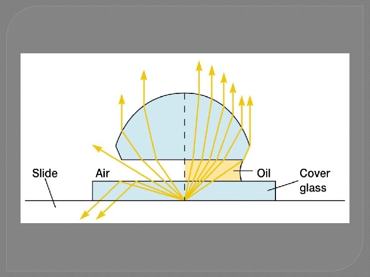
Using a light microscope: Immersion Oil Microscope specimen is on a glass slide. • Light passes through glass slide air lens gets refracted • At high magnification, this refraction (bending) of the light blurs the image • To eliminate refraction between slide and lens: Eliminate the air, replace with immersion oil (Immersion oil same index of refraction as glass)
The Dark-Field Microscope � Produces a bright image of the object against a dark background. � A special condenser condenses the light on specimen but of the objective lens � A cell or particle will deflect the light into the objective lens thus seen as bright shape against dark background. � It is used to observe living, unstained preparations especially to examine motility. 16
Dark Field Microscope
The Phase-Contrast Microscope �Excellent way to observe living cells in wet preparations. �It has special condenser and phaseplate which retards light waves that go through cells in specimen. This makes contrast between cells and background. The cells appear darker against a brighter background. 18
Dissecting Microscope �It is also called stereo- type microscope �It has oculars and stage only. �It magnifies x 10 only �It is useful in Mycology and Parasitology.
Dissecting Microscope
The Fluorescent Microscope �Exposes specimen to ultraviolet, or blue light �Specimens usually stained with fluorochromes �Shows a bright image of the object resulting from the fluorescent light emitted by the specimen. �It is mostly in immunology for detection of antigen and antibody. 21
22
Inverted microscope �Parts of this microscope are similar to bright field microscope except condenser is located above stage while objectives are below the stage. �There is a bigger working distance to allow use of cell culture flasks. �It is mostly used in virology.
Electron Microscope • • Electrons are used instead of light waves. 2 types: 1. Transmission electron microscope 2. Scanning electron microscope • • • Total magnification 100, 000 -300, 000 Resolution. 0003 um It is used to examine viruses and internal cell components.
TEM
SEM
Caring for a Microscope �Clean only with a soft cloth/tissue �Make sure it’s on a flat surface �Be gentle with the microscope �Carry it with 2 HANDS…one on the arm and the other on the base