Microscopes and Other Tools DISSECTING MICROSCOPE Allows you


- Slides: 10
Microscopes and Other Tools
DISSECTING MICROSCOPE • Allows you to see the surface of the specimen • Light reflects off the surface of specimen • Organisms can be alive • Image is in color • Seen in 3 -D • Not for viewing cells
COMPOUND MICROSCOPE • • • Used to view cells Magnification up to 1500 X (Most up to 400 X) Image appears upside down and backwards We can look at living cells and small organisms Specimen must be cut very thin because light must shine through
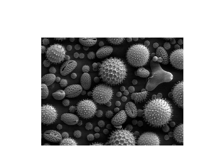
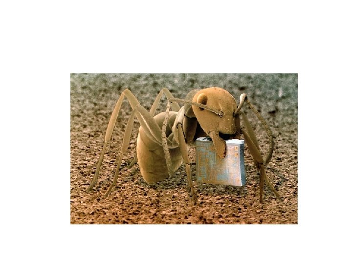
SCANNING ELECTRON MICROSCOPE • Allows you to see surface of specimen • Specimen is covered in a thin layer of gold which allows electrons to “bounce off” • Viewed in a vacuum (all air is removed) • Specimen is NOT alive • Seen in black and white and in 3 D
PHASE CONTRAST MICROSCOPE • Widely used for examining such specimens as biological tissues. • Enhances contrasts of transparent and colorless objects • Able to show parts in a cell or bacteria, which would be very difficult to see in an ordinary light microscope. • Enables viewer to see in 3 D
CENTRIFUGE • Spins test tubes at a very high rotation speed • Separates out the parts of the liquid by density • Ex. Separates blood into RBC, WBC, Plasma and Platelets
ULTRACENTRIFUGE • Separates all of the organelles in a cell
GEL ELECTROPHORESIS • Used to analyze DNA • DNA is cut into pieces and “banding pattern” is seen – Pieces of DNA move through an “electric” field