MICROSCOPE Parts of the Microscope a b c

MICROSCOPE

Parts of the Microscope a. b. c. d. e. f. g. h. i. Eyepiece Coarse Adjustment Fine Adjustment Objectives (LP, HP) Arm Stage Light source Base Diaphragm
 b.")
FUNCTIONS – FILL IN AT BOTTOM OF SHEET a. Magnifies image (10 X) b. Used for rough focus (use with low power) c. Used for fine adjustments (use with high power) d. Enlarges image (scanning 4 x, low power 10 x, high power 40 x) e. Used for carrying microscope f. Platform for holding the slide g. Can be a mirror or light bulb h. Used for carrying the microscope i. Adjusts the amount of light

B. The compound light microscope can be used to view specimens that have dimensions of less than 0. 1 millimeter. You can calculate the total magnification of a specimen by multiplying the magnification of the eyepiece lens by the magnification of the objective lens. • 10 x x 4 x = 40 x magnification of eyepiece lens magnification of obj. lens total magnification The specimen being viewed is magnified 40 times. The greater the total magnification, the smaller the field of view (FOV) or area that you see. The lower the total magnification, the larger the field of view (FOV).

Question 1: • Calculate the total magnification: Eyepiece Objective Total Lens Magnification a. 10 x 4 x 40 x b. 10 x c. 10 x 40 x 100 x 400 x

Question 2: Questions 2 & 3: Question 3: • With which combination • Which set of lenses would you be would you use to locate able to see the largest a specimen on a slide? area of a specimen? Eye & 4 x(scanning) or Eyepiece & 10 x (low power) scanning(4 x) (largest • Which set would you FOV) or eye & low power ( 10 x) use to examine the if scanning is not there details of a specimen? • The smallest area? Eyepiece & 40 x (high Eyepiece 10 x & High power) power 40 x (smallest FOV)

FIELD OF VIEW -How much can you see? In High Power we see 25% of the low power FOV (low power 100 x is 25% of high power 400 x) We see less of the specimen but we see more details of the specimen under high power LOW POWER HIGH POWER

Answer the following Questions • 1. After switching from high power to low power the area of the field of view will appear • a. larger and brighter • b. Smaller and brighter • c. larger and darker • d. smaller and darker • 2. What should a student adjust if the field of view seems too dark? • ________________ • 3. Is the field of vision smaller or larger under low • power? ___________________

• 4. To locate and observe a specimen under a slide, a student should begin by using what objective and what adjustment knob? • 5. What adjustment knob should you use if you are using high power? • 6. Why should a specimen be centered in the middle of the field of view when focusing under low power?

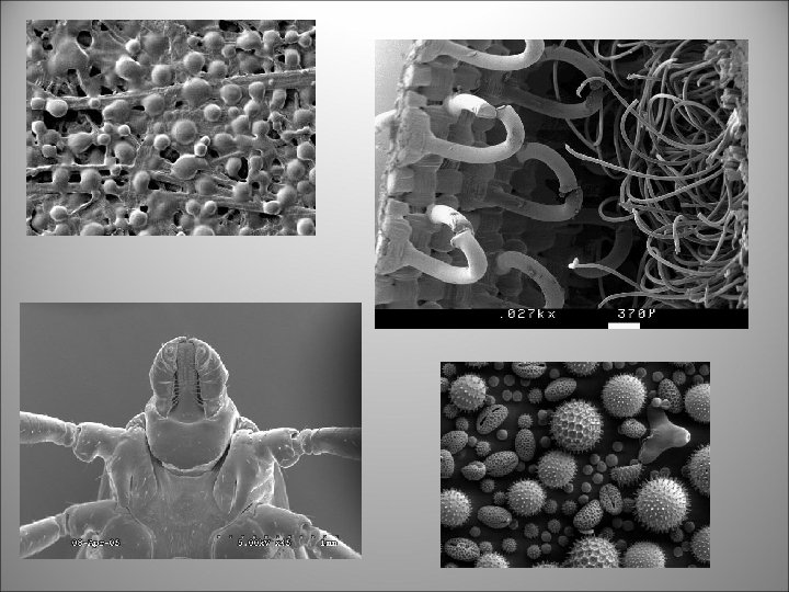
Electron Microscope The limit of resolution restricts the usefulness of light microscopes for studying VERY small specimens such as viruses. • Electron microscopes use a stream of electrons to view these specimens. • Electron microscopes have a limit of resolution more than 1000 times finer than light microscopes.

FIELD OF VIEW How do we find the FOV of a microscope? Low Power (100 x) 1. Find the diameter of the LP FOV 2. Use the clear metric ruler (mm side) 3. Be sure to line up the first mm mark with the left side of the field. LP FOV = 1. 5 mm

The mm is too large to measure microscopic objects, so you need to use the micron (micrometer) µm • 1 mm = 1, 000 µm • 1 µm = 1/1000 mm or 0. 001 mm LP FOV = 1500µm

So how do we determine how big something is in the microscope? • Let’s see how it works: Use the following formula: FOV # of cells (that can fit across diameter) FOV = 2 mm =. 5 mm # of 4 cells OR. 5 mm x 1000 =500 microns
- Slides: 14