MICROSCOPE LAB WHAT DO YOU SEE WHAT CAN

MICROSCOPE LAB WHAT DO YOU SEE? WHAT CAN YOU SEE?

Parts of the Microscope Fill and Chill http: //www. biologycorner. com/microquiz/

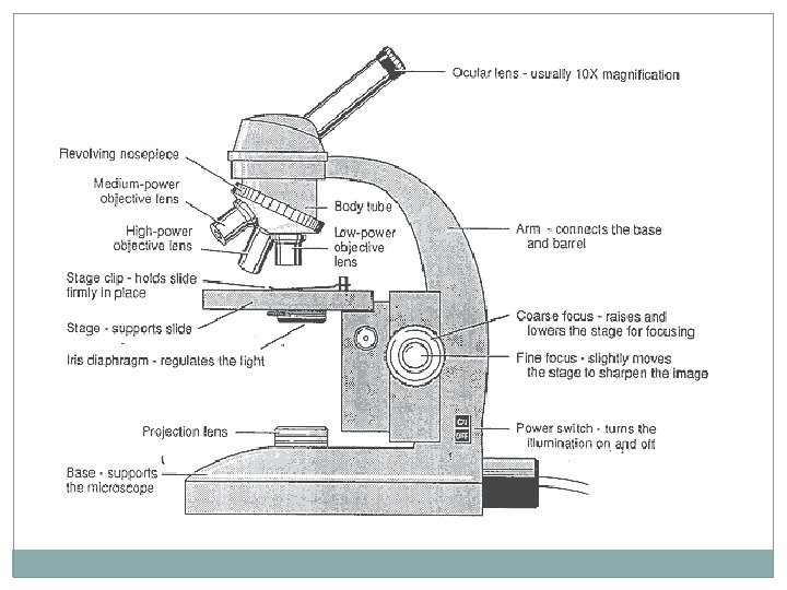
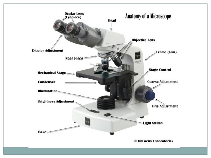
PARTS OF THE MICROSCOPE Stage: Supports the slide. The central opening in the stage allows light to pass through. Stage Clips: Keep the slide in position Diaphragm: Regulates the amount of light reaching the object being viewed. Ocular Lens: Viewing (10 X)/Eyepiece Coarse Adjustment knob: Moves the body tube up or down so you can get the specimen in focus. It is used with the low power objective only! Fine Adjustment Knob: Used with med or high power to get the specimen in sharper focus Arm: Part of the microscope you carry the microscope with Base: Supports the microscope Light Source: Provides light making it easier to view the specimen Objective: Low (4 X), Medium (10 X) and High (40 X) Attached to the revolving nosepiece Body Tube: The long tube that holds the eyepiece and connects it to the objective

CALCULATIONS 1. What is the MAGNIFICATION of the OCULAR LENS? The magnification of the ocular lens is ____X 2. Calculating the MAGNIFICATION of the MICROSCOPE. Magnification of microscope = ocular lens x objective lens Magnification of microscope (4 X objective) ___ x ___ = _____ Magnification of microscope (10 X objective) ___ x ___ = _____ Magnification of microscope (40 X objective) ___ x ___ = _____

3. What is the ACTUAL SIZE of the SPECIMEN you looked at with the microscope? Actual size of specimen (mm) = Field of view (see table above) # of specimens to fit across field of view Magnification of Microscope 40 X (low) 100 X (medium) 400 X (high) Field of view 4. 2 mm 1. 72 mm 0. 42 mm

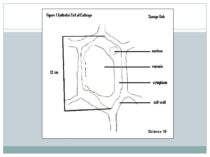
Example of Calculations for Science 10 Using the 4 X objective of a compound light microscope, you view the picture at the side. What is the magnification of the microscope? What is the actual size of the specimen?

Rules for Biological Diagrams � Unlined paper and a sharp pencil � Leave an empty margin of about 1 cm all around your page � When drawing cells, choose only one cell (unless otherwise required) and show the edge of neighboring cells to show the connection � Draw outline of your subject with clear and unbroken lines. � Your drawing should be about ½ of the page � Use Figure # and name as your heading � Always indicate the magnification of the illustration below your drawing � Use stipples for darkened areas � Always label to the right with a ruler � Measurement to the left � Never cross lines � Do little or no erasing � Small letters, no writing

Using a clean sheet of paper, complete a biological drawing. Submit before leaving. Thanks
- Slides: 11