MICROCIRCULATION Structure of the Microcirculation and Capillary System


- Slides: 19
MICROCIRCULATION
Structure of the Microcirculation and Capillary System The microcirculation of each organ is organized specifically to serve that organ’s needs. In general, each nutrient artery entering an organ branches six to eight times before the arteries become small enough to be called arterioles, which generally have internal diameters of only 10 to 15 micrometres. Then the arterioles themselves branch two to five times, reaching diameters of 5 to 9 micrometres at their ends where they supply blood to the capillaries. The arterioles are highly muscular, and their diameters can change many fold. The metarterioles (the terminal arterioles) do not have a continuous muscular coat, but smooth muscle fibres encircle the vessel at intermittent points, as shown in Figure by the black dots on the sides of the metarteriole.
At the point where each true capillary originates from a metarteriole, a smooth muscle fiber usually encircles the capillary. This is called the precapillary sphincter. This sphincter can open and close the entrance to the capillary. The venules are larger than the arterioles and have a much weaker muscular coat. Yet the pressure in the venules is much less than that in the arterioles, so that the venules still can contract considerably despite the weak muscle. Most important, the metarterioles and the precapillary sphincters are in close contact with the tissues they serve. Therefore, the local conditions of the tissues —the concentrations of nutrients, end products of metabolism, hydrogen ions, and so forth—can cause direct effects on the vessels in controlling local blood flow in each small tissue area.
Structure of the Capillary Wall. Figure shows the ultramicroscopic structure of typical endothelial cells in the capillary wall as found in most organs of the body, especially in muscles and connective tissue. Note that the wall is composed of a unicellular layer of endothelial cells and is sur-rounded by a very thin basement membrane on the outside of the capillary. The total thickness of the capillary wall is only about 0. 5 micrometer. The internal diameter of the capillary is 4 to 9 micrometers, barely large enough for red blood cells and other blood cells to squeeze through.
“Pores” in the Capillary Membrane. In last figure there are two very small passageways connecting the interior of the capillary with the exterior. One of these is an intercellular cleft, which is the thin-slit, curving channel that lies between adjacent endothelial cells. Each cleft is interrupted periodically by short ridges of protein attachments that hold the endothelial cells together, but between these ridges fluid can percolate freely through the cleft. The cleft normally has a uniform spacing with a width of about 6 to 7 nanometres (60 to 70 angstroms), slightly smaller than the diameter of an albumin protein molecule. Because the intercellular clefts are located only at the edges of the endothelial cells, they usually represent no more than 1/1000 of the total surface area of the capillary wall. Nevertheless, the rate of motion of water molecules as well as most water-soluble ions and small solutes is so rapid that all of these diffuse with ease between the interior and exterior of the capillaries through these “slit-pores, ” the intercellular clefts.
Also present in the endothelial cells are many minute plasmalemmal vesicles. These form at one surface of the cell by consuming small packets of plasma or extracellular fluid. They can then move slowly through the endothelial cell. It also has been postulated that some of these vesicles combine to form vesicular channels all the way through the endothelial cell
Special Types of “Pores” Occur in the Capillaries of Certain Organs. The “pores” in the capillaries of some organs have special characteristics to meet the peculiar needs of the organs. Some of these characteristics are as follows: 1. In the brain, the junctions between the capillary endothelial cells are mainly “tight” junctions that allow only extremely small molecules such as water, oxygen, and carbon dioxide to pass into or out of the brain tissues. 2. In the liver, the opposite is true. The clefts between the capillary endothelial cells are wide open, so that almost all dissolved substances of the plasma, including the plasma proteins, can pass from the blood into the liver tissues. 3. The pores of the gastrointestinal capillary membranes are midway between those of the muscles and those of the liver. 4. In the glomerular tufts of the kidney, numerous small oval windows called fenestrae penetrate all the way through the middle of the endothelial cells, so that tremendous amounts of very small molecular and ionic substances (but not the large molecules of the plasma proteins) can filter through the glomeruli without having to pass through the clefts between the endothelial cells.
Flow of Blood in the Capillaries—Vasomotion Blood usually does not flow continuously through the capillaries. Instead, it flows intermittently, turning on and off every few seconds or minutes. The cause of this intermittency is the phenomenon called vasomotion, which means intermittent contraction of the metarterioles and precapillary sphincters (and sometimes even the very small arterioles as well). Regulation of Vasomotion. The most important factor found to affect the degree of opening and closing of the metarterioles and precapillary sphincters is the concentration of oxygen in the tissues. When the rate of oxygen usage by the tissue is great so that tissue oxygen concentration decreases below normal, the intermittent periods of capillary blood flow occur more often, and the duration of each period of flow lasts longer, thereby allowing the capillary blood to carry increased quantities of oxygen (as well as other nutrients) to the tissues. This effect, along with multiple other factors that control local tissue blood flow.
Exchange of Water, Nutrients, and Other Substances Between the Blood and Interstitial Fluid
Diffusion Through the Capillary Membrane By far the most important means by which substances are transferred between the plasma and the interstitial fluid is diffusion. fig. demonstrates this process, showing that as the blood flows along the lumen of the capillary, tremendous numbers of water molecules and dissolved particles diffuse back and forth through the capillary wall, providing continual mixing between the interstitial fluid and the plasma. Diffusion results from motion of the water molecules and dissolved substances in the fluid, the different molecules and ions moving first in one direction and then another, bouncing randomly in every direction.
Lipid-Soluble Substances Can Diffuse Directly Through the Cell Membranes of the Capillary Endothelium. If a substance is lipid soluble, it can diffuse directly through the cell membranes of the capillary without having to go through the pores. Such substances include oxygen and carbon dioxide. Because these substances can permeate all areas of the capillary membrane, their rates of transport through the capillary membrane are many times faster than the rates for lipid-insoluble substances, such as sodium ions and glucose that can go only through the pores
Water-Soluble, Non-Lipid-Soluble Substances Diffuse Only Through Intercellular “Pores” in the Capillary Membrane. Many substances needed by the tissues are soluble in water but cannot pass through the lipid membranes of the endothelial cells; such substances include water molecules themselves, sodium ions, chloride ions, and glucose. Despite the fact that not more than 1/1000 of the surface area of the capillaries is represented by the intercellular clefts between the endothelial cells, the velocity of molecular motion in the clefts is so great that even this small area is sufficient to allow tremendous diffusion of water and water-soluble substances through these cleft-pores. , the rate at which water molecules diffuse through the capillary membrane is about 80 times as great as the rate at which plasma itself flows linearly along the capillary. That is, the water of the plasma is exchanged with the water of the interstitial fluid 80 times before the plasma can flow the entire distance through the capillary.
Effect of Molecular Size on Passage Through the Pores. The width of the capillary intercellular cleft-pores, 6 to 7 nanometres, is about 20 times the diameter of the water molecule, which is the smallest molecule that normally passes through the capillary pores. Conversely, the diameters of plasma protein molecules are slightly greater than the width of the pores. Other substances, such as sodium ions, chloride ions, glucose, and urea, have intermediate diameters. Therefore, the permeability of the capillary pores for different substances varies according to their molecular diameters. The capillaries in different tissues have extreme differences in their permeabilities. For instance, the mem-brane of the liver capillary sinusoids is so permeable that even plasma proteins pass freely through these walls, almost as easily as water and other substances. Also, the permeability of the renal glomerular mem-brane for water and electrolytes is about 500 times the permeability of the muscle capillaries, but this is not true for the plasma proteins; for these, the capillary permeabilities are very slight, as in other tissues and organs.
Effect of Concentration Difference on Net Rate of Diffusion Through the Capillary Membrane. The “net” rate of diffusion of a substance through any membrane is proportional to the concentration difference of the substance between the two sides of the membrane. That is, the greater the difference between the concentrations of any given substance on the two sides of the capillary membrane, the greater the net movement of the sub-stance in one direction through the membrane. For instance, the concentration of oxygen in capillary blood is normally greater than in the interstitial fluid. Therefore, large quantities of oxygen normally move from the blood toward the tissues. Conversely, the concentration of carbon dioxide is greater in the tissues than in the blood, which causes excess carbon dioxide to move into the blood and to be carried away from the tissue
The rates of diffusion through the capillary membranes of most nutritionally important substances are so great that only slight concentration differences suffice to cause more than adequate transport between the plasma and interstitial fluid. For instance, the concentration of oxygen in the interstitial fluid immediately outside the capillary is no more than a few per cent less than its concentration in the plasma of the blood, yet this slight difference causes enough oxygen to move from the blood into the interstitial spaces to provide all the oxygen required for tissue metabolism, often as much as several liters of oxygen per minute during very active states of the body.
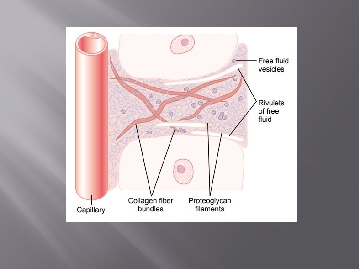
The Interstitium and Interstitial Fluid About one sixth of the total volume of the body consists of spaces between cells, which collectively are called the interstitium. The fluid in these spaces is the interstitial fluid. It contains two major types of solid structures: (1) collagen fiber bundles and (2) proteoglycan filaments. The collagen fiber bundles extend long distances in the interstitium. They are extremely strong and therefore provide most of the tensional strength of the tissues. The proteoglycan filaments, however, are extremely thin coiled or twisted molecules composed of about 98 per cent hyaluronic acid and 2 per cent protein. These molecules are so thin that they can never be seen with a light microscope and are difficult to demonstrate even with the electron microscope. Nevertheless, they form a mat of very fine reticular filaments aptly described as a “brush pile. ”
Gel” in the Interstitium. The fluid in the interstitium is derived by filtration and diffusion from the capillaries. It contains almost the same constituents as plasma except for much lower concentrations of proteins because proteins do not pass outward through the pores of the capillaries with ease. The interstitial fluid is entrapped mainly in the minute spaces among the proteoglycan filaments. This combination of proteoglycan filaments and fluid entrapped within them has the characteristics of a gel and therefore is called tissue gel. Because of the large number of proteoglycan filaments, it is difficult for fluid to flow easily through the tissue gel. Instead, it mainly diffuses through the gel; that is, it moves molecule by molecule from one place to another.
Free” Fluid in the Interstitium. Although almost all the fluid in the interstitium normally is entrapped within the tissue gel, occasionally small rivulets of “free” fluid and small free fluid vesicles are also present, which means fluid that is free of the proteoglycan molecules and therefore can flow freely. When a dye is injected into the circulating blood, it often can be seen to flow through the interstitium in the small rivulets, usually coursing along the surfaces of collagen fibres or surfaces of cells. The amount of “free” fluid present in normal tissues is slight, usually much less than 1 per cent. Conversely, when the tissues develop edema, these small pockets and rivulets of free fluid expand tremendously until one half or more of the edema fluid becomes freely flowing fluid independent of the proteoglycan filaments.