MICROBIOLOGY INTRODUCTION Dr Sahar Mahdi Introduction Is the

MICROBIOLOGY INTRODUCTION Dr. Sahar Mahdi
 those being")
Introduction Is the study of microscopic organisms, (from Greek "small "life") those being unicellular (single cell), multicellular (cell colony), or acellular (lacking cells). Microbiology including virology, mycology, parasitology, and bacteriology. Eukaryotic micro-organisms possess membrane-bound cell organelles and include fungi and protists, whereas prokaryotic organisms —all of which are microorganisms— are conventionally classified as lacking membrane-bound organelles and include eubacteria and archaebacteria. Microbiologists traditionally relied on culture, staining, and microscopy. However, less than 1% of the microorganisms present in common environments can be cultured in isolation using current means. Microbiologists often rely on extraction or detection of nucleic acid, either DNA or RNA sequences.

Applied microbiology Medical microbiology: The study of the pathogenic microbes and the role of microbes in human and animal illness. Includes the study of microbial pathogenesis and epidemiology and is related to the study of disease pathology and immunology. Pharmaceutical microbiology: The study of microorganisms that are related to the production of antibiotics, enzymes, vitamins, vaccines, and other pharmaceutical products and that cause pharmaceutical contamination and spoil. Industrial microbiology: The exploitation of microbes for use in industrial processes. Examples include industrial fermentation and wastewater treatment. Closely linked to the biotechnology industry. . Microbial biotechnology: The manipulation of microorganisms at the genetic and molecular level to generate useful products.

Food microbiology: The study of microorganisms causing food spoilage and food borne illness. Using microorganisms to produce foods, for example by fermentation. Agricultural microbiology: The study of agriculturally relevant microorganisms. This field can be further classified into the following: Soil microbiology: The study of those microorganisms that are found in soil. Veterinary microbiology: The study of the role of microbes in veterinary medicine or animal taxonomy. Environmental microbiology: The study of the function and diversity of microbes in their natural environments.

Benefits Many microbes are responsible for numerous beneficial processes such as industrial fermentation (e. g. the production of alcohol, vinegar and dairy products), antibiotic production . Scientists have also exploited their knowledge of microbes to produce biotechnologically important enzymes such as Taq polymerase, reporter genes for use in other genetic systems and novel molecular biology techniques such as the yeast two-hybrid system. Bacteria can be used for the industrial production of amino acids. Corynebacterium glutamicum is one of the most important bacterial species with an annual production of more than two million tons of amino acids, mainly Lglutamate and L-lysine Since some bacteria have the ability to synthesize antibiotics, they are used for medicinal purposes, such as Streptomyces to make aminoglycoside antibiotics.

Symbiotic microbial communities are known to confer various benefits to their human and animal hosts health including aiding digestion, production of beneficial vitamins and amino acids, and suppression of pathogenic microbes. Some benefit may be conferred by consuming fermented foods, probiotics (bacteria potentially beneficial to the digestive system) and/or probiotics (substances consumed to promote the growth of probiotic microorganisms). Research has suggested that microorganisms could be useful in the treatment of cancer. Various strains of non-pathogenic clostridia can infiltrate and replicate within solid tumors.

History Ancient time The Roman Marcus Terentius Varro made references to microbes when he warned against locating a homestead in the vicinity of swamps "because there are bred certain minute creatures which cannot be seen by the eyes, which float in the air and enter the body through the mouth and nose and there by cause serious diseases.

Medieval Islamic world Avicenna "ibn Sina" At the golden age of Islamic civilization, some scientists had knowledge about microorganisms, such as Ibn Sina in his book " The Canon of Medicine", Ibn Zuhr (also known as Avenzoar) who discovered scabies mites, and Al-Razi who gave the earliest known description of smallpox in his book The Virtuous Life (al-Hawi).


Modern times Antonie van Leeuwenhoek, is considered to be the one of the first to observe microorganisms using a microscope. In 1665 Robert Hooke was made the first recorded microscopic observation, of the fruiting bodies of moulds. In 1676, Anton van Leeuwenhoek, who lived most of his life in Delft, Holland, observed bacteria and other microorganisms in teeth scrapings and rain water, using a single-lens microscope of his own design. While Van Leeuwenhoek is often cited as the first to observe microbes, Joseph Lister was the first person who said infectious diseases are caused by micro-organism and was first person who used phenol as disinfectant on the open wounds of patients.

Antonie van Leeuwenhoek

Innovative laboratory glassware and experimental methods developed by Louis Pasteur and other biologists contributed to the young field of bacteriology in the late 19 th century

The field of bacteriology was founded in the 19 th century by Ferdinand Cohn, a botanist whose studies on algae and photosynthetic bacteria led him to describe several bacteria including Bacillus. Cohn was also the first to formulate a scheme for the taxonomic classification of bacteria and discover spores. Louis Pasteur and Robert Koch are often considered to be the father of microbiology and medical microbiology, respectively. Pasteur is most famous for his series of experiments designed to disprove widely held theory of spontaneous generation, thereby solidifying identity as a biological science. Pasteur also designed methods for food preservation (pasteurization) and vaccines against several diseases such as anthrax, fowl cholera and rabies. Koch is best known for his contributions to the germ theory of disease,

proving that specific diseases were caused by specific pathogenic micro-organisms. He developed a series of criteria that have become known as the Koch's postulates. Koch was one of the first scientists to focus on the isolation of bacteria in pure culture resulting in his description of several novel bacteria including Mycobacterium tuberculosis, the causative agent of tuberculosis. While Pasteur and Koch are often considered the founders of microbiology, their work did not accurately reflect the true diversity of the microbial world because of their exclusive focus on microorganisms having direct medical relevance.
 - in")
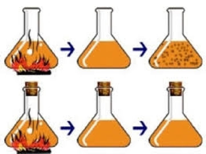
Many believed Spontaneous generation: mean: life can arise from non- living matter) - in 1668, the Italian physician Francesco Redi performed an experiment to disprove Spontaneous generation. ۞ Can you think of an experiment that could disprove spontaneous generation? Redi filled Six jars with decaying meat. Conditions Results 3 jars covered with fine net No maggots 3 open jars maggots appeared From where did the maggots come? What was the purpose of the sealed jars? Spontaneous generation or biogenesis? Rudolf Virchow (German) presented biogenesis that mean : living cells can arise only from pre existing cells.

So there are two hypothesis : The hypothesis that living organisms arise from non living matter is called (spontaneous generation ). The alternative hypothesis that the living organisms arise from pre existing life is called " Biogenesis". Conditions Results ۞ Nutrient broth placed in flask, Microbial growth heated, not sealed ۞Nutrient broth placed in flask No microbial growth heated, then sealed Spontaneous generation or biogenesis? In 1861 : Louis Pasteur demonstrated that microorganisms are present in the air.

The Germ theory of Disease The first proof that bacteria actually cause diseases came from Robert Koch in 1876 , he discovered Anthrax bacilli in blood of cattle that had died of anthrax. He cultured the bacteria on nutrient and then injected samples of the culture in to healthy animals. When these animals became sick and died Koch isolated the bacteria in their blood and compared them with the bacteria originally isolated and found that they were identical. Koch thus established as sequence of experimental steps for relating a specific microbe to a specific disease, now is known as Koch's postulates( ﻣﻔﺎﻫﻴﻢ )ﻛﻮﺥ.

Koch's postulates 1 - The bacteria must be present in every case of the disease. 2 - The bacteria must be isolated from the host with the disease and grown in pure culture. 3 - The specific disease must be reproduced when a pure culture of the bacteria is inoculated into a healthy susceptible host. 4 - The bacteria must be recoverable from the experimentally infected host. •

Cell structure and function Cell Structures There are many cells in an individual, which performs several functions throughout the life. The different types of cell include- prokaryotic cell, plant and animal cell. The size and the shape of the cell range from millimeter to microns, which are generally based on the type of function that it performs. A cell generally varies in their shapes. A few cells are in spherical, rod, flat, concave, curved, rectangular, oval and etc. These cells can only be seen under microscope.
 (or)")
Cell Theory Every living organism is made up of a single cell (unicellular) (or) many cells (multicellular) and all types of cells have certain structures in common like: genetic material and plasma membrane. Cell is the smallest living thing. Each cell arises only from pre-existing cells. Cells are divided into 2 major groups depend on the basis structure: 1 - Prokaryotic cells 2 - Eukaryotic cells

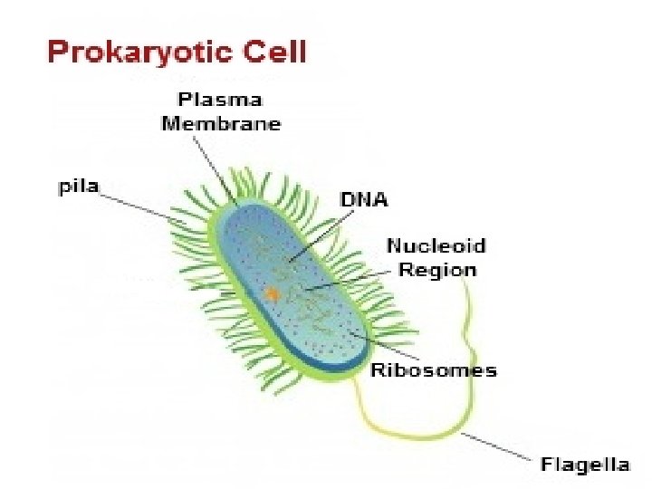
Prokaryotic Cell Structure They are the first organisms to be present on our planet earth. Organisms, with this cell type are known by the term prokaryotic organisms (or) prokaryotes. Bacteria and blue green algae are few examples of this category. Prokaryotic cells are single-celled organisms, with the absence of nucleus and comprises of capsule, cell wall, cell membrane, cytoplasm, nucleiod, ribosome, plasmids, pili and flagella.

Prokaryotic Cell General Features The size of a cell ranges from 1 -10 microns. Few prokaryotic cells vary in their size. They are single-celled (unicellular), which forms a colony or filamentous. The shape of the cell includes spherical, rod and flat shaped organisms. Mode of nutrients-- few organisms are photosynthetic (performing food with the help of sunlight), feed on living things and dead things. They reproduce asexually by the process called binary fission, transformation, conjugation, transduction

Structure and Functions of a Prokaryotic Cell Capsule: It is the slimy outer coating of the cell wall. It is composed of the polypeptide. The main function of the capsule is to protect the cell from getting dry and also helps in protecting cells from external pressures. Cell wall: It is the tougher and a rigid structure, which provides the shape and protects the internal organelles of a cell. It is the middle layer, which is present in between the capsule and cell membrane. Cell membrane: It is the inner delicate structure, which plays a vital role in regulating the entry and exits of materials in the cell. It acts a permeable membrane and separates the cell from its environment. It is of about 5 -10 nm in thickness, which helps in the secretion of proteins and elimination of waste products. It is also called by a name plasma membrane. Cytoplasm: It is the liquid membrane, which is present in between the cell membrane and nucleiod. It plays a vital role in storing all types of materials, which are required for an organism to sustain the life. Nucleiod: It is the cytoplasm region containing genetic material. The DNA of a prokaryotic organism is one big loop or a circular, which is located inside the nucleiod. It plays a vital role in cell division.

Ribosome: It comprises of both RNA and proteins. It helps in protein synthesis in the cell. They are smallest membrane present inside the cytoplasm. Plasmids: They are smallest membrane of a cell with double stranded DNA. Plasmids are rarely present in prokaryotic organisms. The main role of plasmids is it helps in DNA exchanging between the bacterial cells. Pilli: It is the thinnest membrane of a prokaryotic cell. They are composed of protein complex called pilin and are mainly involved in sticking to the objects especially during sexual reproduction. Flagella: It is the helical shaped membrane, whose sizes ranges from 19 -20 nm in diameter and plays a vital role in motility of an organism from one place to another place. It also helps in swimming, gliding, spinning and rotating both in clockwise and anti clockwise directions.

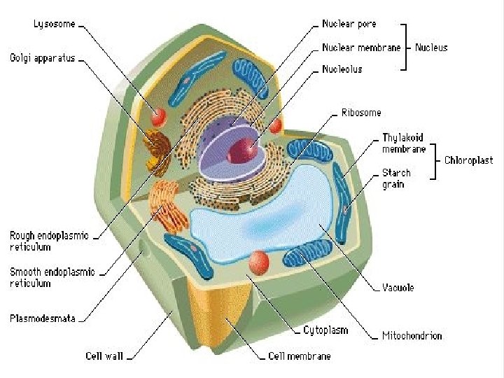
Eukaryotic cells: are more complex than prokaryotic cells, these organisms have membrane bound nucleus with many cell organelles to perform several cellular functions within the system

Eukaryotic Cell General Features The size of a eukaryotic cell ranges from 10 -100 microns. Few eukaryotic cells vary in their size. They are large, advanced, multicellular and have membrane bound organelles. They reproduce both by sexually and by asexually. Mode of nutrients - Autotrophic and heterotrophic. Kingdom protozoa, algae, fungi, Plantae and Animalia are organisms with eukaryotic cell.

Structure and Functions of a Eukaryotic Cell Plasma membrane: They are semi permeable membrane that acts as a boundary of a cell, which protects and separates the cell from the external environment. Nucleus: It is surrounded by nuclear envelope. They are the store house for the cell genetic materials in the form of DNA and store all the necessary information, which are required for a cell to control all types of activities. Nuclear membrane: It is the double membrane layer that surrounds the nucleus and it plays a role of entry and exits of materials within the nucleus. Nucleolus: It is the non membrane bound organelles, which is present within the nucleus and is mainly involved in controlling all types of cellular activities including cellular reproduction. •

Mitochondria: They are the double smooth membrane, which are present in all eukaryotic cells. They are the powerhouse of the cell. It plays a vital role in the synthesis of ATP and converts glucose to ATP. Endoplasmic reticulum: They are the double membrane organelle, which divides the cell into compartments. It is connected to the nuclear membrane of the cell. It plays a vital role in protein synthesis, biosynthesis of lipids and steroids, stores and regulates calcium and metabolism of carbohydrates. Endoplasmic reticulum is of two types“ rough and smooth Endoplasmic reticulum. Ribosome: It is present in the cytoplasm. They are the site for cell protein synthesis, which are composed of ribosomal RNA and proteins.

Golgi Bodies: It is the flattened membrane, which are mainly used to store the substances made by the cell. This membrane also helps in preserving, transporting materials within the cell. Lysosomes: They are the membrane bound organelles, which contains digestive enzymes to break down macromolecules. Lysosome plays a vital role in protecting cell by engulfing or destroying foreign bodies entering the cell. Cytoplasm: They are the jelly types of organelles, which are present in the inner region of a cell. It plays a vital role in keeping a cell in a stable and keeps the cell organelles separate from each other. Chromosomes: The rod shaped structures, which are composed of proteins and DNA. Chromosomes also play a vital role in determining a sex of an individual. All human cells contain 46 numbers of chromosomes.

The Morphological study of bacteria Microscopical examination is usually the first step taken in the identification of unknown bacterium. The bacterium may be allocated to one or other of the major groups when its (morphology) and (staining reactions) have been observed. The morphological features of importance are the size, shape and grouping of the cell and their possession of any distinctive structure such as endospore, flagella, capsules and intracellular granules. Staining reaction are observed after treatment by special procedures such as the Gram and Ziehl. Neelsen stains, The different kinds of bacteria being shown in separate colours due to their different permeability to certain decolorizing agents.

The bacterial cell Bacteria differ in their shapes and sizes depending mainly on their environment. They might be coccoid, filamentous, bacilli, vibriod, …. etc. such shapes were observed in stained and unstained films under the light microscope- earlier and later electron and phase-contrast microscopes which aided also in a more understanding of the bacterial and other organisms structures. The size of bacteria are measured as follows: 1 micrometer (µm) =1 micron(µ) = 1/1000 mm 1 nanometer (nm) = 1 mill micron (mµ) = 1/1000 micrometer (micron) Morphologically bacteria are divided into :
 µm long and (1 -2)")
1 - Bacilli: rod-shape or cylindrical, straight (2 -10) µm long and (1 -2) µm wide. like : Clostridium , bacillus, Brucella , Escherichia coli, etc…. There are fewer grouping : Diplobacilli: appear in pairs after division Streptobacilli: occur in chains. Bacilli may show coccobacilli (pleomorphic shaped), Listeria Cocci…… coccus circular or spherical or lanceolate……shaped, about (1) µm in diameter. occur either as: Clusters……. . Staphylococci or Chain………. . Streptococci Diplococci…… S. pneumoniae Cubical groups (sarcinae)
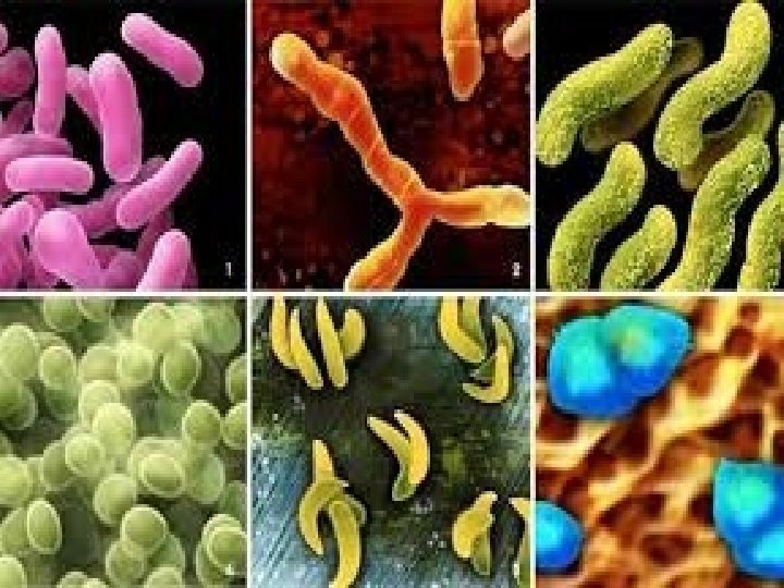

3 - Vibrio : Curved or comma shaped like: Vibrio, Campylobacter …. etc 4 - Spirilla : spiral shaped …. . Spirilla move by flagella. 5 - Spirochaetes……. spiral shaped , filamentous actively motile liptospira, move by means of an axial filament. 6 - mycelia ……. Branching and some times branching filaments Actinomyces , Nocardia There also star- shaped cells Square cell
- Slides: 38