Microbiology Department General Bacteriology Bacterial cell structure Dr

Microbiology Department General Bacteriology Bacterial cell structure Dr Amany Mostafa

Intended Learning Outcomes By the end of this lecture , the student will be able to: 1. . To Identify term( Microbiology ) 2. . Differentiate between prokaryotic and Eukaryotic cell. 3. . Recognize different system of bacterial Bacterial classification 4. . Describe Bacterial Cell Structure ©

INTRODUCTION • Microbiology is the study of living microorganisms which are microscopic &can’t be seen by the naked eye. • Micro-organisms are widely distributed in nature • The agent of human infectious disease include: - bacteria, fungi, protozoa, helminthes and viruses

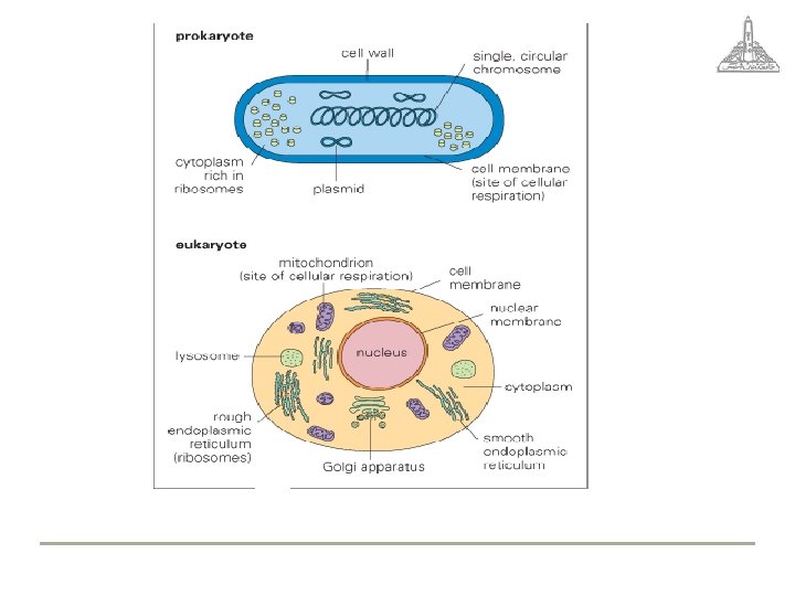
prokaryotic Cell Bacteria are prokaryotic cell: Have • Single naked chromosome • Rigid cell wall (peptidoglycan) • 70 S ribosome • Replicate by simple binary fission

Eukaryotic Cell • • • Contain true nucleus. Cytoplasmic membrane contain sterols: Mitochondria, lysosome 80 S ribosome Rigid cell wall contain chitin mitosis

Example Eukaryotic prokaryotic Fungi, plant, animal cell Bacteria True nucleus Nuclear membrane Nuclear Material nucleoli Multiple chromosome Single chromosome Histones associated with DNA No histones mitosis Simple binary fission Mitochondria Present Absent lysosymes present absent Division Cytop lasmic Struct ure Absent Ribosome Cytoplasmic membrane Cell Wall 80 S 70 S Contain sterol No mesosome Mesosome Contain chitin Contain peptidoglycan

Bacterial Classification The old system of classification Higher bacteria: e. g. Actinomyces (filamentous branching organism) Lower bacteria: Simple unicellular organisms

Lower bacteria • classified on the basis of some traits as: • Shape of the bacterial cell Fig. (2): • cocci: spherical. • bacilli: rod- shaped. • Vibrios: comma- shaped. • spirilla: spiral- shaped.

Lower Bacteria curved bacilli cocci Spiral

Other basis of classification • Method of energy production: glycolysis for anaerobes, cellular respiration for aerobes. • Nutritional requirements: • Reaction to the Gram stain: It differentiates bacteria into Grampositive bacteria (violet in color) and Gram -negative bacteria (pink

Pathogenicity: Ø Saprophytes: live on dead material, soil, water, dust … etc. They almost never cause disease. Ø Parasitic: live in the body of living creatures: § Pathogenic: cause disease. e. g. Mycobacterium tuberculosis, Neisseria gonorrhoeae. § Non pathogenic commensal bacterial flora: Bacteria which don't normally cause diseases

The new system of classification • Nucleotide base composition: the parameter most often used is the mol percent of guanine plus cytosine (G + C) in the total DNA. For any one species, the G + C content is relatively fixed and this provides a basis for classification.

Nucleic base homology • organisms can be classified into groups on the basis of the homology of their DNA base sequences. When a mixture of DNA from two related species is used, hybrid pairs of DNA strands are produced.
 • More recently, (nucleotide base sequences) especially of their 16 S")
Genome sequencing (Ribotyping) • More recently, (nucleotide base sequences) especially of their 16 S ribosomal RNA (r. RNA) is being used in the classification. This technique is based on base sequence homologies in ribosomal RNA. It has provided new insights into the evolutionary relationships among the

Bacterial Cell Structure • The bacterial cell is composed of the following structure

Cell Wall • The cell wall is the outermost component of bacterial cell. • Functions: 1 - maintain shape (rigid) 2 - support cytoplasmic membrane 3 - Role in cell division 4 - Staining reaction to Gram

Composition of cell wall I- Gram positive celll wall Two layers 1 - Peptidoglycan: constitute 50% of cell wall thickness. It is a polymer of N-acetyl muramic acid and N-acetyl glucose amine, joined together by a tetrapeptides side chain. It is responsible for rigidity of cell wall and for maintaining the shape 2 - Teichoic acid: it is a polymer of glycerol or ribitol. It is located in the outer layer of the Gram positive cell wall. It is antigenic.
 N-acetylglucosamine (NAG) (c) Fig. (5) Components and structure of bacterial cell")
N-acetylmuramic acid (NAM) N-acetylglucosamine (NAG) (c) Fig. (5) Components and structure of bacterial cell wall (a) Components of Gram positive bacterial cell wall. (b) Components of Gram negative bacterial cell

2 - Gram negative cell wall • . Peptidoglycan: constitutes up to 5 -10% of Gram negative cell wall. It is formed of 1 - 2 sheets of N- acetyl muramic acid and N- acetyl glucosamine connected by identical tetrapeptide.

Outer membrane: It is formed of bilayered phospholipids that resemble in composition that of cell membrane. It has special channels, consisting of proteins called porins which allow passive diffusion of low molecular weight compounds like sugar, and amino acids.

• Periplasmic space: a space between the inner cytoplasmic membrane and outer membrane where the single sheet of peptidoglycan layer is present. contains gel-like solution of protein.
 layer: It is outer layer. consists of 3 parts: • Inner")
Lipo polysaccharides (LPS) layer: It is outer layer. consists of 3 parts: • Inner lipid A (endotoxin of Gramnegative bacteria). • Middle polysaccharides Core. • Outer polysaccharides side chains (somatic O antigen). This layer is barrier to hydrophobic molecules

Wall Deficient Bacteria 1 - Mycoplasma - the only group of bacteria that exist naturally without cell wall. - It has no defined shape due to lacking the rigid cell wall. - It is resistant to antibiotics which destroy bacterial cell wall, e. g. penicillin

Spheroplasts: • they are bacteria with weakened or damaged cell wall (which retain remnant of peptidoglycan). They are readily produced by growing the organism in the presence antibiotics that specifically inhibit synthesis of the peptidoglycan component of cell wall

Protoplasts: • they are bacteria from which all the cell wall material has been removed. This occurs in bacteria due to the action of lysozyme, which destroys the peptidoglycan layer. Protoplasts and spheroplasts are osmotically sensitive;
. induced by inhibition of cell wall synthesis")
L-forms: • These are abnormal growth forms). induced by inhibition of cell wall synthesis in bacteria of normal morphology. They are produced more readily with Antibiotics than with lysozymes suggesting the need of residual peptidoglycan which acts as primer in its own synthesis

L-forms • L-forms can revert to the normal form on removal of the inhibitor producing relapses of infection. They differ from parent bacteria in lacking rigid cell wall (vary in size and shape), but they are viable, capable of growth and multiplication.

Summary of important points Bacteria is prokaryote Fungi is eukaryotes Old system of classification depend on phenotypic character New method of classification depends on genotypic character Bacterial cell structure Cell wall of Gram positive cell Cell wall of Gram negative cell Cell Wall defiant Bacteria ©

Examples of questions to assess the ILOs Prokaryotic cell lacks: a. Ribosomes. membrane. b. Nuclear membrane. c. Cytoplamic d. Cell wall. ©

A. B. C. D. • The new method of classification of bacteria depends on: Molecular biology. Bacterial Morphology. . Bacterial Pathogenecity Bacterial respiration

• • • Which of these bacteria are naturally occurring with defective cell wall: Mycoplasma. Chlamydia. Rickettsia Actinomyces.

• • • Failure to synthesize cell wall in bacteria leads to production of: Mycoplasma. Spheroplast. Protoplast. None of the above.
- Slides: 33