
Microbiology an Introduction Thirteenth Edition Chapter 24 Microbial
Microbiology an Introduction Thirteenth Edition Chapter 24 Microbial Diseases of the Respiratory System Copyright © 2019 Pearson Education, Inc. All Rights Reserved
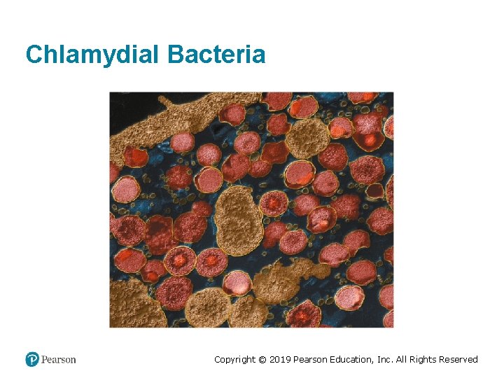
Chlamydial Bacteria Copyright © 2019 Pearson Education, Inc. All Rights Reserved
Structure and Function of the Respiratory System (1 of 2) Learning Objective 24 -1 Describe how microorganisms are prevented from entering the respiratory system. Copyright © 2019 Pearson Education, Inc. All Rights Reserved
Structure and Function of the Respiratory System (2 of 2) • Upper respiratory system – Nose, pharynx, middle ear, and eustachian tubes – Saliva and tears protect mucosal surfaces • Lower respiratory system – Larynx, trachea, bronchial tubes, and alveoli – Ciliary escalator moves particles toward the throat via ciliary action – Alveolar macrophages destroy microorganisms in the lungs – Respiratory mucus protects mucosal surfaces Copyright © 2019 Pearson Education, Inc. All Rights Reserved
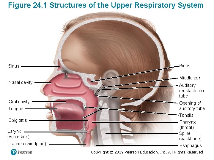
Figure 24. 1 Structures of the Upper Respiratory System Sinus Nasal cavity Oral cavity Tongue Sinus Middle ear Auditory (eustachian) tube Opening of auditory tube Tonsils Epiglottis Larynx (voice box) Pharynx (throat) Spine (backbone) Trachea (windpipe) Esophagus Copyright © 2019 Pearson Education, Inc. All Rights Reserved
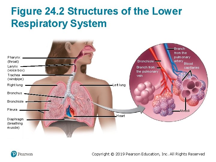
Figure 24. 2 Structures of the Lower Respiratory System Pharynx (throat) Larynx (voice box) Trachea (windpipe) Right lung Bronchiole Branch from the pulmonary vein Branch from the pulmonary artery Blood capillaries Left lung Bronchus Bronchiole Pleura Diaphragm (breathing muscle) Alveoli Heart Copyright © 2019 Pearson Education, Inc. All Rights Reserved
Check Your Understanding-1 Check Your Understanding ü 24 -1 What is the function of hairs in the nasal passages? Copyright © 2019 Pearson Education, Inc. All Rights Reserved
Normal Microbiota of the Respiratory System (1 of 2) Learning Objective 24 -2 Characterize the normal microbiota of the upper and lower respiratory systems. Copyright © 2019 Pearson Education, Inc. All Rights Reserved
Normal Microbiota of the Respiratory System (2 of 2) • Normal microbiota suppress pathogens by competing for nutrients and producing inhibitory substances. • Lower respiratory system is nearly sterile Copyright © 2019 Pearson Education, Inc. All Rights Reserved
Check Your Understanding-2 Check Your Understanding ü 24 -2 Normally, the lower respiratory tract is nearly sterile. What is the primary mechanism responsible? Copyright © 2019 Pearson Education, Inc. All Rights Reserved
Microbial Diseases of the Upper Respiratory System (1 of 2) Learning Objective 24 -3 Differentiate pharyngitis, laryngitis, tonsillitis, sinusitis, and epiglottitis. Copyright © 2019 Pearson Education, Inc. All Rights Reserved
Microbial Diseases of the Upper Respiratory System (2 of 2) • Pharyngitis – Sore throat • Laryngitis • Tonsillitis • Sinusitis – Usually self-limiting • Epiglottitis – Most life-threatening disease of the upper respiratory system Copyright © 2019 Pearson Education, Inc. All Rights Reserved
Check Your Understanding-3 Check Your Understanding ü 24 -3 Which one of the following is most likely to be associated with a headache: pharyngitis, laryngitis, sinusitis, or epiglottitis? Copyright © 2019 Pearson Education, Inc. All Rights Reserved
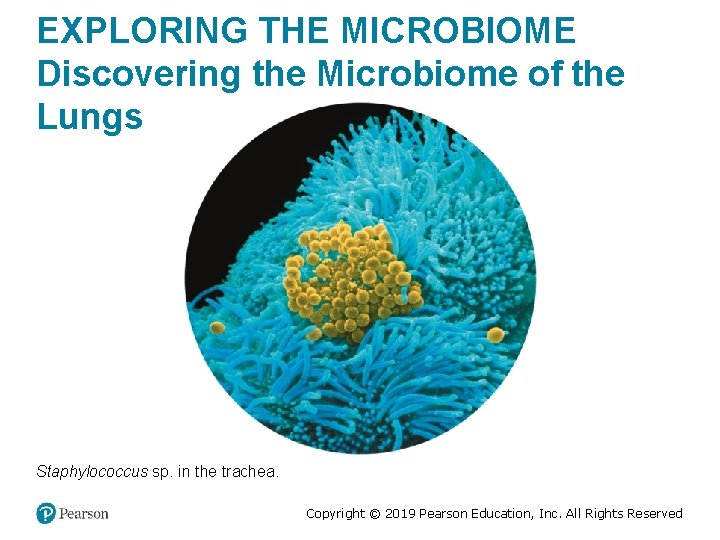
EXPLORING THE MICROBIOME Discovering the Microbiome of the Lungs Staphylococcus sp. in the trachea. Copyright © 2019 Pearson Education, Inc. All Rights Reserved
Bacterial Diseases of the Upper Respiratory System (1 of 2) Learning Objective 24 -4 List the causative agent, symptoms, prevention, preferred treatment, and laboratory identification tests for streptococcal pharyngitis, scarlet fever, diphtheria, cutaneous diphtheria, and otitis media. Copyright © 2019 Pearson Education, Inc. All Rights Reserved
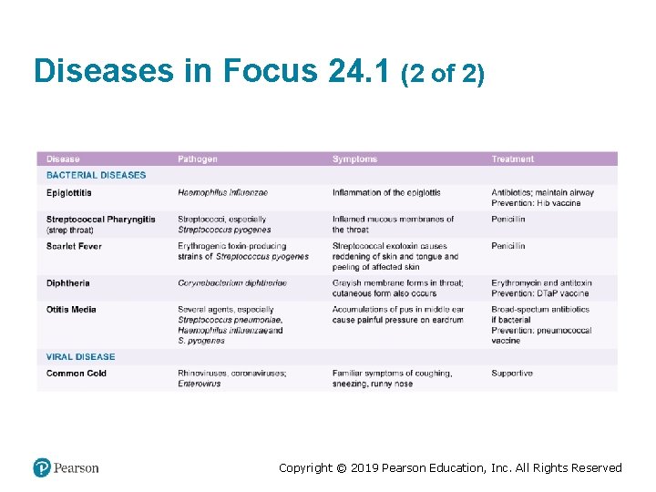
Bacterial Diseases of the Upper Respiratory System (2 of 2) • Streptococcal pharyngitis (strep throat) – Caused by group A streptococci (GAS) § Streptococcus pyogenes – Resistant to phagocytosis – Streptokinases lyse clots – Streptolysins are cytotoxic – Local inflammation, fever, tonsillitis, enlarged lymph nodes – Diagnosis by rapid antigen detection tests • Scarlet fever – Erythrogenic toxin produced by lysogenized S. pyogenes Copyright © 2019 Pearson Education, Inc. All Rights Reserved
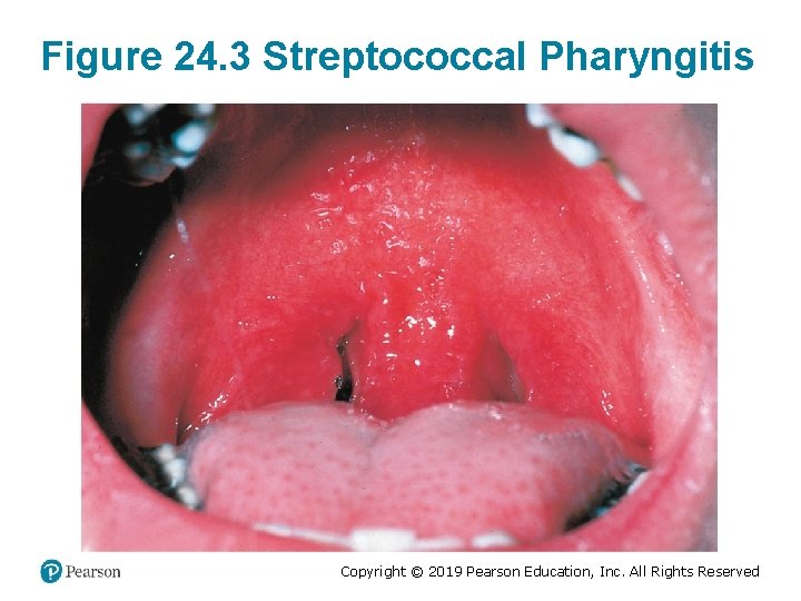
Figure 24. 3 Streptococcal Pharyngitis Copyright © 2019 Pearson Education, Inc. All Rights Reserved
Diphtheria • Caused by Corynebacterium diphtheriae – Gram-positive rod; pleomorphic • Forms a tough grayish membrane in the throat – Fibrin and dead tissue – Blocks passage of air to the lungs • Exotoxin produced by lysogenized bacteria – Circulates in the blood; damages the heart and kidneys • Cutaneous diphtheria – Forms skin ulcer • Prevented by DTa. P vaccine – Diphtheria toxoid Copyright © 2019 Pearson Education, Inc. All Rights Reserved
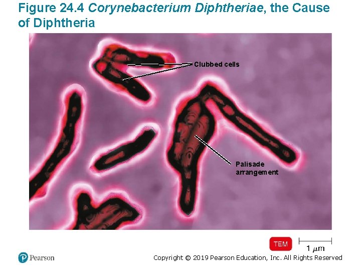
Figure 24. 4 Corynebacterium Diphtheriae, the Cause of Diphtheria Clubbed cells Palisade arrangement Copyright © 2019 Pearson Education, Inc. All Rights Reserved
Check Your Understanding-4 Check Your Understanding ü 24 -4 Among streptococcal pharyngitis, scarlet fever, or diphtheria, which two diseases are usually caused by the same genus of bacteria? Copyright © 2019 Pearson Education, Inc. All Rights Reserved
Otitis Media • Infection of the middle ear – Formation of pus puts pressure on the eardrum • Causes – Streptococcus pneumoniae – Nonencapsulated Haemophilus influenzae – Moraxella catarrhalis – S. pyogenes – respiratory syncytial viruses • Common in childhood due to smaller auditory tube • Treated with broad-spectrum penicillins Copyright © 2019 Pearson Education, Inc. All Rights Reserved
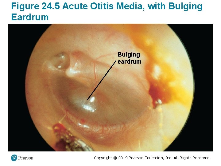
Figure 24. 5 Acute Otitis Media, with Bulging Eardrum Bulging eardrum Copyright © 2019 Pearson Education, Inc. All Rights Reserved
Viral Disease of the Upper Respiratory System Learning Objective 24 -5 List the causative agents and treatments for the common cold. Copyright © 2019 Pearson Education, Inc. All Rights Reserved
The Common Cold • Over 200 different viruses – Rhinoviruses (30– 50%) § Thrive in temperatures lower than body temperature – Coronaviruses (10– 15%) – Enterovirus D 68 (EV-D 68) • Sneezing, nasal secretion, congestion – Can lead to laryngitis and otitis media – Not accompanied by fever • Antibiotics are of no use. – Relief via cough suppressants and antihistamines Copyright © 2019 Pearson Education, Inc. All Rights Reserved
Check Your Understanding-5 Check Your Understanding ü 24 -5 Which viruses, rhinoviruses, or coronaviruses cause about half of the cases of the common cold? Copyright © 2019 Pearson Education, Inc. All Rights Reserved
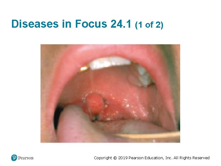
Diseases in Focus: Microbial Diseases of the Upper Respiratory System • A patient presents with fever and a red, sore throat. Later, a grayish membrane appears in the throat. Gram-positive rods are cultured from the membrane. • Can you identify infections that could cause these symptoms? Copyright © 2019 Pearson Education, Inc. All Rights Reserved
Diseases in Focus 24. 1 (1 of 2) Copyright © 2019 Pearson Education, Inc. All Rights Reserved
Diseases in Focus 24. 1 (2 of 2) Copyright © 2019 Pearson Education, Inc. All Rights Reserved
Microbial Diseases of the Lower Respiratory System • Caused by many of the same bacteria and viruses as the upper respiratory system – Bronchitis – Bronchiolitis – Pneumonia § Pulmonary alveoli are involved Copyright © 2019 Pearson Education, Inc. All Rights Reserved
Bacterial Diseases of the Lower Respiratory System Learning Objectives 24 -6 List the causative agent, symptoms, prevention, preferred treatment, and laboratory identification tests for pertussis and tuberculosis. 24 -7 Compare and contrast the seven bacterial pneumonias discussed in this chapter. 24 -8 List the etiology, method of transmission, and symptoms of melioidosis. Copyright © 2019 Pearson Education, Inc. All Rights Reserved
Pertussis (Whooping Cough) (1 of 2) • Caused by Bordetella pertussis – Gram-negative coccobacillus • Produces a capsule – Allows attachment to ciliated cells in the trachea § Destroys ciliated cells and shuts down the ciliary escalator • Tracheal cytotoxin of cell wall damages ciliated cells. • Pertussis toxin enters the bloodstream. Copyright © 2019 Pearson Education, Inc. All Rights Reserved
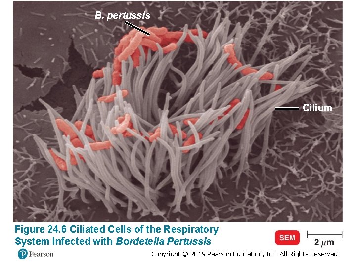
B. pertussis Cilium Figure 24. 6 Ciliated Cells of the Respiratory System Infected with Bordetella Pertussis Copyright © 2019 Pearson Education, Inc. All Rights Reserved
Pertussis (Whooping Cough) (2 of 2) • Stage 1: catarrhal stage, like the common cold • Stage 2: paroxysmal stage, violent coughing, gasping for air • Stage 3: convalescence stage, may last for months • Prevented by DTa. P vaccine • Treated with erythromycin or other macrolides Copyright © 2019 Pearson Education, Inc. All Rights Reserved
Check Your Understanding-6 Check Your Understanding ü 24 -6 Another name for pertussis is whooping cough. This symptom is caused by the pathogens’ attack on which cells? Copyright © 2019 Pearson Education, Inc. All Rights Reserved
Big Picture: Bioterrorism (1 of 5) • Biological weapons: pathogens intentionally used for hostile purposes • History of Bioweapons – 1346: plague-ridden bodies used by the Tartar army during the Siege of Kaffa (Ukraine) § Spread to the rest of Europe causing the “Black Death” pandemic of 1348– 1350 – Eighteenth century: smallpox-contaminated blankets used against Native Americans during the French and Indian War – 1937– 1945: plague-carrying flea bombs used in the Sino-Japanese War Copyright © 2019 Pearson Education, Inc. All Rights Reserved
Big Picture: Bioterrorism (2 of 5) • Biological weapons banned in the twentieth century – 1920: The Geneva Conventions § Prohibited deploying bioweapons – 1975: The Biological Weapons Convention § Banned the possessions and development of bioweapons • Emergence of Bioterrorism – Radical groups and individuals – 2001 bioterrorism anthrax attacks in the United States Copyright © 2019 Pearson Education, Inc. All Rights Reserved
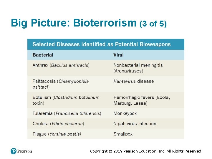
Big Picture: Bioterrorism (3 of 5) Copyright © 2019 Pearson Education, Inc. All Rights Reserved
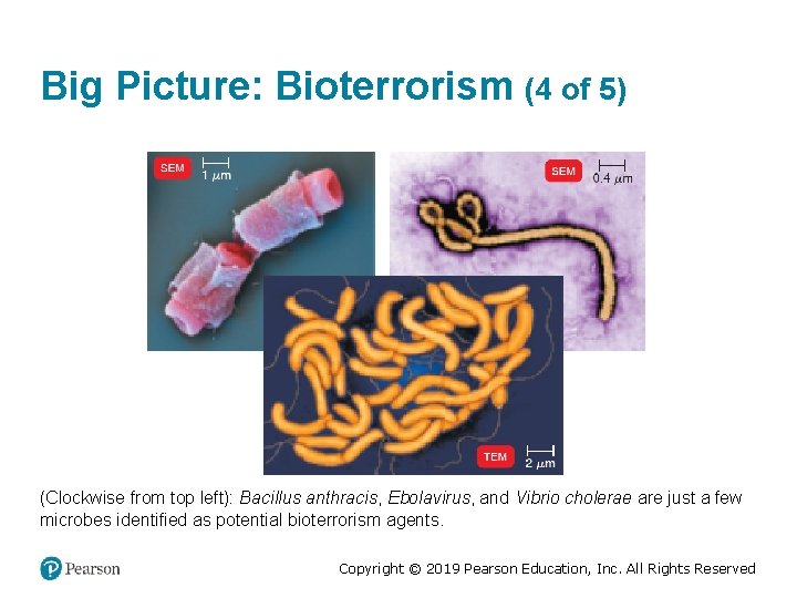
Big Picture: Bioterrorism (4 of 5) (Clockwise from top left): Bacillus anthracis, Ebolavirus, and Vibrio cholerae are just a few microbes identified as potential bioterrorism agents. Copyright © 2019 Pearson Education, Inc. All Rights Reserved
Big Picture: Bioterrorism (5 of 5) • Public health authorities implemented protocols to deal with potential bioterrorism. – New technologies and techniques to identify bioweapons § DNA chips § Recombinant cells § Pro Strips Rapid Screening System – Tests for anthrax, ricin toxin, botulinum toxin, plague, and SEB (staphylococcal enterotoxin B) – Vaccination Copyright © 2019 Pearson Education, Inc. All Rights Reserved
Tuberculosis (1 of 2) • Caused by Mycobacterium tuberculosis – Acid-fast rod; obligate aerobe – 20 -hour generation time – Lipids in the cell wall make it resistant to drying and antimicrobials. • Other causes – Mycobacterium bovis § Bovine tuberculosis; < 1% of U. S. cases – Mycobacterium avium-intracellulare complex § Infects people with late-stage HIV infection Copyright © 2019 Pearson Education, Inc. All Rights Reserved
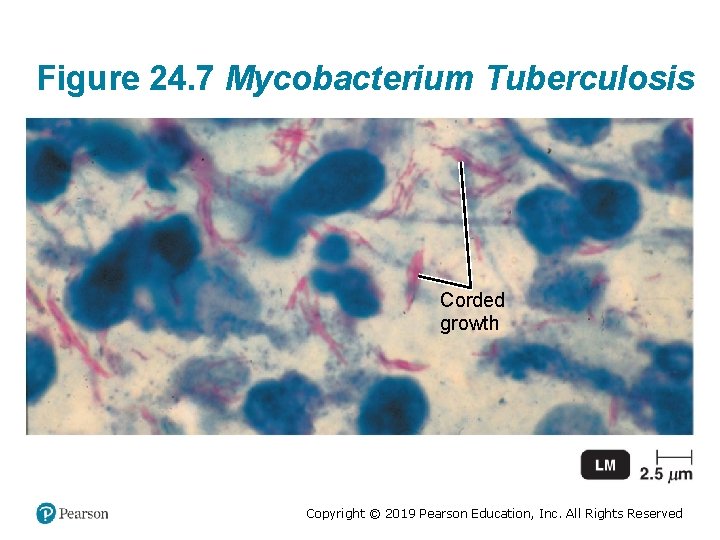
Figure 24. 7 Mycobacterium Tuberculosis Corded growth Copyright © 2019 Pearson Education, Inc. All Rights Reserved
Pathogenesis of Tuberculosis • Inhaled organisms are phagocytized by alveolar macrophages. • Mycolic acids in the cell wall stimulate an inflammatory response. • Organisms are isolated in the walled-off tubercle. • Tubercles heal and become calcified (Ghon's complexes) – Latent TB (tubercle bacilli remain dormant). • Tubercle breaks down, releasing bacteria into the lungs and cardiovascular and lymphatic systems – Miliary tuberculosis: disseminated infection Copyright © 2019 Pearson Education, Inc. All Rights Reserved
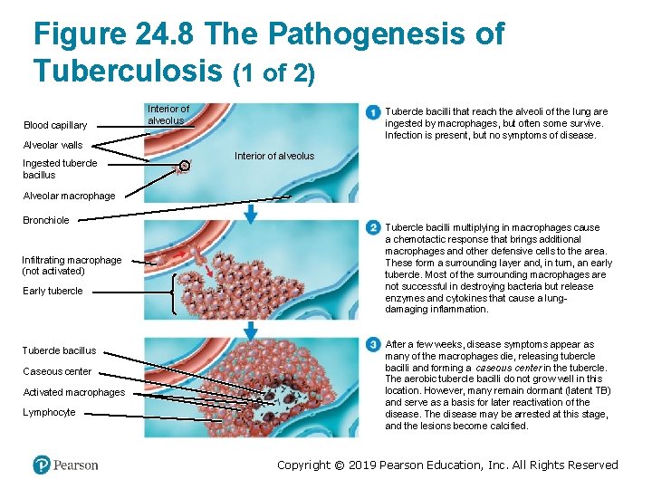
Figure 24. 8 The Pathogenesis of Tuberculosis (1 of 2) Blood capillary Interior of alveolus Tubercle bacilli that reach the alveoli of the lung are ingested by macrophages, but often some survive. Infection is present, but no symptoms of disease. Alveolar walls Ingested tubercle bacillus Interior of alveolus Alveolar macrophage Bronchiole Infiltrating macrophage (not activated) Early tubercle Tubercle bacillus Caseous center Activated macrophages Lymphocyte Tubercle bacilli multiplying in macrophages cause a chemotactic response that brings additional macrophages and other defensive cells to the area. These form a surrounding layer and, in turn, an early tubercle. Most of the surrounding macrophages are not successful in destroying bacteria but release enzymes and cytokines that cause a lungdamaging inflammation. After a few weeks, disease symptoms appear as many of the macrophages die, releasing tubercle bacilli and forming a caseous center in the tubercle. The aerobic tubercle bacilli do not grow well in this location. However, many remain dormant (latent TB) and serve as a basis for later reactivation of the disease. The disease may be arrested at this stage, and the lesions become calcified. Copyright © 2019 Pearson Education, Inc. All Rights Reserved
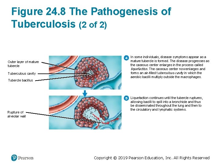
Figure 24. 8 The Pathogenesis of Tuberculosis (2 of 2) Outer layer of mature tubercle Tuberculous cavity Tubercle bacillus Rupture of alveolar wall In some individuals, disease symptoms appear as a mature tubercle is formed. The disease progresses as the caseous center enlarges in the process called liquefaction. The caseous center now enlarges and forms an air-filled tuberculous cavity in which the aerobic bacilli multiply outside the macrophages. Liquefaction continues until the tubercle ruptures, allowing bacilli to spill into a bronchiole and thus be disseminated throughout the lung and then to the circulatory and lymphatic systems. Copyright © 2019 Pearson Education, Inc. All Rights Reserved
Diagnosis of Tuberculosis • Tuberculin skin test – Positive reaction means a current or previous infection. – T cells react with purified protein derivative from the TB bacterium. § Delayed hypersensitivity induration • Followed by an X-ray or CT exam, acid-fast staining of sputum, and culturing of bacteria • Rapid blood test for IFN-γ and Nucleic acid amplification test (NAAT) – Higher specificity and less cross-reactivity Copyright © 2019 Pearson Education, Inc. All Rights Reserved
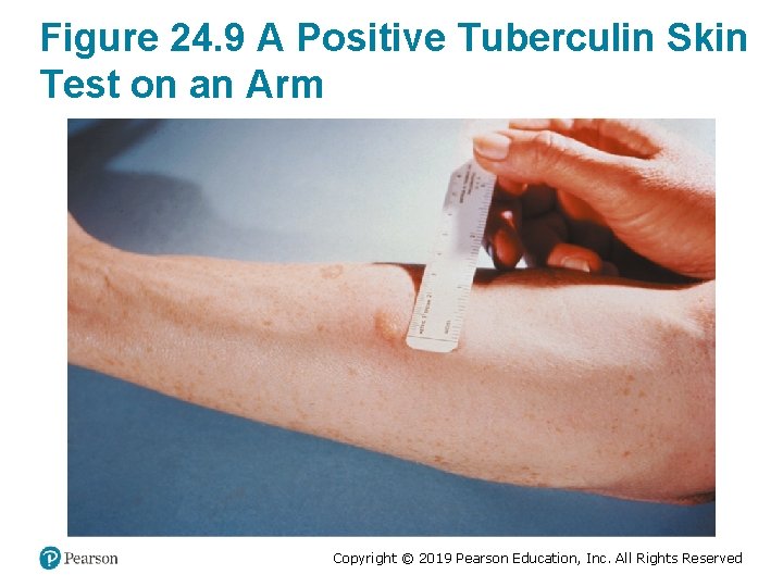
Figure 24. 9 A Positive Tuberculin Skin Test on an Arm Copyright © 2019 Pearson Education, Inc. All Rights Reserved
Treatment of Tuberculosis • Minimum of 6 months of drug therapy due to slow growth and dormancy • First-line drugs: isoniazid, rifampin, ethambutol, pyrazinamide • Second-line drugs: aminoglycosides, fluoroquinolones, streptomycin, para-aminoslicyclic acid (PAS) • Multi-drug-resistant (MDR) strains: resistant to firstline drugs • Extensively drug-resistant (XDR) strains: resistant to second-line drugs Copyright © 2019 Pearson Education, Inc. All Rights Reserved
Tuberculosis (2 of 2) • Copyright © 2019 Pearson Education, Inc. All Rights Reserved
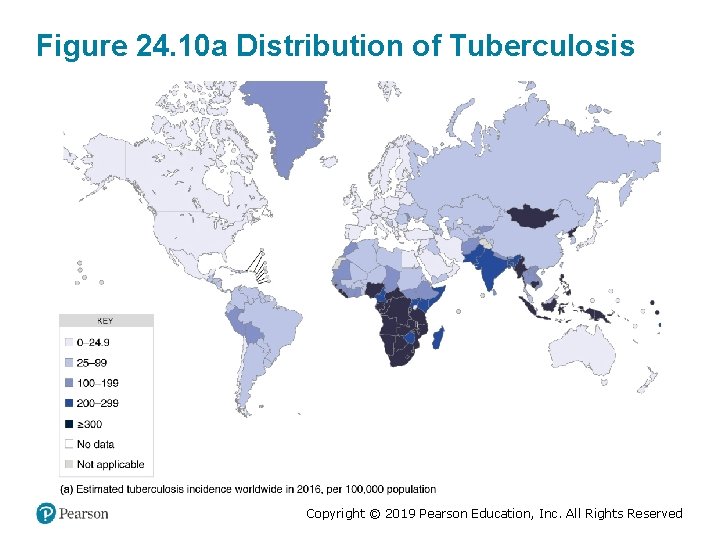
Figure 24. 10 a Distribution of Tuberculosis Copyright © 2019 Pearson Education, Inc. All Rights Reserved
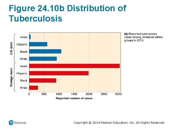
Figure 24. 10 b Distribution of Tuberculosis Copyright © 2019 Pearson Education, Inc. All Rights Reserved
Bacterial Pneumonias • Typical pneumonia – Caused by S. pneumoniae • Atypical pneumonia – Caused by other microorganisms • Lobar pneumonia – Infects the lobes of the lungs • Bronchopneumonia – Infects the alveoli adjacent to the lungs • Pleurisy – Pleural membranes inflamed Copyright © 2019 Pearson Education, Inc. All Rights Reserved
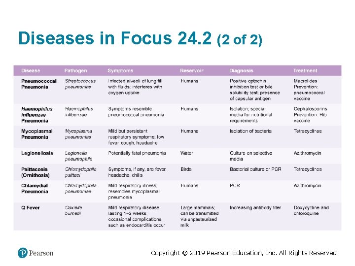
Pneumococcal Pneumonia • Caused by S. pneumoniae – Gram-positive; encapsulated diplococci – 90 serotypes • Infected alveoli of the lung fill with fluids and RBCs; interferes with oxygen uptake • Diagnosis: optochin-inhibition test, bile solubility test, or presence of capsular antigen in urine • Treated with macrolides • Prevented with conjugated pneumococcal vaccine Copyright © 2019 Pearson Education, Inc. All Rights Reserved
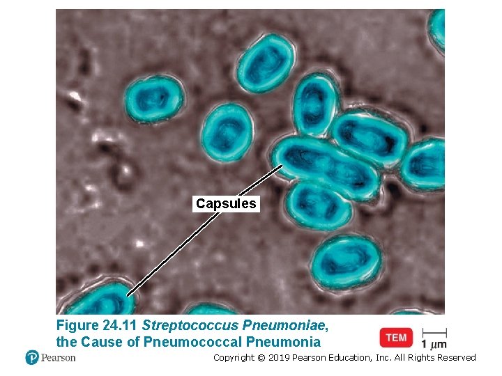
Capsules Figure 24. 11 Streptococcus Pneumoniae, the Cause of Pneumococcal Pneumonia Copyright © 2019 Pearson Education, Inc. All Rights Reserved
Haemophilus Influenzae Pneumonia • Gram-negative coccobacillus • Symptoms resemble those of pneumococcal pneumonia • Risk group: Children under 5 and adults over 65 – The Hib vaccine reduced the incidence in children by 99% • Diagnosis: isolation on special media for nutritional requirements (X and V factors) • Treated with cephalosporins Copyright © 2019 Pearson Education, Inc. All Rights Reserved
Mycoplasmal Pneumonia • Also called primary atypical pneumonia or walking pneumonia • Caused by Mycoplasma pneumoniae – No cell wall • Mild but persistent respiratory symptoms; low fever, cough, headache – Common in children and young adults • “Fried-egg” appearance on media • Diagnosis: PCR • Treated with tetracyclines Copyright © 2019 Pearson Education, Inc. All Rights Reserved
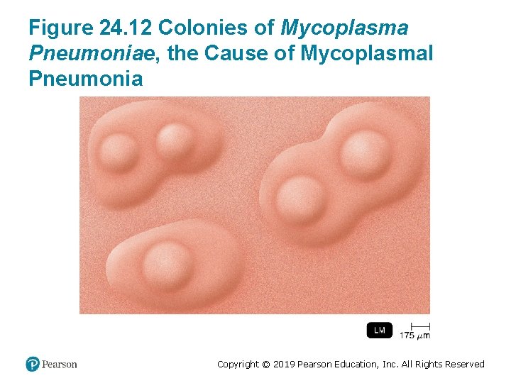
Figure 24. 12 Colonies of Mycoplasma Pneumoniae, the Cause of Mycoplasmal Pneumonia Copyright © 2019 Pearson Education, Inc. All Rights Reserved
Legionellosis • Also called Legionnaires’ disease • Caused by Legionella pneumophila – Aerobic, gram-negative rod – Grows in water and air conditioning, biofilms, and waterborne amebae • Transmitted by inhaling aerosols; not transmitted person to person • Symptoms: high fever and cough – Similar to symptoms of Pontiac fever • Treated with azithromycin and macrolides Copyright © 2019 Pearson Education, Inc. All Rights Reserved
Psittacosis (Ornithosis) • Caused by Chlamydophila psittaci – Gram-negative intracellular bacterium • Transmitted to humans by elementary bodies from bird droppings transmitted through air • Fever, headache, chills, disorientation • Diagnosis: growth of bacteria in eggs, cell culture or PCR • Treated with tetracyclines Copyright © 2019 Pearson Education, Inc. All Rights Reserved
Chlamydial Pneumonia • Caused by Chlamydophila pneumoniae • Transmitted person to person • Mild respiratory illness common in young people; resembles mycoplasmal pneumonia • Possible association with artherosclerosis • Diagnosis: PCR • Treated with azithromycin Copyright © 2019 Pearson Education, Inc. All Rights Reserved
Q Fever • Caused by Coxiella burnetii – Obligately parasitic, intracellular gammaproteobacteria • Acute Q fever – High fever, muscle aches, headache, coughing • Chronic Q fever – Endocarditis (may occur years after infection) • Transmitted to farm animals from tick bites – Transmitted to humans from the inhalation of aerosols from animals and unpasteurized milk • Treated with doxycycline; chloroquine for chronic infections Copyright © 2019 Pearson Education, Inc. All Rights Reserved
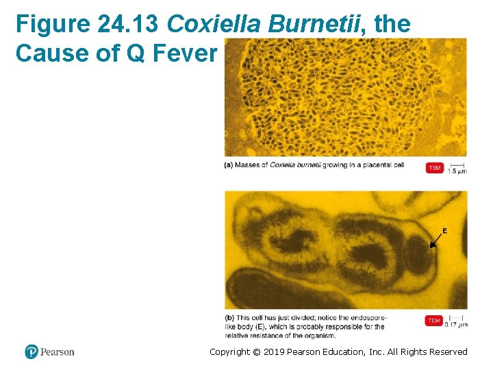
Figure 24. 13 Coxiella Burnetii, the Cause of Q Fever Copyright © 2019 Pearson Education, Inc. All Rights Reserved
Melioidosis • Caused by Burkholderia pseudomallei – Gram-negative rod • Occurs mostly in Southeast Asia and northern Australia (in moist soils) • Commonly affects those with lowered immune systems – Pneumonia or tissue abscesses (necrotizing fasciitis) and severe sepsis • Transmission by inhalation, puncture wounds, and ingestion • Treated with ceftazidime Copyright © 2019 Pearson Education, Inc. All Rights Reserved
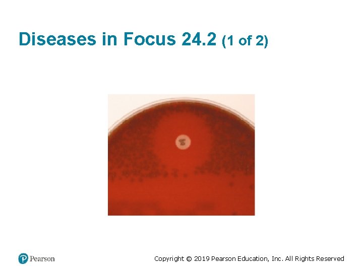
Diseases in Focus: Common Bacterial Pneumonias • A 27 -year-old man with a history of asthma is hospitalized with a 4 -day history of progressive cough and 2 days of spiking fevers. Gram-positive cocci in pairs are cultured from a blood sample. • Can you identify infections that could cause these symptoms? Copyright © 2019 Pearson Education, Inc. All Rights Reserved
Diseases in Focus 24. 2 (1 of 2) Copyright © 2019 Pearson Education, Inc. All Rights Reserved
Diseases in Focus 24. 2 (2 of 2) Copyright © 2019 Pearson Education, Inc. All Rights Reserved
Check Your Understanding-7 Check Your Understanding ü 24 -7 What group of bacterial pathogens causes what is informally called “walking pneumonia”? ü 24 -8 The bacterium causing melioidosis in humans also causes a disease of horses known as what? Copyright © 2019 Pearson Education, Inc. All Rights Reserved
Viral Diseases of the Lower Respiratory System Learning Objective 24 -9 List the causative agent, symptoms, prevention, and preferred treatment for viral pneumonia, RSV, and influenza. Copyright © 2019 Pearson Education, Inc. All Rights Reserved
Viral Pneumonia • Viral pneumonia occurs as a complication of influenza, measles, or chickenpox • Few labs are equipped to test clinical samples properly for viruses • SARS-associated coronavirus (SARS) – Emerged in Asia in 2003 • Middle East respiratory syndrome coronavirus (MERS-Co. V) – Reported in Saudi Arabia in 2012 Copyright © 2019 Pearson Education, Inc. All Rights Reserved
Respiratory Syncytial Virus (RSV) • Most common viral respiratory disease in infants and life-threatening pneumonia in older adults – Almost all children are infected by age 2 – 14, 000 deaths annually, mostly in older adults • Causes cell fusion (syncytium) in cell culture • Coughing and wheezing for more than a week • Diagnosis: serological test for viruses and antibodies • Treated with palivizumab Copyright © 2019 Pearson Education, Inc. All Rights Reserved
Influenza (Flu) (1 of 3) • Influenzavirus – Contains eight RNA segments and an outer lipid bilayer • Chills, fever, headache, and muscle aches – No intestinal symptoms – 3, 000 to 50, 000 deaths in the United States annually • Avian, swine, and mammalian strains – Swine serve as “mixing vessels” for new strains Copyright © 2019 Pearson Education, Inc. All Rights Reserved
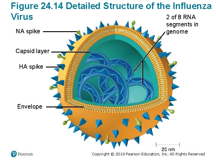
Figure 24. 14 Detailed Structure of the Influenza 2 of 8 RNA Virus NA spike segments in genome Capsid layer HA spike Envelope Copyright © 2019 Pearson Education, Inc. All Rights Reserved
Influenza (Flu) (2 of 3) • Hemagglutinin (HA) spikes – Recognize and attach to host cells • Neuraminidase (NA) spikes – Help the virus separate from the infected cell • Antigenic drift – Minor antigenic changes in HA and NA – Allow the virus to elude some host immunity • Antigenic shifts – Changes great enough to evade most immunity – Lead to pandemics – Involve the reassortment of the eight RNA segments Copyright © 2019 Pearson Education, Inc. All Rights Reserved
Influenza (Flu) (3 of 3) • 1% mortality; usually the very young and very old • Multivalent vaccine for the most important strains – Composition of the vaccine determined annually by the identification of circulating viruses § Labor-intensive to produce § Does not provide long-term immunity • Difficult to diagnose from clinical symptoms • Treated with zanamivir (Relenza) and oseltamivir (Tamiflu) – Inhibits neuraminidase Copyright © 2019 Pearson Education, Inc. All Rights Reserved
Check Your Understanding-8 Check Your Understanding ü 24 -9 Is reassortment of the RNA segments of the influenza virus the cause of antigenic shift or antigenic drift? Copyright © 2019 Pearson Education, Inc. All Rights Reserved
Fungal Diseases of the Lower Respiratory System Learning Objective 24 -10 List the causative agent, mode of transmission, preferred treatment, and laboratory identification tests for four fungal diseases of the respiratory system. Copyright © 2019 Pearson Education, Inc. All Rights Reserved
Histoplasmosis • Caused by Histoplasma capsulatum – Dimorphic fungus – Yeast-form grows intracellularly in macrophages • Forms lung lesions; 0. 1% of cases become a severe, generalized disease • Acquired from airborne conidia in areas with bird or bat droppings – Limited geographical range in the United States • Treated with itraconazole Copyright © 2019 Pearson Education, Inc. All Rights Reserved
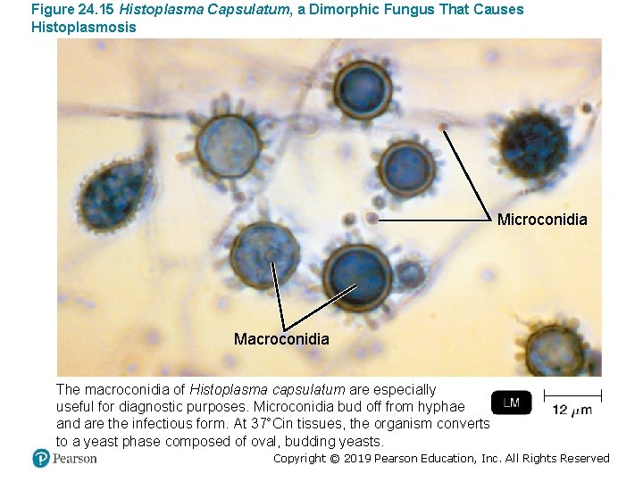
Figure 24. 15 Histoplasma Capsulatum, a Dimorphic Fungus That Causes Histoplasmosis Microconidia Macroconidia The macroconidia of Histoplasma capsulatum are especially useful for diagnostic purposes. Microconidia bud off from hyphae and are the infectious form. At 37°Cin tissues, the organism converts to a yeast phase composed of oval, budding yeasts. Copyright © 2019 Pearson Education, Inc. All Rights Reserved
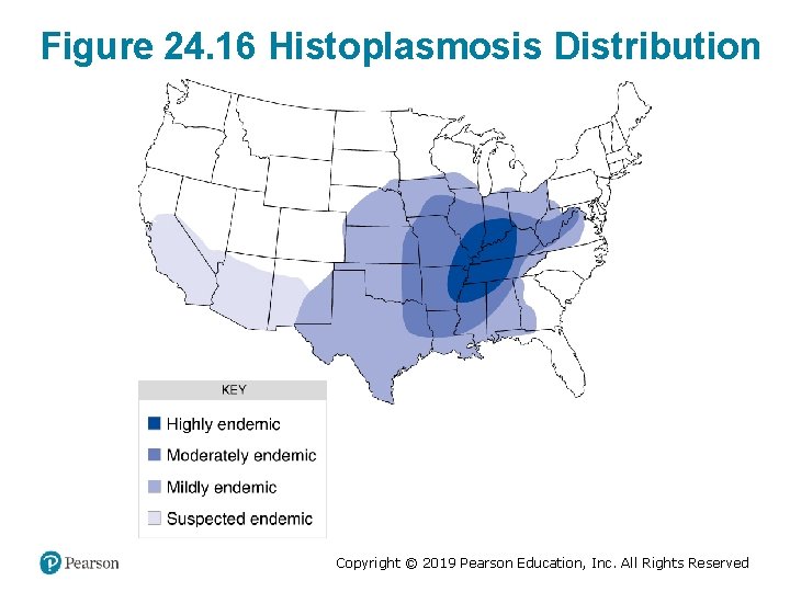
Figure 24. 16 Histoplasmosis Distribution Copyright © 2019 Pearson Education, Inc. All Rights Reserved
Coccidioidomycosis • Also known as Valley fever or San Joaquin fever • Caused by Coccidioides immitis – Dimorphic fungus • Arthroconidia found in alkaline desert soils of the American Southwest • Form a spherule filled with endospores in tissues • Most infections are not apparent; fever, coughing, weight loss – < 1% of cases resemble tuberculosis • Treated with fluconazole or itraconazole Copyright © 2019 Pearson Education, Inc. All Rights Reserved
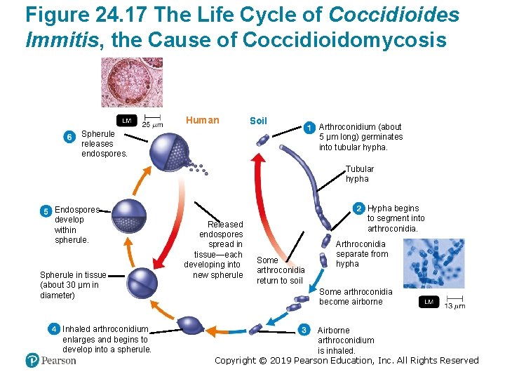
Figure 24. 17 The Life Cycle of Coccidioides Immitis, the Cause of Coccidioidomycosis Human Soil Spherule releases endospores. Arthroconidium (about 5 μm long) germinates into tubular hypha. Tubular hypha Endospores develop within spherule. Spherule in tissue (about 30 μm in diameter) Inhaled arthroconidium enlarges and begins to develop into a spherule. Released endospores spread in tissue—each developing into new spherule Hypha begins to segment into arthroconidia. Some arthroconidia return to soil Arthroconidia separate from hypha Some arthroconidia become airborne Airborne arthroconidium is inhaled. Copyright © 2019 Pearson Education, Inc. All Rights Reserved
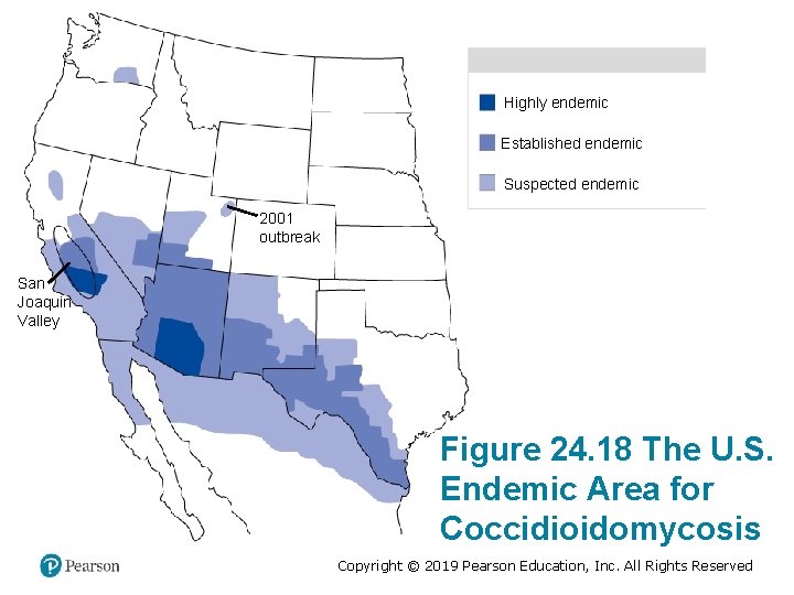
Highly endemic Established endemic Suspected endemic 2001 outbreak San Joaquin Valley Figure 24. 18 The U. S. Endemic Area for Coccidioidomycosis Copyright © 2019 Pearson Education, Inc. All Rights Reserved
Pneumocystis Pneumonia (PCP) • Caused by Pneumocystis jirovecii – No universal agreement if it is a protozoan or fungus • Asymptomatic in the immunocompetent; causes pneumonia in the immunocompromised – Primary indicator of AIDS • Found in the lining of the alveoli – Forms a cyst – Cysts rupture, releasing eight trophozoites • Treated with trimethoprim-sulfamethoxazole Copyright © 2019 Pearson Education, Inc. All Rights Reserved
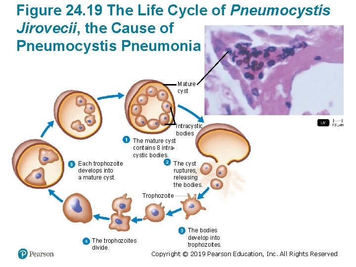
Figure 24. 19 The Life Cycle of Pneumocystis Jirovecii, the Cause of Pneumocystis Pneumonia Mature cyst Intracystic bodies Each trophozoite develops into a mature cyst. The mature cyst contains 8 intracystic bodies. The cyst ruptures, releasing the bodies. Trophozoite The trophozoites divide. The bodies develop into trophozoites. Copyright © 2019 Pearson Education, Inc. All Rights Reserved
Blastomycosis (North American Blastomycosis) • Caused by Blastomyces dermatitidis – Dimorphic fungus – Grows in soil • Symptoms resemble bacterial pneumonia; cutaneous abscesses; extensive tissue damage • Reported ~100 cases annually • Treated with itraconazole and amphotericin B Copyright © 2019 Pearson Education, Inc. All Rights Reserved
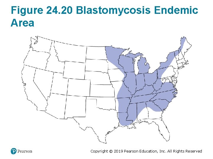
Figure 24. 20 Blastomycosis Endemic Area Copyright © 2019 Pearson Education, Inc. All Rights Reserved
Other Fungi Involved in Respiratory Disease • Aspergillus fumigatus – Causes aspergillosis – Airborne conidia; grows in compost piles • Rhizopus and Mucor – Mold spores • Predisposing factors: – Immunocompromised state – Cancer – Diabetes Copyright © 2019 Pearson Education, Inc. All Rights Reserved
Check Your Understanding-9 Check Your Understanding ü 24 -10 The droppings of both blackbirds and bats support the growth of Histoplasma capsulatum; which of these two animal reservoirs is normally actually infected by the fungus? Copyright © 2019 Pearson Education, Inc. All Rights Reserved
Diseases in Focus: Microbial Diseases of the Lower Respiratory System • Three weeks after working on the demolition of an abandoned building in Kentucky, a worker is hospitalized for acute respiratory illness. At the time of demolition, a colony of bats inhabited the building. An X-ray examination reveals a lung mass. A purified protein derivative test is negative; a cytological examination of the mass reveals ovoid yeast cells. • Can you identify infections that could cause these symptoms? Copyright © 2019 Pearson Education, Inc. All Rights Reserved
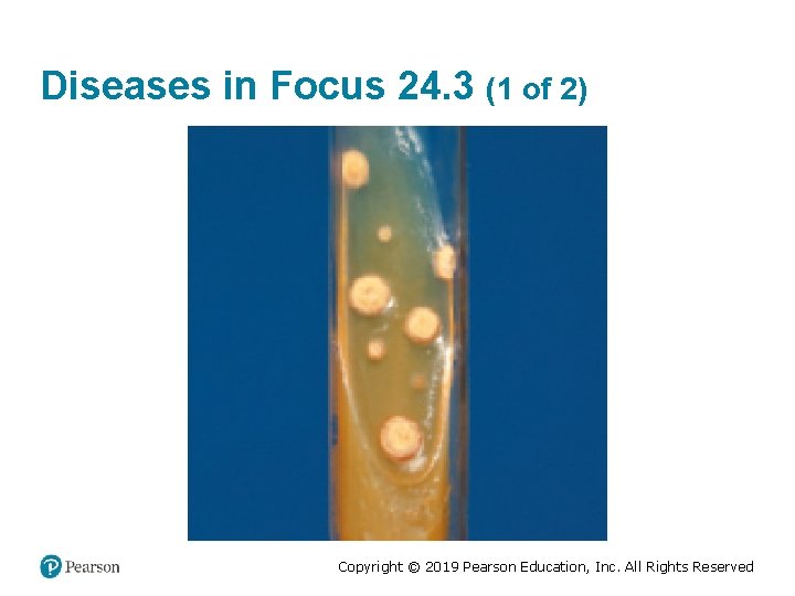
Diseases in Focus 24. 3 (1 of 2) Copyright © 2019 Pearson Education, Inc. All Rights Reserved
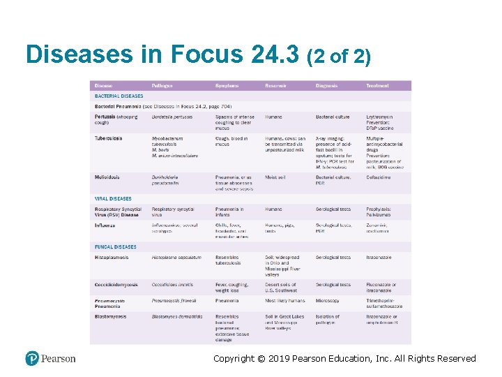
Diseases in Focus 24. 3 (2 of 2) Copyright © 2019 Pearson Education, Inc. All Rights Reserved
Copyright © 2019 Pearson Education, Inc. All Rights Reserved
- Slides: 91