Microbiology A nd Systems Approach 2 ed Chapter

Microbiology: A nd Systems Approach, 2 ed. Chapter 6: An Introduction to the Viruses

6. 1 The Search for the Elusive Viruses were too small to be seen with the first microscopes Ø The cause of viral infections was unknown for years Ø Louis Pasteur first proposed the term virus Ø 1890 s Ø l l Ø Ivanovski and Beijerinck showed that a disease in tobacco was caused by a virus Loeffler and Frosch discovered an animal virus that causes foot –and-mouth disease in cattle Many years of experimentation showed what we know today and by the 1950 s virology had grown

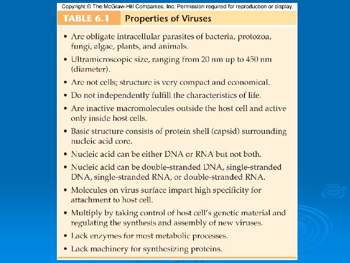
6. 2 The Position of Viruses in the Biological Spectrum Ø Ø Ø Can infect every type of cell Cannot exist independently from the host cell, so aren’t considered living things However, since they can direct life processes they are often considered more than lifeless molecules Referred to as infectious particles, either active or inactive Obligate intracellular parasites Cannot multiply unless they invade a specific host cell and instruct its genetic and metabolic machinery to make and release new viruses

6. 3 The General Structure of Viruses Figure 6. 1

Size Range Smallest infectious agents Most are so small, they can only be seen with an electron microscope Ø Animal viruses Ø Ø l l Ø Proviruses- around 20 nm in diameter Mimiviruses- up to 450 nm in length Viewing viruses l l Special stains and an electron microscope Negative staining outlines the shape Positive staining shows internal details Shadowcasting technique

Figure 6. 2

Viral Components: Capsids, Nucleic Acids, and Envelopes Molecular structure- composed of regular, repeating subunits that give rise to their crystalline appearance Ø Contain only those parts needed to invade and control a host cell Ø l External coating • Capsid • Envelope- in 13 of the 20 families of animal viruses • If no envelope, called naked virus l Core • • l Ø DNA RNA The capsid and the nucleic acid together are called the nucleocapsid Fully formed virus that is able to establish an infection in a host cell- virion

Figure 6. 4

The Viral Capsid: The Protective Outer Shell Ø Constructed from identical subunits called capsomers Ø Made up of protein molecules Ø Two different types l Helical • Rod-shaped capsomers • Assemble in to helical nucleocapsid

Figure 6. 5

Figure 6. 6

Ø Icosahderal l l Three-dimensional, 20 -sided figure with 12 evenly spaced corners Although they all display this symmetry, there are wide variations

Figure 6. 7

Figure 6. 8

Figure 6. 9

Figure 6. 10

The Viral Envelope Ø Enveloped viruses take a bit of the host cell membrane in the form of an envelope Ø In the envelope, some or all of the regular membrane proteins are replaced with viral proteins Ø Some proteins form a binding layer between the envelope and the capsid Ø Glycoproteins remain exposed as spikes (peplomers)- essential for attachment

Functions of the Viral Capsid/Envelope Ø Protects nucleic acids Ø Help introduce the viral DNA or RNA into a suitable host cell Ø Stimulate the immune system to produce antibodies that can protect the host cells against future infections

Nucleic Acids: At the Core of a Virus Ø Genome- the sum total of the genetic information carried by an organism Ø Number of viral genes compared with a callquite small Ø They only have the genes necessary to invade host cells and redirect their activity Ø Some viruses are exceptions to the rules re: DNA and RNA l l Parvoviruses contain single-stranded DNA Reoviruses contain double-stranded RNA

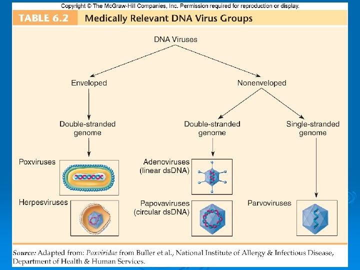
DNA Viruses Ø ss. DNA Ø ds. DNA l l linear circular

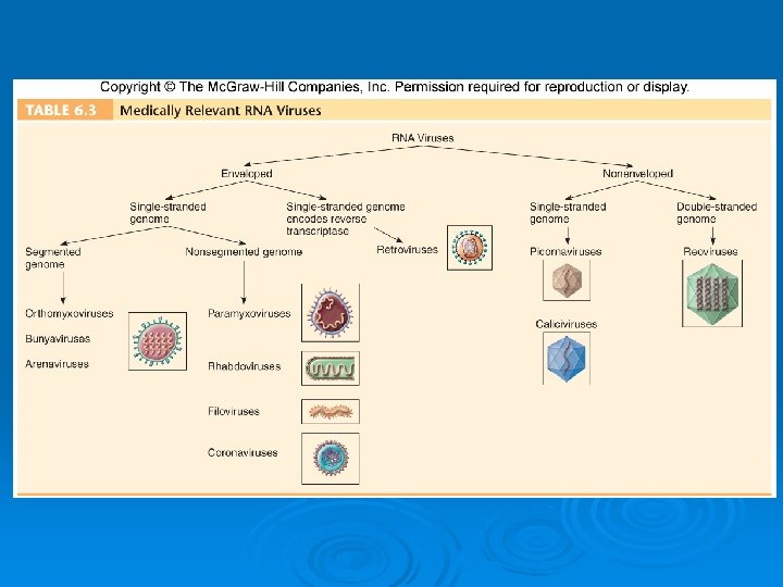
RNA Viruses Ø Mostly single-stranded l l Positive-sense RNA: genomes that are ready for immediate translation into proteins Negative-sense RNA: genomes have to be converted into the proper form to be made into proteins Ø Segmented- individual genes exist on separate pieces of RNA

Other Substances in the Virus Particle Ø Other Substances in the Virus Particle l l l Can contain enzymes for specific operations within the host cell Polymerases to synthesize DNA and RNA Replicases to copy RNA

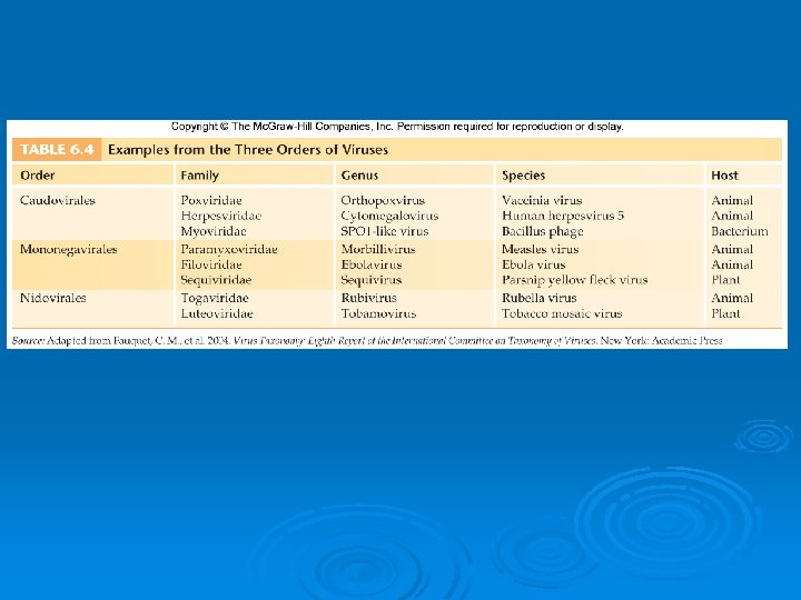
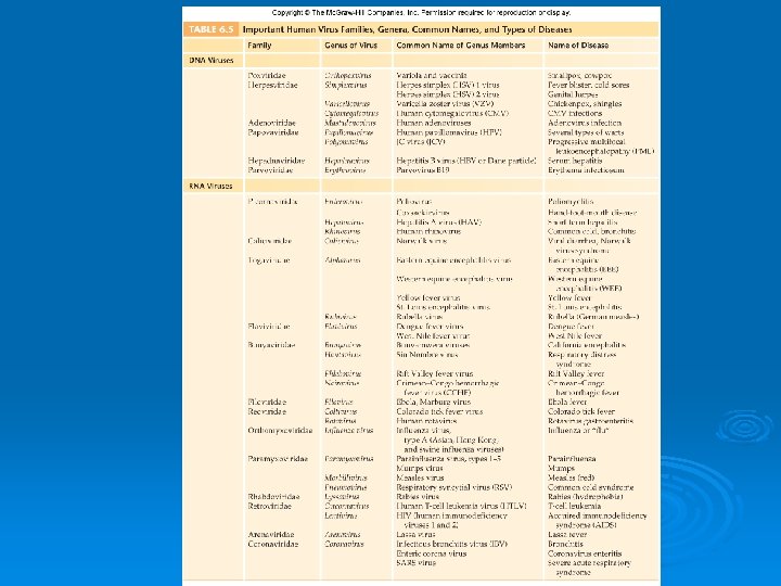
6. 4 How Viruses are Classified and Named Ø Main criteria l l l Structure Chemical composition Similarities in genetic makeup Ø International Committee on the Taxonomy of Viruses, 2000 l l l 3 orders 63 famillies “-viridae” 263 genera “-virus” Ø Some virologists use a species naming system, but it is not an official designation

6. 5 Modes of Viral Multiplication Ø The host cell is absolutely necessary for viral multiplication

Figure 6. 11

Multiplication Cycles in Animal Viruses Ø Adsorption Ø Penetration Ø Uncoating Ø Synthesis Ø Assembly Ø Release

Adsorption Ø Virus encounters susceptible host cells Ø Adsorbs specifically to receptor sites on the cell membrane • Because of the exact fit required, viruses have a limited host range

Figure 6. 12

Penetration Ø Flexible cell membrane of the host is penetrated by the whole virus or its nucleic acid Ø Endocytosis: entire virus engulfed by the cell and enclosed in a vacuole or vesicle Ø The viral envelope can also directly fuse with the host cell membrane

Figure 6. 13

Uncoating Ø Enzymes in the vacuole dissolve the envelope and capsid Ø The virus is now uncoated

Synthesis Ø Free viral nucleic acid exerts control over the host’s synthetic and metabolic machinery Ø DNA viruses- enter host cell’s nucleus where they are replicated and assembled l l l DNA enters the nucleus and is transcribed into RNA The RNA becomes a message for synthesizing viral proteins (translation) New DNA is synthesized using host nucleotides Ø RNA viruses- replicated and assembled in the cytoplasm

Assembly Ø Mature virus particles are constructed from the growing pool of parts

Release Ø Nonenveloped and complex viruses are released when the cell lyses or ruptures Ø Enveloped viruses are liberated by budding or exocytosis Ø Anywhere from 3, 000 to 100, 000 virions may be released, depending on the virus Ø Entire length of cycle- anywhere from 8 to 36 hours

Figure 6. 15

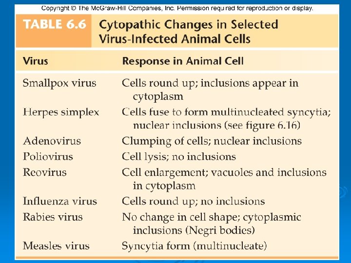
Damage to the Host Cell and Persistent Infections Ø Cytopathic effects- virus-induced damage to the cell that alters its microscopic appearance Ø Inclusion bodies- compacted masses of viruses or damaged cell organelles

Figure 6. 16

Important for the diagnosis of viral infections Ø Some viral infections maintain a carrier relationship Ø l l The cell harbors the virus and is not immediately lysed Persistent infections- from a few weeks to the remainder of the host’s life Some viruses remain in a chronic latent state, periodically becoming activated Ø Some viruses enter their host cell and permanently alter its genetic material, leading to cancer Ø l l l Oncogenic viruses Their effect is called transformation Oncoviruses- mammalian viruses capable of initiating tumors

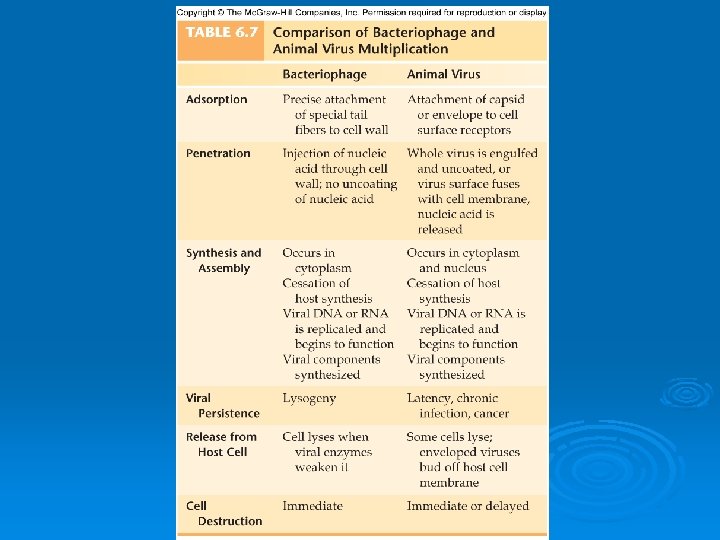
Viruses that Infect Bacteria Ø Bacteriophage Ø Most contain ds. DNA Ø Often make the bacteria they infect more pathogenic for humans

T-even Phages Ø Ø Ø Ø Icosahedral capsid head containing DNA Central tube surrounded by a sheath Collar Base plate Tail pins Fibers Similar stages as animal viruses l l l Adsorb to host bacteria The nucleic acid penetrates the host after being injected through a rigid tube inserted through the bacterial membrane and wall Entry of the nucleic acid causes the cessation of host cell DNA replication and protein synthesis The host cell machinery is then used for viral replication and synthesis of viral proteins As the host cell produces new parts, they spontaneously assemble

Figure 6. 17

Figure 6. 18

Figure 6. 19

Lysogeny: The Silent Virus Infection Ø Temperate phages- special DNA phages that undergo adsorption and penetration but are not replicated or released immediately Ø Instead the viral DNA enters an inactive prophage stage Ø Lysogeny: the cell’s progeny will also have the temperate phage DNA Ø Lysogenic conversion: when a bacterium acquires a new trait from its temperate phage

Figure 6. 20

6. 6 Techniques in Cultivating and Identifying Animal Viruses Ø Primary purposes of viral cultivation l l l Ø Using Live Animal Inoculation l l l Ø To isolate and identify viruses in clinical specimens To prepare viruses for vaccines To do detailed research on viral structure, multiplication cycles, genetics, and effects on host cells Specially bred strains of white mice, rats, hamsters, guinea pigs, and rabbits Occasionally invertebrates or nonhuman primates are used Animal is exposed to the virus by injection Using Bird Embryos l l l Enclosed in an egg- nearly perfect conditions for viral propagation Chicken, duck, and turkey are most common Egg is injected through the shell using sterile techniques

Figure 6. 21
 Culture Techniques Ø Most viruses are propagated in some sort of")
Using Cell (Tissue) Culture Techniques Ø Most viruses are propagated in some sort of cell culture Ø The cultures must be developed and maintained Ø Animal cell cultures are grown in sterile chambers with special media Ø Cultured cells grow in the form of a monolayer Ø Primary or continuous

Figure 6. 22

6. 7 Medical Importance of Viruses Ø Most common cause of acute infections that do not result in hospitalization Ø Most do not cause death but those that do can have very high mortality rates Ø Others can lead to long-term debility

6. 8 Other Noncellular Infectious Agents Ø Spongiform encephalopathies l l l Ø Satellite viruses l l Ø Chronic, persistent diseases Long period of latency Deposition of protein fibrils in the brain tissue- prions Defective forms of viruses Dependent on other viruses for replication Viroids l l l Parasitize plants Very small Composed only of naked strands of RNA

6. 9 Treatment of Animal Viral Infections Ø Because they are not bacteria, antibiotics are ineffective Ø Antiviral drugs block virus replication by targeting one of the steps in the viral life cycle Ø Interferon shows potential for treating and preventing viral infections Ø Vaccines stimulate immunity
- Slides: 59