Microbiology A nd Systems Approach 2 ed Chapter

Microbiology: A nd Systems Approach, 2 ed. Chapter 4: Prokaryotic Profilesthe Bacteria and Archaea

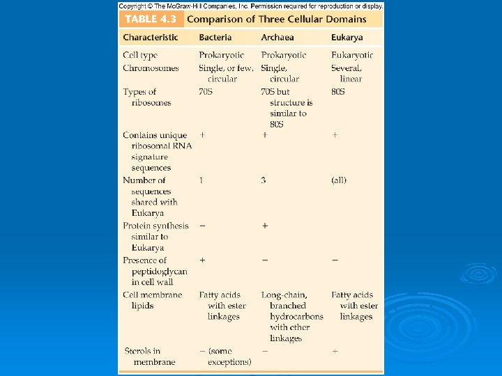
How are Prokaryotes Different from Eukaryotes? Ø The way their DNA is packaged l l No nucleus Not wrapped around histones Ø The makeup of their cell wall l l Bacteria- peptidoglycan Archae- tough and made of other chemicals, distinct to them Ø Their internal structures l No complex, membrane-bound organelles

4. 1 Prokaryotic Form and Function

Structures common to all bacterial cells Ø Cell membrane Ø Cytoplasm Ø Ribosomes Ø One (or a few) chromosomes

Structures found in most bacterial cells Ø Cell wall Ø Surface coating or glycocalyx

Structures found in some bacterial cells Ø Flagella Ø Pili Ø Fimbriae Ø Capsules Ø Slime layers Ø Inclusions Ø Actin cytoskeleton Ø Endospores

Figure 4. 1

4. 2 External Structures Ø Appendages: l l l Cell extensions Common but not present on all species Can provide motility (flagella and axial filaments) Can be used for attachment and mating (pili and fimbriae)
, and basal body Ø Vary in both")
Flagella Ø Three parts: Filament, hook (sheath), and basal body Ø Vary in both number and arrangement l Polar arrangement: flagella attached at one or both ends of the cell • Monotrichous- single flagellum • Lophotrichous- small bunches or tufts of flagella emerging from the same site • Peritrichous- dispersed randomly over the structure of the cell

Figure 4. 2

Figure 4. 3

Flagellar Function Ø Chemotaxis- positive and negative Ø Phototaxis Ø Move by runs and tumbles

Figure 4. 4

Figure 4. 5

Axial Filaments Ø AKA periplasmic flagella Ø In spirochetes Ø A type of internal flagellum that is enclosed in the space between the cell wall and the cell membrane

Figure 4. 6

Pili Ø Elongate, rigid tubular structures Ø Made of the protein pilin Ø Found on gram-negative bacteria Ø Used in conjugation

Figure 4. 8

Fimbriae Ø Small, bristlelike fibers Ø Most contain protein Ø Tend to stick to each other and to surfaces

Figure 4. 7

The Glycocalyx Ø Develops as a coating of repeating polysaccharide units, protein, or both Ø Protects the cell Ø Sometimes helps the cell adhere to the environment Ø Differ among bacteria in thickness, organization, and chemical composition l l Slime layer- a loose shield that protects some bacteria from loss of water and nutrients Capsule when the glycocalyx is bound more tightly to the cell and is denser and thicker

Figure 4. 9

Functions of the Glycocalyx Many pathogenic bacteria have glycocalyces Ø Protect the bacteria against phagocytes Ø Important in formation of biofilms

4. 3 The Cell Envelope: The Boundary layer of Bacteria Ø Majority of bacteria have a cell envelope Ø Lies outside of the cytoplasm Ø Composed of two or three basic layers l l l Cell membrane Cell wall In some bacteria, the outer membrane

Differences in Cell Envelope Structure Ø The differences between gram-positive and gram-negative bacteria lie in the cell envelope Ø Gram-positive l l Two layers Cell wall and cytoplasmic membrane Ø Gram-negative l l Three layers Outer membrane, cell wall, and cytoplasmic membrane

Figure 4. 12

Structure of the Cell Wall Ø Helps determine the shape of a bacterium Ø Provides strong structural support Ø Most are rigid because of peptidoglycan content

Figure 4. 13

Structure of the Cell Wall, cont. Ø Keeps cells from rupturing because of changes in pressure due to osmosis Ø Target of many antibiotics- disrupt the cell wall, and cells have little protection from lysis Ø Gram-positive cell wall l l A thick (20 to 80 nm), homogeneous sheath of petidoglycan Contains tightly bound acidic polysaccharides Ø Gram-Negative Cell Wall l l Single, thin (1 to 3 nm) sheet of peptidoglycan Periplasmic space surrounds the peptidoglycan

Figure 4. 14

Nontypical Cell Walls Ø Some aren’t characterized as either gram- positive or gram-negative Ø For example, Mycobacterium and Nocardia- unique types of lipids (acid-fast) Ø Archaea - unusual and chemically distinct cell walls Ø Some don’t have a cell wall at all Ø Mycoplasmas- lack cell wall entirely

Mycoplasmas and Other Cell-Wall. Deficient Bacteria Ø Mycoplasma cell membrane is stabilized by sterols and is resistant to lysis l l l Very small bacteria (0. 1 to 0. 5 µm) Range in shape from filamentous to coccus Not obligate parasites Can be grown on artificial media Found in many habitats Important medical species: Mycoplasma pneumoniae

Ø Some bacteria lose their cell wall during part of their life cycle l l l L-forms Arise naturally from a mutation in the wallforming genes Can be induced artificially by treatment with a chemical that disrupts the cell wall • When this occurs with gram-positive cells, the cell becomes a protoplast • With gram-negative cells, the cell becomes a spheroplast

Figure 4. 16

The Gram-Negative Outer Membrane Similar to the cell membrane, except it contains specialized polysaccharides and proteins Ø Outermost layer- contains lipopolysaccharide Ø Innermost layer- phospholipid layer anchored by lipoproteins to the peptidoglycan layer below Ø Outer membrane serves as a partial chemical sieve Ø l l Only relatively small molecules can penetrate Access provided by special membrane channels formed by porin proteins

Cell Membrane Structure Ø Also known as the cytoplasmic membrane Ø Very thin (5 -10 nm) Ø Contain primarily phospholipids and proteins Ø The exceptions: mycoplasmas and archaea Ø Functions l l l Provides a site for functions such as energy reactions, nutrient processing, and synthesis Regulates transport (selectively permeable membrane) Secretion

Practical Considerations of Differences in Cell Envelope Structure Ø Outer membrane- an extra barrier in gram- negative bacteria l l Makes them impervious to some antrimicrobial chemicals Generally more difficult to inhibit or kill than grampositive bacteria Ø Cell envelope can interact with human tissues and cause disease l l Corynebacterium diphtheriae Streptococcus pyogenes

4. 4 Bacterial Internal Structure Ø Contents of the Cell l l Cytoplasm Gelatinous solution Site for many biochemical and synthetic activities 70%-80% water Also contains larger, discrete cell masses (chromatin body, ribosomes, granules, and actin strands)

Bacterial Chromosome Single circular strand of DNA Ø Aggregated in a dense area of the cell- the nucleoid Ø Figure 4. 17

Plasmids Ø Nonessential, double-stranded circles of DNA Ø Present in cytoplasm but may become incorporated into the chromosomal DNA Ø Often confer protective traits such as drug resistance or the production of toxins and enzymes
")
Ribosomes Made of RNA and protein Ø Special type of RNAribosomal RNA (r. RNA) Ø Characterized by S (for Svedberg) unitsthe prokaryotic ribosome is 70 S Ø Figure 4. 18

Inclusions Ø Inclusions- also known as inclusion bodies l l Some bacteria lay down nutrients in these inclusions during periods of nutrient abundance Serve as a storehouse when nutrients become depleted Some enclose condensed, energy-rich organic substances Some aquatic bacterial inclusions include gas vesicles to provide buoyancy and flotation

Granules A type of inclusion body Contain crystals of inorganic compounds Are not enclosed by membranes Example- sulfur granules of photosynthetic bacteria Ø Polyphosphate granules of Corynebacterium and Mycobacterium are called metachromatic granules because they stain a contrasting color in methylene blue Ø Magnetotactic bacteria contain granules with iron oxide- give magnetic properties to the cell Ø Ø

Figure 4. 19

The Actin Cytoskeleton Ø Long polymers of actin Ø Arranged in helical ribbons around the cell just under the cell membrane Ø Contribute to cell shape

Figure 4. 20

Bacterial Endospores: An Extremely Resistant Stage Ø Dormant bodies produced by Bacillus, Clostridium, and Sporosarcina Figure 4. 21

Endospore-Forming Bacteria Ø These bacteria have a two-phase life cycle l Phase One- Vegetative cell • Metabolically active and growing • Can be induced by the environment to undergo spore formation (sporulation)


Phase Two: Endospore Stimulus for sporulation- the depletion of nutrients Ø Vegetative cell undergoes a conversion to a sporangium Ø Sporangium transforms in to an endospore Ø Hardiest of all life forms Ø l l l Withstand extremes in heat, drying, freezing, radiation, and chemicals Heat resistance- high content of calcium and dipicolinic acid Some viable endospores have been found that were more than 250 million years old

Ø Germination l l Breaking of dormancy In the presence of water and a specific germination agent Quite rapid (1 ½ hours) The agent stimulates the formation of hydrolytic enzymes, digest the cortex and expose the core to water Ø Medical Significance l Several bacterial pathogens • • l Bacillus anthracis Clostridium tetani Clostridium perfingens Clostridium botulinum Resist ordinary cleaning methods

4. 5 Bacterial Shapes, Arrangements, and Sizes Ø Three general shapes l l Coccus- roughly spherical Bacillus- rod-shaped • Coccobacillus- short and plump • Vibrio- gently curved l l Spirillum- curviform or spiral-shaped Pleomorphism- when cells of a single species vary to some extent in shape and size

Figure 4. 22

Figure 4. 23

Figure 4. 24

Arrangement, or Grouping Ø Cocci- greatest variety in arrangement l l l Ø Bacilli- less varied l l Ø Single Pairs (diplococci) Tetrads Irregular clusters (staphylococci and micrococci) Chains (streptococci) Cubical packet (sarcina) Single Pairs (diplobacilli) Chain (streptobacilli) Row of cells oriented side by side (palisades) Spirilla l Occasionally found in short chains

Figure 4. 25

4. 6 Classification Systems in the Kingdom Prokaryotae One of the original classification systems- shape, variations in arrangement, growth characteristics, and habitat Ø Currently by comparing sequence of nitrogen bases in r. RNA Ø Definitive published source for bacterial classification Ø l l Bergey’s Manual Since 1923 Early classification- the phenotypic traits of bacteria Current version- combines phenotypic information with r. RNA sequencing

Taxonomic Scheme Ø Kingdom Prokaryotae- 4 divisions based upon the nature of the cell wall l l Gracilicutes- gram-negative Firmicutes- gram-positive Tenericutes- lack cell wall Mendosicutes- the archae

Diagnostic Scheme Ø Many medical microbiologists prefer Ø Informal working system Ø See Table 4. 2

Species and Subspecies Common definition of species used for animals (can produce viable offspring only when it mates with others of its own kind) does not work for bacteria Ø Bacteria do not exhibit a typical mode of sexual reproduction Ø For bacteria- a species is a collection of bacterial cells, all of which share an overall similar pattern of traits Ø Individual members of a bacterial species can show variations Ø l l Subspecies, strain, or type- bacteria of the same species that have differing characteristics Serotype- representatives of a species that stimulate a distinct pattern of antibody responses in their hosts

Obligate Intracellular Parasites Ø Rickettsias l l l Very tiny Gram-negative Atypical in lifestyle and other adaptations • Most-pathogens that alternate between a mammalian host and blood-sucking arthorpods • Cannot survive or multiply outside a host cell • Cannot carry out metabolism completely on their own l Human diseases • Rocky Mountain Spotted Fever by Rickettsia rickettsii • Endemic typhus by Rickettsia typhi

Ø Chlamydias l l l Genera Chalmydia and Chalmydophila Require host cells for growth and metabolism Not closely related Not transmitted by arthropods Human diseases • Chlamydia trachomatis- causes a severe eye infection (trachoma) and the chlamidial STD • Chlamydophila pneumonia- causes lung infections

Free-Living Nonpathogenic Bacteria Ø Photosynthetic Bacteria l l Produce oxygen during photosynthesis Some produce other substances during photosynthesis, such as sulfur granules or sulfates

Ø Cyanobacteria: l l l the Blue-Green Bacteria For many years, called Blue-Green Algae Gram-negative cell wall General prokaryotic structure Can be unicellular or can occur in colonial or filamentous groupings Specialized adaptation- thylakoids • Chlorophyll a • Other photosynthetic pigments l l Gas inclusions Widely distributed in nature

Figure 4. 27

Green and Purple Sulfur Bacteria Ø Green and Purple Sulfur Bacteria l l l Photosynthetic Contain pigments Different chlorophyll than cyanobacteriabacteriochlorophyll Do not give off oxygen Live in areas deep enough for anaerobic conditions but yet where their pigments can absorb light • • • Sulfur springs Freshwater lakes Swamps

Archae: The Other Prokaryotes Ø Domain Archaea Ø Prokaryotic in general structure Ø Share many bacterial characteristics Ø Evidence may be pointing to them being more closely related to Domain Eukarya than to bacteria

Ø How they differ from other cell types l l Certain genetic sequences are found only in their r. RNA Unique membrane lipids and cell wall construction Ø The most primitive of all life forms Ø Most closely related to the first cells that originated on earth Ø Modern archaea live in habitats that share conditions with the ancient earth l l Methane producers Hyperthermophiles Extreme halophiles Sulfur reducers
- Slides: 70