Microbiology 5 Prokaryotic Cells and Microorganisms Prokaryote Cells

Microbiology 5 Prokaryotic Cells and Microorganisms

Prokaryote Cells – Bacteria and Archaea although relatively simple, cellular structure and function is incredibly versatile and adaptable general structural plan can be represented in the following flow chart External Appendages flagella pili fimbriae Glycocalyx capsule, slime layer Cell Envelope cell wall cell membrane Internal cytoplasmic matrix ribosomes inclusions nucleoid / chromosome actin cytoskeleton endospore copy into notes Prokaryote Cell (Bacteria / Archaea)

structures essential to the functions of all prokaryotic cells – cell membrane, cytoplasm, ribosomes, chromosome most have a cell wall with a surface coating (glycocalyx) specific structures for some but not all – pili, flagella, fimbriae, capsules, slime layers, inclusions, actin cytoskeleton, and endospores draw and label bacterial cell (p-91 text)

Structure of a Bacterial Cell bacteria – prokaryotes with peptidoglycan in cell wall 1. EXTERNAL STRUCTURES surface structures / cell extensions bacteria often have accessory appendages coming from their surface appendages divided into two groups provide motility (flagella / axial filaments) provide attachments / channels (fimbriae / pili) flagella – bacterial propellers provide the power of motility / self-propulsion three distinct parts filament – helical structure composed of the protein flagellin hook (sheath) – the filament is inserted into a curved tubular hook basal body – the hook in anchored into the cell by the basal body (stack of rings firmly anchored through the cell wall to the cell membrane

the hook and filament are free to rotate 360 o (eukaryote cells - a flagella undulates back and forth) all spirilla, 50% of bacilli and small number of cocci are flagillated flagella vary in number and arrangement (2 general patterns) polar arrangement – flagella are attached at one / both ends of the cell (3 subtypes: monotrichous – single falgellum / lophotrichous – small bunches (tufts) of flagella emerging from the same site / amphitrichous – flagella at both ends) peritrichous arrangement – flagella are dispersed randomly over the surface of the cell

flagellar responses locomotion sensory appendages that detect and respond to environmental signals chemotaxis –signal is chemical in nature (positive chemotaxis – movement toward stimulus (nutrient) / negative chemotaxis – movement away from stimulus (harmful) system for detecting chemicals is linked to the mechanism that drives the flagellum (receptor cells in the cell membrane) - flagella rotates to induce movement flagella rotates counterclockwise – produces a run - cell moves (swims) in a smooth linear direction toward the stimulus runs are interrupted by tumbles – flagella reversing its direction it is believed that attractant molecules inhibit tumbles (movement in one direction) – repellants cause numerous tumbles, keeping the bacteria away fro the stimulus (redirection)
 a few")
some photosynthetic bacteria – phototaxis (response to light rather than chemicals) a few pathogenic bacteria use their flagella to invade the surface of mucous membranes during infections (Helicobacter pylori)
 show a worm-like / serpentine mode of locomotion")
periplasmic flagella corkscrew shaped bacteria (spirochetes) show a worm-like / serpentine mode of locomotion caused by two or more long, coiled threads – periplasmic flagella (axial filament) internal flagellum that is enclosed in the space between the outer sheath and the cell wall peptidoglycan filaments curl around the spirochete coils – impart a twisting / flexing motion to the cell

Nonflagellar Appendages – Fimbriae and Pili structures fimbria and pilus – common surface appendages that are involved in interactions with other cells – do not provide locomotion fimbirae – small, bristle-like fibers emerging from the surface of many types of bacterial cells – most contain protein tendency stick to each other and surfaces – mutual clinging of cells that may lead to biofilms, microbial colonization of rocks and glass some pathogens can colonize tissue because of tight adhesion between fimbriae and epithelial cells (Escherichia coli)

pili – come in several varieties bacteria with pili participate in mating process – conjugation (transfer of DNA) type IV (only gram neg bacteria) – flexible tube made of protein (pilin) contribute to the infectiousness of Neisseria gonorrhoeae – mechanism for binding to epithelial cells of reproductive tract and Pseudomonas – twitching motility

glycocalyx – bacterial surface coating bacterial cell surface exposed to severe environmental conditions glycocalyx develops as a coating of macromolecules to protect the cell glycocalycies differ in thickness, organization and chemical comp some bacteria covered in a lose shield – slime layer (protects from dehydration, loss of nutrients, serve as adhesion) some bacteria produce capsules (repeating polysaccharides / proteins or both) – bound more tightly to cell – mucoid (thick, gummy consistency)

PART 2 – CELL ENVELOPE

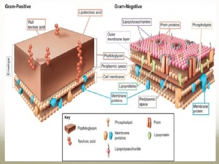
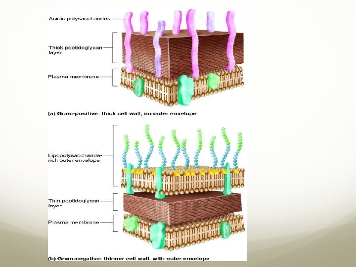
2. CELL ENVELEOPE the outer boundary layer of bacteria cell envelope – chemically complex external covering that encloses the cytoplasm of a bacterial cell 2 layers – cell wall and cell essential for the cell’s normal function and integrity basic types of cell envelopes Hans Christian Gram (Danish physician) developed a bacterial staining technique – gram stain – differentiates between gram+ (positive) and gram- (negative) bacteria gram+ cells (purple) two layers – thick cell wall with peptidogycan and a cell membrane gram- cells (red/pink) three layers outer membrane, thin peptidoglycan layer and the cell membrane
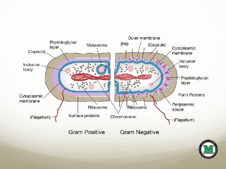

the reaction of the gram stain are due to the differences in the cell wall gram+ - the thick peptidoglycan traps the crystal violet-mordent complex and makes it inaccessible to the decolorizer – leaves the cells purple gram- - the cell walls are thinner – crystal violet is removed with the decolorizer (alcohol dissolves the outer membrane - the loss of dye) – leaves a colorless cell that can be stained with a red counterstain. I would copy this into my notes…

structure of cell walls accounts for important bacterial characteristics determines shape provides strong structural support (peptidoglycan (PG) – repeating framework of long glycan chains cross-linked by short peptide fragments)

many bacterial live in aqueous habitats with low solute concentration constantly absorb excess water by osmosis – peptidolgycan in cell wall prevents cells from rupturing (internal pressure) some drugs used to treat bacterial infection (penicillin, cephalosporins) are effective b/c they target the peptide cross-links in the peptidoglycan – disrupts cell integrity – cause lysis of the bacterial cell
 contains tightly bound")
Gm+ cell wall thick, homogeneous sheath of peptidoglycan (20 -80 nm) contains tightly bound polysaccharides (teichoic acid directly attached to peptidoglycan and lipopolysaccharide attached to lipids in the plasma membrane) appear to function in cell wall maintenance, enlargement during cell division and binding of some pathogens to tissue cell wall loosely adheres to cell membrane – at the junction is the periplasmic space site for temporary storage of enzymes released by cell membrane site of peptidoglycan synthesis copy into notes

Gm- cell wall more complex in morphology than gm+ cell wall composed of an outer membrane (OM) and a thin section of peptidoglycan OM similar to the cell membrane except contains specialized types of lipopolysaccharide (LPS) and lipoproteins lipids form the matrix of top layer OM and polysaccharide strands project from the lipid surface lipids may become toxic when released during infection saccharides function as receptors and interfere with host defenses contains two types of proteins – porins and structural proteins porins – inserted in the upper later of the OM – regulatory control over molecules that leave and enter the cell Structural proteins also embedded in the upper layer of the OM bottom layer of the OM is similar to the cell membrane in structure – composed of phospholipids and lipoproteins
 sheet")
bottom layer of gm- cell wall – single, thin (1 -3 nm) sheet of PG rigid protective structure – thinness provides flexibility and sensitivity to lysis well developed periplasmic space above and below the PG site of metabolic rxns – synthesis and transport of proteins, actions of enzymes, energy release copy into notes

nontypical cell walls bacteria that lack the structure of gm+ / gm- cell wall / no wall can stain gm+ or gm- or variable staining Mycobacterium and Nocardia have PG and stain gm+ - bulk of cell walls contain unique lipids (very long-chain fatty acid – mycolic acid) thick, waxy nature of cell wall responsible for resistance to chemicals and dyes acid-fast stain – hot carbol fuchsin dye become tenaciously attached (held-fast) to cells so an acid-alcohol solution will not remove the dye used to dx TB and leprosy

Mycoplasmas and other cell-wall-deficient bacteria Mycoplasmas – bacteria that usually lack a cell wall – cell membrane contains sterols to prevent lysis extremely tiny bacteria (0. 1 -0. 5 μm) show pleomorphism (extreme variations in shape) – filamentous, coccus or doughnut shaped require sterols added to artificial media to grow found in plans, soil and animals most important medical species – Mycoplasma pneumoniae – adheres to epithelial cells in the lung and causes an atypical pneumonia in humans (walking pneumonia) attachment organelles

some bacteria can lose their cell wall during a part of their life cycle wall-deficient forms – L forms / L phase variants arise naturally from a mutation in the wall-forming genes OR induced artificially by treatment with chemicals (lysozyme / penicillin) that disrupts the cell wall Gm+ cell wall treated with lysozyme / penicillin – cell wall completely lost and becomes a protoplast (fragile cell bound by only a cell membrane – susceptible to lysis) Gm- cell exposed to lysozyme / penicillin – loses its PG but retains its outer membrane (OM) and becomes a shperoplast (less fragile, but weakened cell)

cell membrane structure cell / cytoplasmic membrane - layer in the cell envelope just beneath the cell wall very thin (5 -10 nm), flexible sheet / sheet that surrounds the cytoplasm structure – lipid bilayer with embedded proteins membranes are dynamic and constantly changing (fluid mosaic model) – lipid phase is in motion and proteins can move depending on fxn lipid phase provides selective permeability - regulates transport of molecules in and out of the cell

functions of the cell membrane site for energy rxns, nutrient processing, and synthesis regulate transport – passage of nutrients into the cell and waste out of the cell selective permeability regulate secretion (enzymes / toxins) – release of metabolic products into the extracellular environment

PART 3 – INTERNAL STRUCTURES

3. Bacterial Internal Structure contents of cell cytoplasm cell membrane surrounds the cytoplasm (cytoplasmic matrix) – complex solution – site for cell’s biochemical and synthetic activities major component is water (70 -80%) – solvent for nutrients including sugars, amino acids, salts and other organic molecules – building blocks for cell synthesis / source of energy cytoplasm holds the chromosome, ribosomes. granules and actin strands
 bacterial chromosome – single circular strand")
bacterial chromosomes and plasmids (source of genetic info) bacterial chromosome – single circular strand of DNA bacteria do not have a true nucleus – DNA (long molecule, tightly coiled) is aggregated in a central area of the cell – nucleoid genes carry information necessary for bacterial maintenance and growth plasmid –nonessential DNA exist apart from the chromosome – sometimes become integrated into it – provide protective traits such as resisting drugs, producing toxins and enzymes important in genetic engineering – can be manipulated in the laboratory and transferred from one bacterial cell to another
 60% and")
ribosomes – site of protein synthesis made of ribosomal RNA (r. RNA) 60% and protein 40% – dispersed throughout the cytoplasm (spheres and in chains (polysomes) and attached to the cell membrane inclusions / granules – storage bodies bacteria store nutrients (glycogen, (PHB) poly β-hydroxybutyrate) as inclusion bodies of varying size, content and number as the environmental source of nutrients becomes depleted, energy is supplied by the inclusion bodies bacterial cytoskeleton protein polymers associated with the cell wall – actin filaments that curl within the body of a cell and help maintain its shape during enlargement / cell division

bacterial endospores – resistance facilitate survival in hostile conditions dormant bodies produced by Bacillus, Clostridium and others two-phase life cycle that shifts between a vegetative cell and an endospore vegetative cell – active and growing phase when exposed to certain environmental conditions, signals endospore formation (sporulation) endospore formation and resistance ▪ major stimulus is lack of nutrients (amino acids) ▪ once stimulus is received by vegetative cell, converts to a committed sporulating cell (sporangium) ▪ transformation from vegetative cell – sporangium – endospore requires 612 h
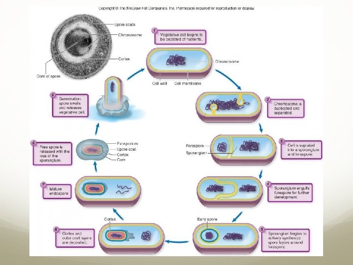

▪ bacterial endospores are the hardiest of all life forms – capable of withstanding extremes in heat, drying, freezing, radiation and chemicals that would kill normal cells germination of endospores ▪ endospores are revitalized when favorable conditions arise ▪ germination (breaking of dormancy) happens in the presence of water and a specific germination agent (varies among species – small organic molecule like and amino acid or inorganic salt) - once initiated, occurs within 1 -1/2 h ▪ the agent stimulates the formation of hydrolytic (digestive) enzymes by the endospore membranes – digest the cortex and expose the core to water ▪ as the core rehydrates and takes up nutrients, the vegetative cell begins to grow outside of the endospore coat – reverts to a fully active vegetative cell Clostridium spp.

PART 4 – Bacterial Shapes, Arrangements and Sizes
 organisms")
Bacterial Shapes, Arrangement and Sizes ▪ generally, bacteria function as independent, single-celled (unicellular) organisms – capable of carrying out necessary life activities (reproduction, metabolism and nutrient processing) considerable variation in shape, size and colonial arrangement three general shapes: coccus (cocci) – spherical (ball-like) - can be oval, bean-shaped or pointed variants bacillus (bacilli) – cylindrical (longer than wide) or rod-shaped – can be blocky, spindle-shaped, round-ended, filamentous (thread-like) or clubbed (drumstick) coccobacillus – round, plump rod vibrio – slightly curved

spirillum – curviform / spiral-shaped cylinder – rigid helix, twisted twice or more along its axis (corkscrew) pleomorphism (varying shape) – due to variations in cell wall structure caused by nutritional or slight hereditary differences
 factors")
bacterial cells can also be categorized according to arrangement (style of grouping) factors influencing arrangement are its pattern of division and how the cells remain attached afterword greatest variety in arrangement are cocci (due to division of coccus in a singe plane, perpendicular plane , in two perpendicular planes or intersecting planes – after division the daughter cells remain attached) diplococci – pairs tetrads – groups of four irregular clusters chains bacilli are less varied in arrangement – divide only in the transverse plane (perpendicular to the axis) – occur in single cells or paired with their ends attached Important Families and Genera of Pathogenic Bacteria TABLE 4. 4 (p-112 textbook)
 independent")
PART 5 Prokaryotic Groups with Unusual Characteristics Free-living Nonpathogenic Bacteria photosynthetic bacteria (phototrophic) independent cells that contain light trapping pigments and can use the energy of sunlight to synthesize required nutrients from inorganic compounds two types: oxygen producers and those that produce other substances (sulfates)

Cyanobacteria Gm- photrophic bacteria most dominant organism on Earth may have converted the atmosphere from anaerobic to aerobic through their production of O 2 may have evolved to eukaryotic cells (algae / plants) with chloroplasts (genetic analysis) diverse distribution and morphology – flourish in all aquatic environments (fresh, salt, hot-springs, Antarctic) and wide range of terrestrial habitats Prochlorococcus and Trichodesmium account for 30 -40% biomass and 50% O 2 production in the oceans can convert nitrogen gas (N 2) to ammonia (NH 4+) that can be used by plants (nitrogen cycle)

primary photosynthetic pigments – green chlorophyll b and bluish phycocyanin (can also produce shades of yellow, orange and red from other pigments)

green and purple bacteria photosynthetic – contain pigments have a different type of chlorophyll (bacteriochlorophyll) than cyanobacteria – co not give off O 2 as a product of photosynthesis live in sulfur springs, freshwater lakes, and swamps deep enough for anaerobic conditions yet where the pigment can absorb wavelengths of light named for predominant colors – can develop brown, pink, purple, blue and orange colorations utilize sulfur compounds (H 2 S / S) in their metabolism
 – many")
• gliding, fruiting bacteria • mixed collection of gm- bacteria (Proteobacteria) – many morphological forms • live in water and soil • tendency to “glide” over moist surfaces – rotation of filaments • myxobacteria (slime bacteria) – complex life cycle – vegetative cells respond to chemotaxic signals by swarming together (many-celled, colored structure – fruiting body - survival structure that makes spores) – can be seen by the unaided eye of tree bark and plant debris

Unusual Forms of Medically Significant Bacteria • most bacteria are free-living / parasitic forms that can metabolize and reproduce by independent means • rickettsias and chlamydias require host cells – obligate intracellular parasites Rickettsias • distinctive, very tiny, gm- bacteria • typical morphology – atypical life cycle and adaptations • most are pathogens – alternate between mammalian host and blood sucking arthropods (fleas, ticks, lice) • cannot survive outside a host cell – cannot carry out metabolism completely on own (closely attached to host) • diseases: Rocky Mountain spotted fever – Rickettsia rickettsii (ticks), endemic typhis – Rickettsia typhi (lice) the mitochondrion – eukaryotic organelle – is genetically related to rickettsias – evolutionary link

Chlamydias genera Chlamydia and Chlamydophila similar to rickettsias – require host cells for growth and metabolism – obligate parasites – not transmitted by arthropods Chlamydia trachomatis – cause severe eye infection (trachoma) that can lead to blindness and common STI Chlamydophila pneumoniae – lung infections

ARCHAEA – the other prokaryotes unusual anatomy, physiology, and genetics single-celled, simple organisms (archaea / archaeons) – prokaryotic in general structure – share bacterial characteristics archaea and eukaryotes share several r. RNA sequences that are not found in bacteria cell walls composed of protein or polysaccharides – lack peptidoglycan (PG) – some lack cell walls altogether extremophiles (love extreme habitiats) – high acidity and high temps; high salt and alkalinity; low temps and high pressure methane producers, hyperthermophiles, extreme halophiles and sulfur reducers ubiquitous – live in all environments – soils, oceans, animal intestines – most common cells on Earth
 – complex")
methanogens convert CO 2 and H 2 into methane gas (CH 4) – complex pathways inhabitants of anaerobic mud and bottom sediments of lakes and oceans some found in the oral cavity and large intestine of humans methane may contribute to “greenhouse effect” and contribute to global warming methane may be a source of alternative fuel

extreme halophiles require salt to grow – can multiply in 36% Na. Cl (high salt tolerance) exist in inland seas, salt lakes and salt mines – not found in the ocean because salt content is not high enough to support them halobacteria – use red pigment to synthsize ATP in presence of light red pigment responsible for “red herrings” (color of Red Sea) and red color of salt ponds

hyperthermophiles flourish at temps between 80 – 120 o. C – volcanic waters and soils; submarine vents – often salt and acid tolerant sampling of the sulfur heat vents in the deep ocean revealed archaea flourishing at temps 250 o. C (150 o. C above boiling water) and 256 atmospheres of pressure (Earth’s surface 1 atmosphere)
- Slides: 50