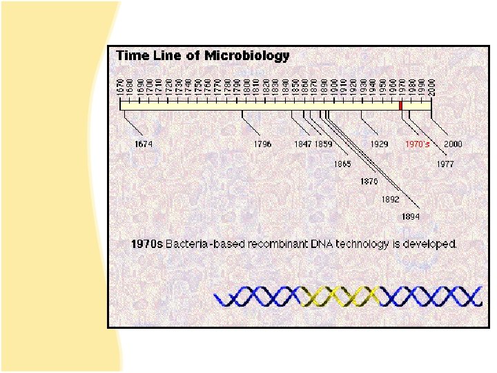
Microbial Genetics 1953 Structure of DNA Revealed Watson

Microbial Genetics

• 1953 Structure of DNA Revealed • Watson & Crick

Genetics The study of heredity The science of genetics explores: • Transmission of biological traits from parent to offspring. • Expression and variation of those traits. • Structure and function of genetic material. • How this material changes.
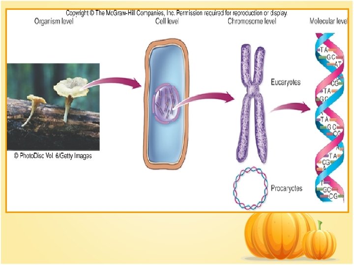

• Genome : Sum total of genetic material of an organism (chromosomes + mitochondria/chloroplasts and/or plasmids) – Genome of cells – DNA – Genome of viruses – DNA or RNA • DNA complexed with protein constitutes the genetic material as chromosomes. • Bacterial chromosomes are a single circular loop. • Eucaryotic chromosomes are multiple and linear.

Chromosome is subdivided into genes, The fundamental unit of heredity responsible for a given trait. – – Site on the chromosome that provides information for a certain cell function. Segment of DNA that contains the necessary code to make a protein or RNA molecule. Three basic categories of genes: 1. Genes that code for proteins - structural genes 2. Genes that code for RNA. 3. Genes that control gene expression - regulatory genes.

• All types of genes constitute the genetic makeup Genotype. • The expression of the genotype creates observable traits Phenotype.

Genomes Vary in Size • Smallest virus – 4 -5 genes • E. coli – single chromosome containing 4, 288 genes; 1 mm; 1, 000 X longer than cell • Human cell – 46 chromosomes containing 31, 000 genes; 6 feet; 180, 000 X longer than cell.
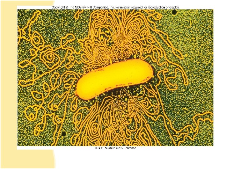

The bacterial genome: Chromosome Plasmids Bacteriophage Insertion sequences Transposons

Structure and Function of Genetic Material ØDNA & RNA ØDNA=Deoxyribonucleic acid ØRNA=Ribonucleic acid ØBasic building blocks: ØNucleotides ØPhosphate group ØPentose sugar ØNitrogenous base
 Chains of nucleotides 5’ to")
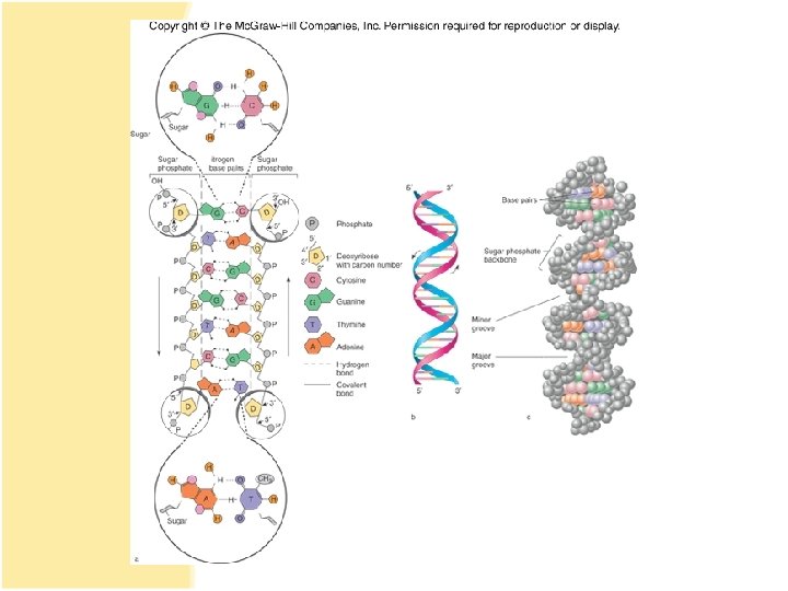
Structure of DNA • • Double stranded (double helix) Chains of nucleotides 5’ to 3’ (strands are anti-parallel) Complimentary base pairing – A-T – G-C

A=T GΞC

DNA • Two strands twisted into a helix. • Basic unit of DNA structure is a nucleotide. • Each nucleotide consists of 3 parts: – A 5 carbon sugar - deoxyribose – A phosphate group – A nitrogenous base – adenine, guanine, thymine, cytosine • Nucleotides covalently bond to form a sugarphosphate linkage – the backbone – each sugar attaches to two phosphates – • 5′ carbon and 3′ carbon

• Nitrogenous bases covalently bond to the 1′ carbon of each sugar and span the center of the molecule to pair with an appropriate complementary base on the other strand. – Adenine binds to thymine with 2 hydrogen bonds – Guanine binds to cytosine with 3 hydrogen bonds • Antiparallel strands 3′ to 5′ and 5′ to 3′. • Each strand provides a template for the exact copying of a new strand. • Order of bases constitutes the DNA code.

Significance of DNA Structure 1. Maintenance of code during reproduction constancy of base pairing guarantees that the code will be retained. 2. Providing variety - order of bases responsible for unique qualities of each organism.

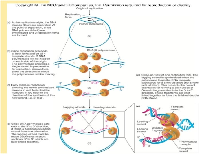
DNA Replication • Making an exact duplicate of the DNA involves 30 different enzymes. • Begins at an origin of replication. • Helicase unwinds and unzips the DNA double helix. • An RNA primer is synthesized by primase. • DNA polymerase III adds nucleotides in a 5′ to 3′ direction. – leading strand – synthesized continuously in 5′ to 3′ direction – lagging strand – synthesized 5′ to 3′ in short segments; overall direction is 3′ to 5′.

• DNA polymerase I removes the RNA primers and replaces them with DNA. • When replication forks meet, ligases link the DNA fragments along the lagging strand to complete the synthesis. • Separation of the daughter molecules is complete. • DNA replication is semiconservative because each chromosome ends up with one new strand of DNA and one old strand.

Enzymes involved in this process are: • Helicases. • DNA polymerase III. • DNA polymerase I. • DNA primase (RNA polymerase). • DNA ligase.

Okazaki fragments

DNA polymerase I and III are able to cut out the ‘wrong’ nucleotide and replace it with the correct one this called proof reading activity

DNA replication is bidirectional. Two replication forks Form simultaneously, moving away from each other and developing a replication bubble

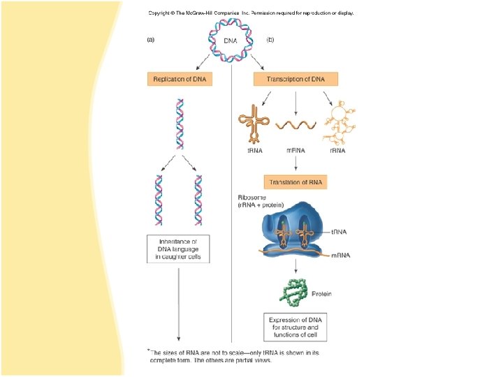
Applications of the DNA code • Information stored on the DNA molecule is conveyed to RNA molecules through the process of transcription. • The information contained in the RNA molecule is then used to produce proteins in the process of translation.

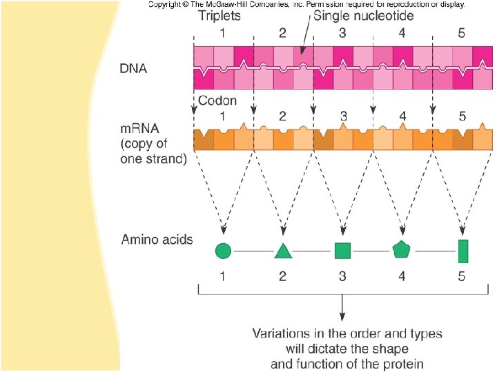
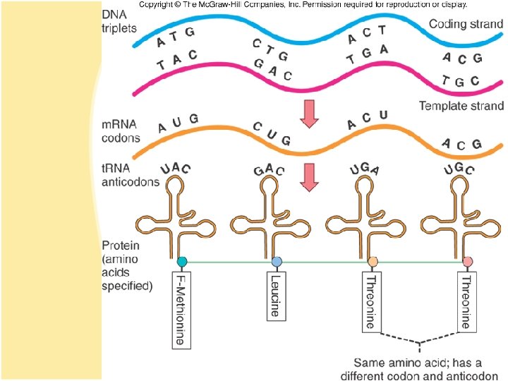
Gene-Protein Connection 1. Each triplet of nucleotides on the RNA specifies a particular amino acid. 2. A protein’s primary structure determines its shape and function. 3. Proteins determine phenotype. Living things are what their proteins make them. 4. DNA is mainly a blueprint that tells the cell which kinds of proteins to make and how to make them.

Protein Synthesis • DNA------ m. RNA------- protein Transcription Translation Central Dogma of Molecular Genetics

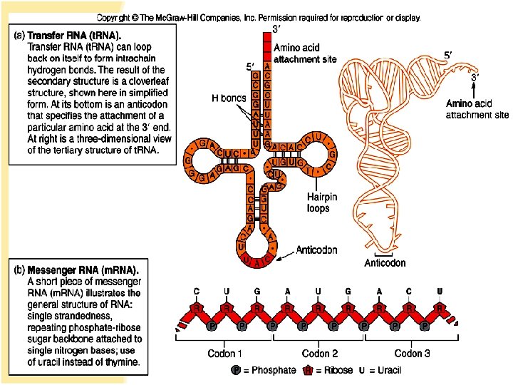
RNAs • Single-stranded molecule made of nucleotides – 5 carbon sugar is ribose. – 4 nitrogen bases – adenine, uracil, guanine, cytosine – phosphate
 – carries DNA")
• 3 types of RNA: – Messenger RNA (m. RNA) – carries DNA message through complementary copy; message is in triplets called codons. – Transfer RNA (t. RNA) – made from DNA; secondary structure creates loops; bottom loop exposes a triplet of nucleotides called anticodon which designates specificity and complements m. RNA; carries specific amino acids to ribosomes – Ribosomal RNA (r. RNA) – component of ribosomes where protein synthesis occurs.

DNA Transcription RNA polymerase RNA Translation Ribosomes PROTEINS

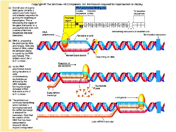
Transcription 1. RNA polymerase binds to promoter region upstream of the gene. 2. RNA polymerase adds nucleotides complementary to the template strand of a segment of DNA in the 5′ to 3′ direction. 3. Uracil is placed as adenine’s complement. 4. At termination, RNA polymerase recognizes signals and releases the transcript. 100 -1, 200 bases long

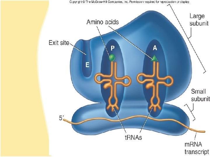
Translation • Ribosomes assemble on the 5′ end of a m. RNA transcript. • Ribosome scans the m. RNA until it reaches the start codon, usually AUG. • A t. RNA molecule with the complementary anticodon and methionine amino acid enters the P site of the ribosome and binds to the m. RNA.

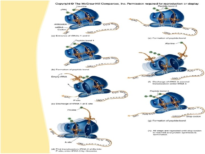
Translation Elongation • A second t. RNA with the complementary anticodon fills the A site. • A peptide bond is formed. • The first t. RNA is released and the ribosome slides down to the next codon. • Another t. RNA fills the A site and a peptide bond is formed. • This process continues until a stop codon is encountered.

Translation Termination • Termination codons – UAA, UAG, and UGA – are codons for which there is no corresponding t. RNA. • When this codon is reached, the ribosome falls off and the last t. RNA is removed from the polypeptide.

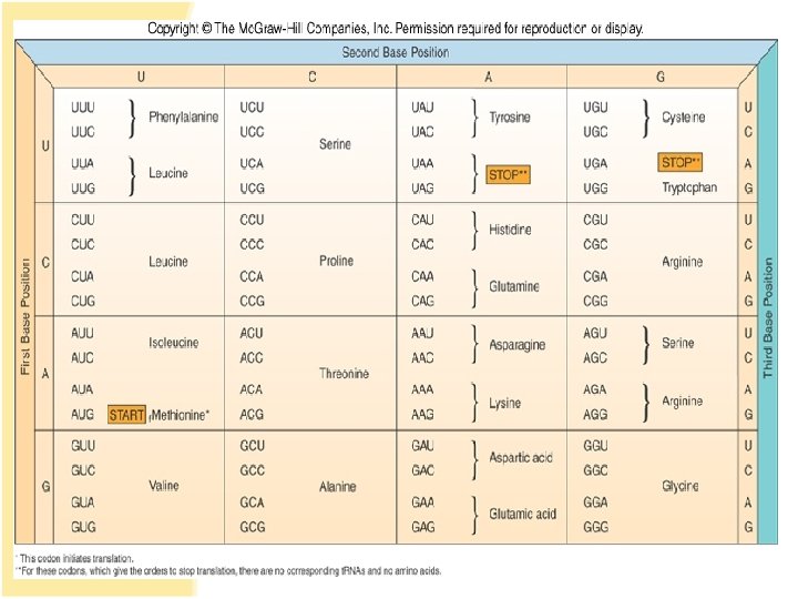
The Master Genetic Code • Represented by the m. RNA codons and the amino acids they specify • Code is universal • Code is redundant

Polyribosomal complex allows for the synthesis of many protein molecules simultaneously from the same m. RNA molecule.

Regulation of Protein Synthesis and Metabolism • Genes are regulated to be active only when their products are required. • In procaryotes this regulation is coordinated by operons, a set of genes, all of which are regulated as a single unit.

Operons • 2 types of operons: 1 -Inducible – operon is turned ON by substrate: catabolic operons- enzymes needed to metabolize a nutrient are produced when needed. 2 -Repressible – genes in a series are turned OFF by the product synthesized; anabolic operon –enzymes used to synthesize an amino acid stop being produced when they are not needed.

Antibiotics That Affect Transcription and Translation • Rifamycin – binds to RNA polymerase. • Actinomycin D - binds to DNA and halts m. RNA chain elongation. • Erythromycin and spectinomycin – interfere with attachment of m. RNA to ribosomes. • Chloramphenicol, linomycin and tetracyclinebind to ribosome and block elongation. • Streptomycin – inhibits peptide initiation and elongation.
- Slides: 48