Mic 101 Lecture 8 Features of Prokaryotic cells


 I. single coccus: II. diplococcus: daughter cells separate remain")
 Ø rods of various length: Ø oval to “hot")


- Slides: 47
Mic 101: Lecture 8 Features of Prokaryotic cells
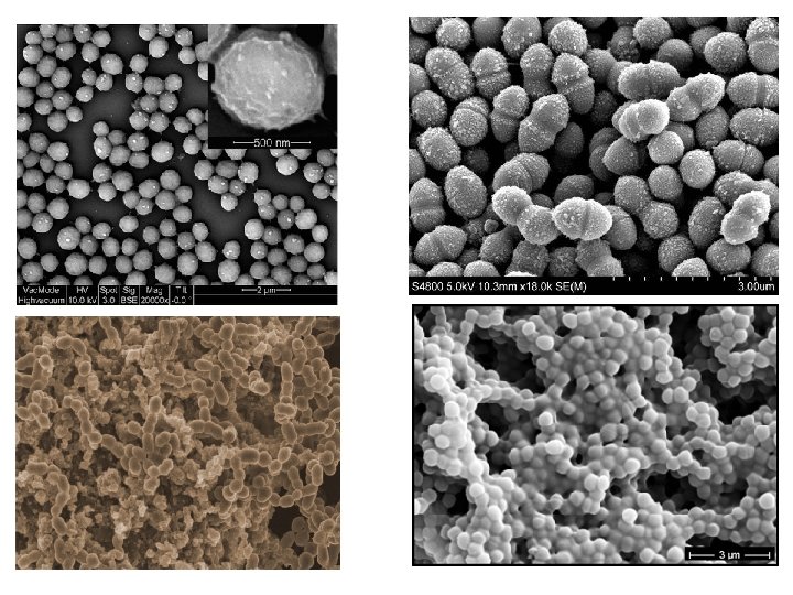
Bacterial Cells. Magnification= 400 x
Bacterial Cells. Magnification=4000 X
Bacterial Cells. Magnification=8, 000 X
Bacterial Cells. Magnification= 30, 000 X
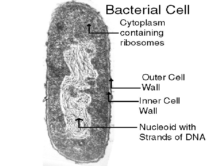
Typical Prokaryotic cell
Morphology • Size: - 0. 2 -2. 0μm diameter - 2 -8 μm length • Shape: three shapes common: 1. coccus = sphere 2. bacillus = rod 3. spiral = twisted • Cell division by binary fission:
Morphology: Arrangement • Cocci(singular Coccus) I. single coccus: II. diplococcus: daughter cells separate remain in pair III. streptococci: after division remain attached in chain IV. tetrad: division occurs in two planes and group as four V. sarcinae: division occurs in three planes and group as eight VI. staphylococci: division occurs in multiple planes and form grape like structure
Morphology • Bacilli (Singular Bacillus) Ø rods of various length: Ø oval to “hot dog” Ø -rods divide only along the short axis I. single bacillus II. diplobacilli: daughter cells separate remain in pair III. streptobacilli: chain after division IV. coccobacillus: short oval, often confused with cocci
Morphology • Spiral one or more twists I. vibrio: curved rod II. spirillum: rigid helical shape, like corkscrew III. spirochete: flexible helical shape, v Most bacteria are monomorphic: always one shape v Some are genetically pleomorphic: have varied shapes within the population of a single species
Examples: Helicobacter pylori Treponema pallidum Campylobacter jejuni Spiral Bacteria
The Cell Wall • The cell wall is the usually semi-rigid layer, that lies just outside the plasma membrane. Functions of Cell wall • • • It helps determine the shape of the cell. It helps protect the cell from osmotic lysis. It can protect the cell from toxic substances. In pathogens it can contribute to pathogenicity. The prokaryotic cell wall also is the site of action of several antibiotics.
Cell wall
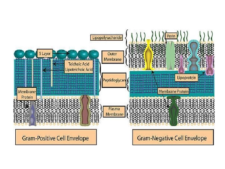
Gram-positive cell walls • Gram-positive bacteria normally have cell walls that are thick and composed primarily of peptidoglycan. Peptidoglycan in gram positive bacteria often contains a peptide inter-bridge • Also, gram-positive cell walls usually contain large amounts of teichoic acids, polymers of glycerol or ribitol joined by phosphate groups. Amino acids such as D-alanine or sugars like glucose are attached to the glycerol and ribitol groups. • Two types of teichoic acid: ü Lipoteichoic acid –bound to membrane lipids ü Wall teichoic acid –linked to peptidoglycan
Gram negative cell walls • Gram-negative bacteria have a cell wall that is thinner than that of Gram-positive bacteria with a few layers of peptidoglycan • An Outer membrane, lies above the peptidoglycan layer. The outer membrane is much thicker than the peptidoglycan layer. • Peptidoglycan is bonded to lipoproteins in the outer membrane. • Does not contain teichoic acids. • More susceptible to mechanical breakage.
Gram positive cell walls contain a thick peptidoglycan layer with techoic acids. Gram negative cell walls contain a thin peptidoglycan layer (without techoic acids) that is surrounded by a thick plasma membrane. Gram positive bacteria will stain purple because of their thick peptidoglycan cell wall. Peptidoglycan is composed of N-acetylglucosamine (NAG) & N-acetylmuramic acid (NAM)
Peptidoglycan, also known as murein, is a polymer consisting of sugars and amino acids that forms a mesh-like layer outside the plasma membrane of most bacteria, forming the cell wall. The sugar component consists of alternating residues of β-(1, 4) linked N-acetylglucosamine (NAG) and N-acetylmuramic acid (NAM). Thick layer in Gram positive bacteria Thin layer in Gram negative bacteria Absent in Archea
Ultrastructure of Gram negative Cell Wall
Ultrastructure of Gram positive Cell Wall
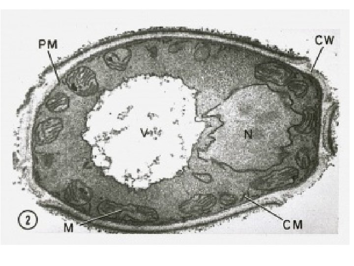
The plasma membrane • Thin structure lying inside the cell wall and enclosing the cytoplasm. • It is 5 -10 nm thick, and consists mainly of lipoprotein. • The cytoplasmic membrane is the physical and metabolic barrier between the interior and exterior of the bacterial cell.
Ultra-structure of Membrane
Arrangement of membrane proteins • The protein molecules in the cell membrane can be arranged in a variety of ways. • Peripheral proteins- not embedded, associated firmly with inner or outer surface of the membrane; may function as enzyme. • Integral proteins-firmly embedded in the membrane some penetrate the membrane completely. Some make channels through which substances enter and exit the cell.
Fluidity of Membranes • Membrane fluidity refers to the viscosity of the lipid bilayer of a cell membrane or a synthetic lipid membrane. . • Viscosity of the membrane can affect the rotation and diffusion of proteins and other bio-molecules within the membrane, there-by affecting the functions of these molecules. The membrane must be viscous to allow membrane protein to move freely enough to perform their functions without destroying the structure of the membrane. This dynamic arrangement of phospholipid and protein is referred to as the fluid mosaic model.
Function of plasma membrane • Act as selective barrier through which materials can transport • Selective permeability • Can produce certain enzymes to break nutrients and produce energy • Sometimes the pigments and enzymes of photosynthetic bacteria found in plasma membrane
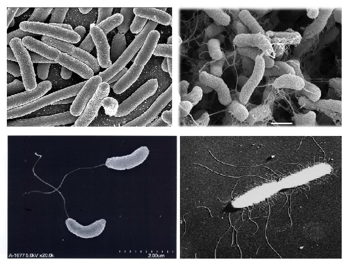
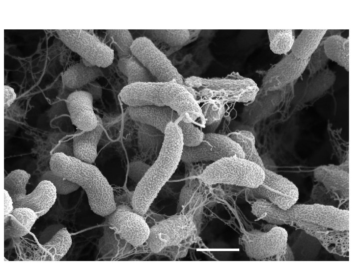
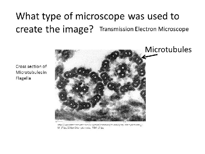
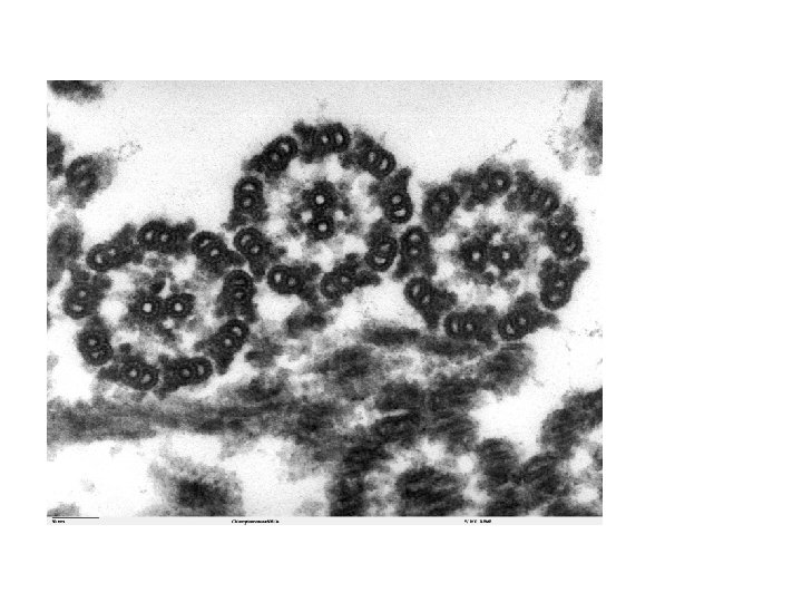
Flagella • Flagella are long, thin, whip-like appendages attached to a bacterial cell that allow for bacterial movement. . Each flagella consists of a filament, composed of a protein called flagellin, and a hook, which attaches the filament to the cell at the motor. • Flagella are found in all three domains of the living world: bacteria, archaea, and eukaryota
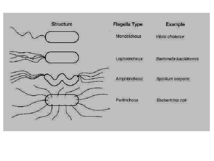
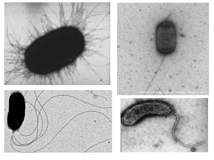
Flagella • -long, filamentous appendages • -used for motility Arrangements: a. monotrichous: one on one end b. amphitrichous: one or more on each end c. lophotrichous: two or more on one end d. peritrichous: all over cell
Ultra-structure of Flagella
Flagella Three basic structure a. filament: v -made up of chains of flagellin protein form helix surrounding a hollow core v -sticks out beyond plasma membrane and cell wall b. hook: Ø Filament is attached to a wider structure called hook Ø -provides rotational movement of flagella Ø -solid, composed of hook protein c. basal body: -rod and disc structure -anchors flagellum to cell wall and plasmamembrane
Pili and Fimbrae v Pili are short, straight and thinner than flagella v composed of pilin protein v Used for attachment rather than movement v Fimbriae: 1. Evenly distributed 2. Adhere with any surfaces even cell 3. Few to several hundreds per cell
Pili
Cytoplasm • • 1. 2. 3. the substance inside the plasma membrane ~80% water Contains proteins (enzymes), carbohydrates, lipids, ions includes some solid structures: nucleoid, ribosomes Inclusions Nuclear Area / Nucleoid • Single long double strand DNA in the bacterial chromosome • Carry cells genetic info of cell • 20% of cell volume is occupied by DNA
Prokaryotic ribosome -site of protein synthesis -composed of r. RNA and protein -consist of 2 subunits: 30 s + 50 s = 70 s prokaryotic ribosome 30 s = one molecule of r. RNA 50 s=two molecule of r. RNA
Binary Fission • 1. Binary fission means formation of two from one • 2. In adequate nutrition and favorable environment, cells gather nutrient material and elongate • The DNA replicates and divides into two copies • The cytoplasmic content divides into two equal parts by formation of septum • Formation of cell wall in the daughter cell completes binary fission
Division of Prokaryotic cells
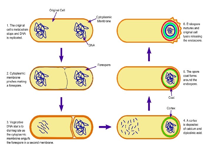
Endospore • Produced under unfavorable condition • Mostly formed by some Gram + bacilli (e. g Clostridium & Bacillus species) • Exception Gram – bacteria Coxiella burnetii Endospore 1. dehydrated, thick wall structure 2. resistant to heat, toxins, radiation, etc 3. Formation of endospore process called sporulation 4. endospores can remain dormant for thousands of years 5. upon return of favorable conditions, endospores germinate into vegetative cells