Methodology in the Biological Level of Analysis Learning

Methodology in the Biological Level of Analysis Learning Objectives: 1. 2. 3. Discuss how and why particular research methods are used at the biological level of analysis. Discuss the use of brain imaging technologies. Discuss ethical considerations related to research studies at the biological level of analysis.

Two-Types of Research Methods Invasive and Non-Invasive • Non-Invasive – Researcher does not invade the body to conduct research • Brain Scans • Experiments • Invasive – Researcher invades the body to conduct research • Brain Surgery • Autopsy

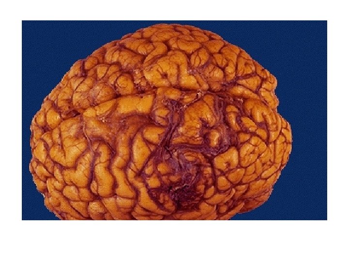
Studying the Brain • Lesion studies: a brain region is destroyed and behavior is observed – Lesions of hypothalamus in animals ---> overeating – Strokes interrupt blood flow in brain ---> damage brain

Experimental Studies • Ablation – Surgical removal of parts of the brain • Using electricity – Electrode (And you are awake) – Deep lesioning • High electrical current destroy tissue • Low electrical current stimulate/activate tissue

Advantages of ablation/lesion in animal research • gives a possibility to understand how the brain functions without having to wait for naturally occurring damages • Under anaesthetic, an animal’s head can be held in a fixed position – apparatus to insert an electrode into a particular location in the brain so that you can investigate an exact correlate of behaviour by comparing between behaviour before the brain damage and afterwards.

Disadvantages of ablation/lesion in animal research: • perhaps limited what such studies can tell about the human brain • You cannot be absolute sure that behavioural changes are only due only to ablation • There may be ethical issues in using this technique

Can you control someone with electricity? • YES…Electrical stimulation can control a person – No radio waves!!!! – Some control but hard to predict • Different from person to person

Brain Imaging Techniques • Usually Non-invasive – Unless scans follow an invasive procedure • Non-Imagine Techniques – EEG – ERP • Imaging Techniques – CT Scan – MRI Scan (plus f. MRI) – PET Scan
")
Studying the Brain • Electrical Recording: overall brain wave activity monitored by electroencephalograph (EEG) • Electrical Stimulation of the Brain (ESB)
 • Measures the")
How do you know what the brain is doing? Electroencephalography (EEG) • Measures the waves of electrical activity • EEG Machine amplifies the brain waves – Used to monitor change in brain activity • Daydreaming • Hypnosis
 · Evoked Response Potential · Electrodes are placed on the skull")
Event-Related Potential (ERP) · Evoked Response Potential · Electrodes are placed on the skull · measure the electric signal of brain activity · Critique · Good temporal resolution · almost instantaneous · Not as good for spatial resolution · cannot get a single cell · only large chunk

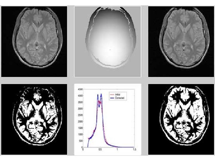
Images of the Brain CT Scan • Computed Tomographic Scanning – Special X-Ray – Can reveal the effects of: • Strokes • Injuries • Tumors – These can be related to behavior

Studying the Brain MRI Scan of Tumor in Speech Center – MRI scans generate 3 D views of the human brain • f. MRI – faster version of MRI for Face Recognition in Normal Volunteer

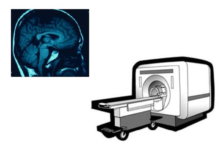
Images of the Brain MRI Scan • Magnetic Resonance Imaging – Use magnetic field • 3 dimensional picture – 2 dimensional slice taken to study – Peer into the brain as if it were transparent • Functional MRI (f. MRI) – Brain activity visible
 · More technical · involves aligning brain atoms with a")
Magnetic Resonance Imaging (MRI) · More technical · involves aligning brain atoms with a magnetic field & then applying radio wave pulses to the sample · Middle-of-the-road method · Okay for temporal resolution · Okay for spatial resolution

CAT vs. MRI • Harmless radio waves are used in MRI – not X-rays and injected dyes (CAT) • more sensitive than the CAT scan – very accurate pictures • still pictures (CAT and MRI) – useful for structure – not possible to deal with function

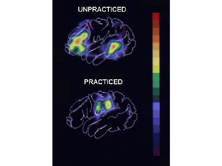
Studying the Brain – PET scans use radioactive glucose to assess brain activity
 • Most Remarkable!!! • Glucose consumption")
Images of the Brain PET Scan (Considered Invasive!!) • Most Remarkable!!! • Glucose consumption by the brain • View changes in brain activity – Color representation • Which areas are active when you read, hear a word, think about meaning.
 used the PET to show the front of")
Some empirical research • Restak (1984) used the PET to show the front of the brain and the part that produces movement became active when a person was asked to move the right hand. When a person is asked to think about moving the hand, only the front part is active (and not the part involved in actual movement). • Martin &Brust (1985) showed that participants asked to listen to and recall a story had activity in the part of the brain responsible for processing auditory information, and also in the hippocampus when asked to recall (hippocampus involved in memory).

• It is possible to use MRI in a functional capacity • MRI and f. MRI have been used to investigate similar functions to those investigated using PET – language, attention, vision, memory • PET and MRI can be used in combination. – good spatial resolution (i. e. images and structures are seen very precisely) – disadvantage of a poor temporal resolution (i. e. it is difficult to match the psychological and neural

Research Methods Charting the Brain’s Inner Realms • Clinical Studies – Changes in personality, behavior or sensory capacity • Experimental Techniques (invasive) – Ablation – Electrical stimulation – Deep lesioning

Methodology Biological Perspective • Traditional Research Methods – – – Correlational Studies Double-blind trials Experiments Interviews Case studies Questionnaires • May use invasive and noninvasive techniques • Reference Packet: Bio Perspective Research Methodology (Pages 1 -2)

Ethics in Bio Analysis
- Slides: 27