Metastatic Gastric Cancer from Pancreatic Ductal Adenocarcinoma Bae

Metastatic Gastric Cancer from Pancreatic Ductal Adenocarcinoma Bae SY, Lee JH, Min BH, Kim JJ, Rhee JC, Kim KM* Departments of Medicine and Pathology*, Sungkyunkwan University School of Medicine, Samsung Medical Center, Seoul, Korea
 - PPI on demand due to intermittent heartburn")
Endoscopy, 2 years ago (M/66) - PPI on demand due to intermittent heartburn
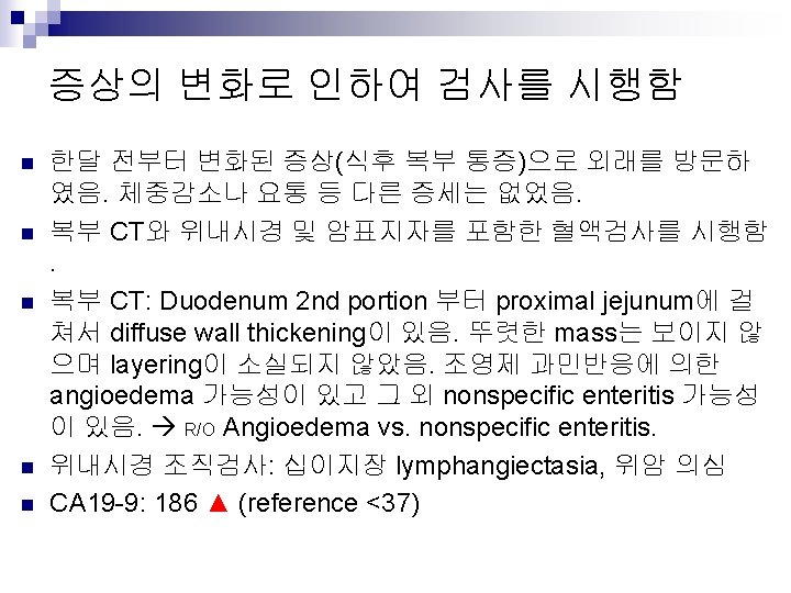

n Duodenum 2 nd portion 부터 proximal jejunum에 걸쳐서 diffuse wall thickening이 있음. 뚜렷한 mass는 보이지 않으며 layering이 소실되지 않았음. 조영제 과민반응에 의한 angioedema 가능성 이 있고 그 외 nonspecific enteritis 가능성이 있음. R/O Angioedema vs. nonspecific enteritis.

Initial EGD: duodenum ♠ Biopsy: duodenum, lymphangiectasia

Initial EGD: stomach ♠ A few atypical glands, consistent with tubular adenocarcinoma, poorly differentiated. Note: After deep sections, a few atypical glands were identified in the deep mucosa adjacent to muscularis mucosa. For confirmation, please perform multiple deep biopsies.
 n Pathology report: after deep sections,")
Biopsy of the initial endoscopy (2009 -12 -31) n Pathology report: after deep sections, a few atypical glands were identified in the deep mucosa adjacent to muscularis mucosa.
 # 1. Tubular adenocarcinoma (M/D), with multiple endolymphatic tumor")
Repeat EGD (2009 -2 -11) # 1. Tubular adenocarcinoma (M/D), with multiple endolymphatic tumor emboli # 2. Atypical glands, highly suggestive of tubular adenocarcinoma (P/D)
 Tumor in the lymphatic space")
Repeat EGD (2009 -2 -11) Tumor in the lymphatic space

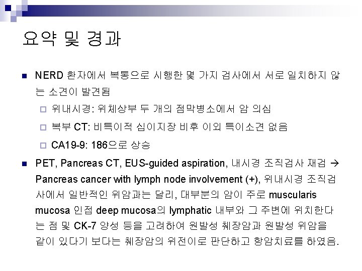
PET-CT - CT에서 특이소견이 없었으나 CA 19 -9이 186으로 상승되어 시행 ♠ Mildly increased FDG uptake (p-SUV = 2. 3)in the pancreatic tail portion and mild hypermetabolic soft tissue lesion(p-SUV = 2. 2) around celiac axis. R/O pancreas cancer with metastasis around celiac axis.

Pancreas CT

EUS-guided aspiration - cytology: ductal adenocarcinoma ♠ ill-defined low echoic mass was seen in the pancreatic tail. FNA was done by using 25 G needle. Low echoic soft tissue was seen around celiac trunk and luminal narrowing was found.

CK-7 Immunostaining of the gastric mucosal lesion - R/O pancreatic origin ?


Sensitivity & Specificity of CK-7 Park et. al Arch Pathol Lab Med. 2007; 131: 1561– 1567

Sensitivity & Specificity of CK-7 Park et. al Arch Pathol Lab Med. 2007; 131: 1561– 1567

Oien. Semin Oncol 2009'36: 8 -37

CA 19 -9
")
Follow up endoscopy due to anemia -PD (pancreas cancer)
 #1 #1 #2 Endoscopy: On the GC side of")
Second endoscopy (2009 -1 -12) #1 #1 #2 Endoscopy: On the GC side of high body a 2. 5 cm sized reddish flat elevated lesion without definite abnormal converging fold was noticed (#1 x 5). On the posterior to number #1 a 2. 0 cm sized reddish flat elevated similar lesion(previous tissue confirmed as adenoca) was noticed (#2 x 5). Two lesions(#1 and #2) were grossly separated. Pathology: #1. Stomach, high body, greater curvature, biopsy: A few atypical glands (see note) #2. Stomach, "posterior aspect of #1", biopsy: Atypical glands (see note) Based on histology, malignancy (tubular adenocarcinoma) is suspected. This case was diagnosed though intradepartment consultation.
 n n #1 & #2 biopsy: atypical glands Based")
Second endoscopy (2009 -1 -12) n n #1 & #2 biopsy: atypical glands Based on histology, malignancy (tubular adenocarcinoma) is suspected.
- Slides: 24