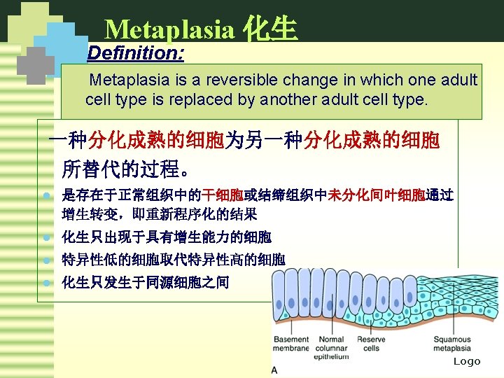
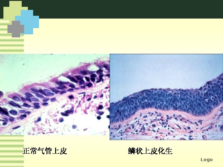
MetaplasiaTypes Epithelial metaplasia Squamous metaplasia columnar epithelium trachea
 transitional epithelium (pelvis)")


 l 线粒体的损伤(Irreversible mitochondria")

 When cellular injury")


 Logo")


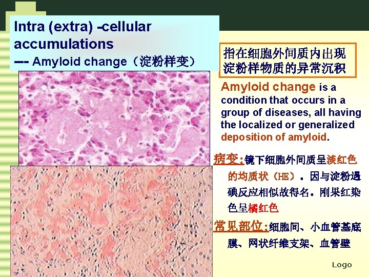
-cellular accumulations ---Proteins 细胞内蛋白积聚,胞质内圆形嗜酸性小滴、空泡 或不规则聚集物 l Reabsorbent droplets :")


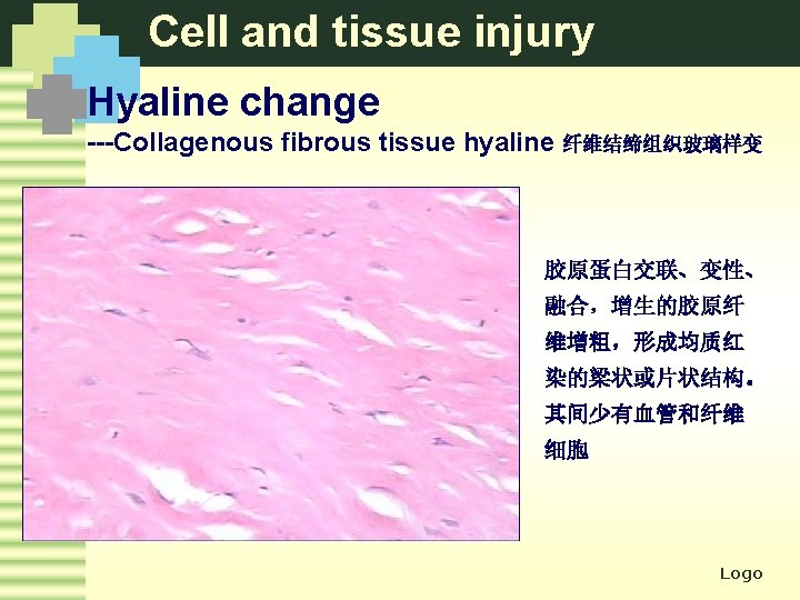
-cellular accumulations --- Hyaline change 玻璃样变 Definition: Various histological or")


 cellular accumulations ---Mucoid change 粘液样变 Change characterized by accumulation")


 Logo")


 蛋白水解酶→溶解液化")

 Logo")


- Slides: 103
Metaplasia-Types Epithelial metaplasia Squamous metaplasia columnar epithelium (trachea, cervix, 鳞状上皮化生 cholecyst) transitional epithelium (pelvis) Intestinal metaplasia Gastric glandular epithelium 肠上皮化生 Pseudo-pyloric gland Metaplasia 假幽门腺化生 corpus and sinus gastric gland Mesenchymal metaplasia Osseous, cartilage and adipose tissue metaplasia 骨、软骨、脂肪组织化生 Fibroblasts Logo
Metaplasia-Types Logo
Metaplasia-Types Squamous metaplasia normal Logo
Metaplasia-Types Squamous metaplasia Logo
Metaplasia-Types villi of normal trachea squamous metaplasia Logo
Metaplasia-Types Logo
Metaplasia-significance Metaplasia---a “double-edged sword” Advantage Disadvantage Defending loss of normal function Cancer transformation Logo
Adaptation Increased demands Growth stimulation hypertrophy hyperplasia adaptation atrophy Diminished nutrition Low stimulation metaplasia Chronic stimulation Pathological Logo
Tissue and Cellular Injury 第 一 Logo 章
Cell and tissue injury ---Causes 1 Hypoxia 2 Chemical agents 3 Physical agents 4 Biologic agents 5 Immunologic reactions 6 Genetic defects 7 Nutritional imbalances 8 Others Logo
Cell and tissue injury ---Mechanisms of cell injury l ATP的耗竭(ATP depletion) l 线粒体的损伤(Irreversible mitochondria damage ) l 膜渗透性的缺陷(Loss of membrane permeability) l 细胞内钙的流入和钙内环境稳定的破坏(Overload of intracellular calcium and loss of calcium homeostasis) l 氧自由基的集聚(Accumulation of oxygen-derived free radicals) Logo
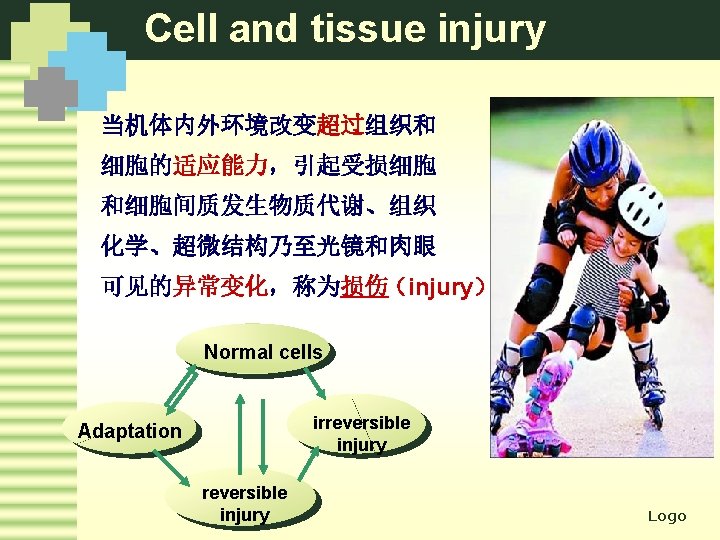
Cell and tissue injury ---Morphologic changes 细胞损伤 可逆性损伤 reversible injury 变性 Degeneration cellular injury 不可逆性损伤 irreversible injury 细胞死亡 cell death 坏死 Necrosis 凋亡 apoptosis Logo
Cell and tissue injury Morphologic changes--Reversible injury 各种细胞损伤的早期改变为ATP减少、细胞膜完整性缺 失、蛋白合成下降、细胞骨架损伤和DNA损伤。在一定 限度内损伤的改变为可逆的,传统形态学上称变性( Degeneration) When cellular injury is sublethal and sustained, cells and tissues tend to accumulate substances in abnormal quantities. These materials may be endogenous or exogenous. 变性指细胞或细胞间质受损伤后,由于代谢障碍,使细胞内或细 胞间质内出现异常物质或正常物质异常蓄积的现象,通常伴有细 胞功能低下。 Logo
Cell and tissue injury ---Morphologic changes 细胞肿胀 Cellular swelling reversible cell injury 脂肪变 Fatty change 脂质 lipids 蛋白质 proteins Intra (extra)-cellular accumulations 淀粉样变 Amyloid change 糖原 glycogen 玻璃样变 hyaline change 黏液样变 mucoid change 病理性色素沉着 Pigments Logo 病理性钙化Pathologic calcification
Cell and tissue injury Reversible injury--cellular swelling The commonest and earliest form of cell injury from almost all causes. Hypoxia infection intoxication mitochondria injury ATP↓ Na+-K+ pump dysfunction Intracellular accumulation of Na+ and H 2 O The common site--- liver, kidney , heart Logo
Cell and tissue injury Reversible injury--cellular swelling Grossly: The affected organ is enlarged due to swelling. The cut surface bulges outwards and is slightly opaque. 肿大,包膜紧张,切面隆起,边缘外翻,混浊无光,如沸水烫过 Cellular swelling of liver Logo
Cell and tissue injury Reversible injury--cellular swelling LM: The cells are swollen. Small clear vacuoles are seen in the cells. 病变初期,细胞质内出现的红染细颗粒状物。若水钠进一步 积聚,细胞基质高度疏松呈空泡状,其极期称为气球样变 Normal cell Granularity change Viral hepatitis Hydropic change Logo
Cell and tissue injury Reversible injury--cellular swelling Logo
Cell and tissue injury Reversible injury--cellular swelling 肝细胞水肿(气球样变) Logo
Cell and tissue injury Reversible injury--cellular swelling Hydropic degeneration of renal tubule Logo
Cell and tissue injury Reversible injury--cellular swelling EM: dilatation of endoplasmic reticulum and mitochondrial swelling 线粒体、内质网肿胀 dilatation of endoplasmic reticulum mitochondrial swelling Logo
Cell and tissue injury Reversible injury--cellular swelling Gross: The affected organ is enlarged due to swelling. The cut surface bulges outwards and is slightly opaque. LM: The cells are swollen. Small clear vacuoles in the cells. EM: swelling of endoplasmic reticulum and mitochondria 肉眼: 肿大,包膜紧张,切面隆起,边缘外翻,混浊无光,如沸水烫过 光镜: 病变初期,细胞质内出现的红染细颗粒状物。若水钠进一步积聚, 细胞基质高度疏松呈空泡状,其极期称为气球样变 电镜: 线粒体、内质网肿胀 Logo
Cell and tissue injury Reversible injury--fatty change Definition: The accumulation of fat in non-fatty parenchymal cells. 脂肪变: 实质细胞内脂肪的异常蓄积 Liver, heart, kidneys and other organs Gross: enlarges, yellow, soft, and greasy. LM: The fatty change appears as clear vacuoles within parenchymal cells. 肉眼: 器官体积增大,淡黄色,边缘圆钝,切面呈油腻感 光镜: 细胞内出现边缘较整齐的大小不等的圆形空泡, 苏丹Ⅲ呈桔红色,锇酸呈黑色 Logo
Cell and tissue injury Fatty change of liver l alcohol abuse l protein malnutrition l obesity l hepatotoxin l diabetes Logo
Cell and tissue injury Intracellular accumulations ---fatty change Logo
Cell and tissue injury Fatty change of liver LM: Small vacuoles around nucleus ↓coalesce large vacuoles that displace the nucleus to the periphery of the cell. Logo
Cell and tissue injury Fatty change of liver Special staining Sudan III : orange red Osmic acid: black Logo
Cell and tissue injury Fatty change of liver congestion: central parts of the lobules toxication: perilobules Logo
Fatty change of the liver Upper Left: gross appearance. Lower left: HE stain. Upper Right: Sudan III Logo stain for fat. Lower Right: electron microscopy
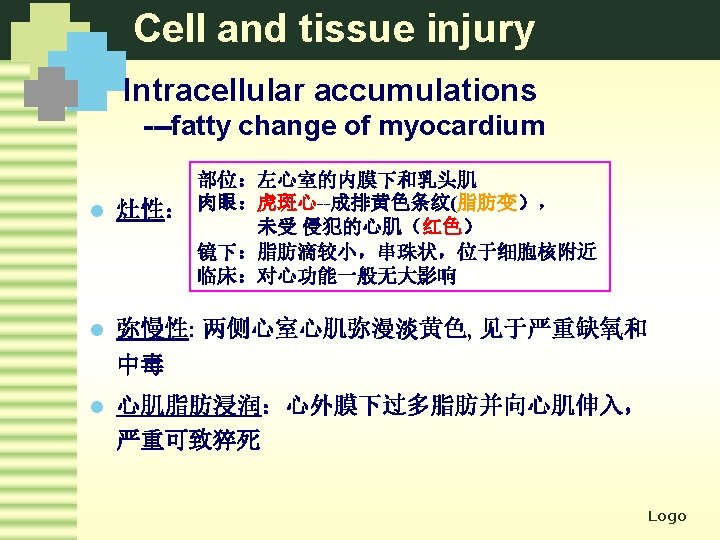
Cell and tissue injury Fatty change of myocardium Tiger stripe heart Bands of yellow streaks alternate with redbrown muscle appearance Fatty change are in yellow Myocardium are in red-brown Logo
Cell and tissue injury fatty change of myocardium Special stain Sudan III : orange red Osmic acid: black Logo
Cell and tissue injury Myocardium fatty infiltration Logo
Cell and tissue injury Lipid—cholesterol and cholesteryl esters 胆固醇和胆固醇酯 l 动脉粥样硬化 Atherosclerosis l 黄色瘤 Xanthomas l 炎症和坏死 inflammation and necrosis Atherosclerosis —foam cells Logo
Cell and tissue injury Intra (extra)-cellular accumulations ---Proteins 细胞内蛋白积聚,胞质内圆形嗜酸性小滴、空泡 或不规则聚集物 l Reabsorbent droplets : renal tubules l Russell bodies: plasma cells l Defect in protein folding Logo
Cell and tissue injury Intracellular accumulations---Proteins Reabsorbent droplets Logo
Cell and tissue injury Intracellular accumulations---Proteins Russell bodies 浆细胞胞浆内可见圆形的嗜伊红小 体, 将核挤向一侧,是免疫球蛋白蓄积的结果 Logo
Cell and tissue injury Intracellular accumulations---Proteins Mallory小体--酒精性肝病时,肝细胞胞质中细胞 中间丝前角蛋白变性 Logo
Cell and tissue injury Intra(extra)-cellular accumulations --- Hyaline change 玻璃样变 Definition: Various histological or cytological alterations characterized by homogeneous, glasslike eosinophilic appearance in HE stained sections 玻璃样变通常用来描述在常规HE切片中细胞内或细胞外 组织变成均质、红染、毛玻璃样。又称透明变 Logo
Cell and tissue injury Hyaline change-Types Intracellular hyaline l Reabsorb droplets : renal tubules l Mallory alcoholic bodies: hepatocytes l Russell bodies: plasma cells Arterioles hyaline Collagenous fibrous tissue hyaline Logo
Cell and tissue injury Hyaline change-- Intracellular hyaline Logo
Cell and tissue injury Hyaline change -- arterioles hyaline细动脉壁玻璃样变 Hypertension and diabetes mellitus l Extravasated plasma protein l Deposition of basement membrane Logo
Cell and tissue injury Arterioles hyaline Logo
Cell and tissue injury Intra(extra) cellular accumulations ---Mucoid change 粘液样变 Change characterized by accumulation of mucin in intracellular or extracellular loci. 指间质内有粘多糖和蛋白质的蓄积 病变: 镜下间质疏松, 多突起的星芒纤维细 胞散在于灰兰色的粘 液样基质中 常见部位: 胶原病, 如 风湿病等, 动脉粥样 硬化及间叶组织肿瘤 基质中 Logo
Cell and tissue injury Pathologic pigmentation 病理性色素沉着 有色物质在细胞内、外的异常蓄积 endogenous 含铁血黄素Hemosiderin l 脂褐素Lipofuscin l 黑色素Melanin l 胆红素bilirubin l exogenous 碳末Carbon l 煤尘 smut l 纹身色素tattooing l Logo
exogenous Logo
Cell and tissue injury Pathologic pigmentation--Hemosiderin Heart failure cell Logo
Cell and tissue injury Pathologic pigmentation--Lipofuscin Logo
Cell and tissue injury Pathologic pigmentation-- Melanin Logo
Cell and tissue injury Pathologic pigmentation-- bilirubin Logo
Cell and tissue injury Pathologic calcification 病理性钙化 Definition: Abnormal deposits of calcium salts occur in any tissues except bones and teeth 骨、牙之外的组织中固态钙盐沉积 Types: dystrophic calcification 继发于局部变性、坏死或其他异物,钙磷代谢正常 metastatic calcification 钙磷代谢障碍, 见于甲状旁腺功能障碍、骨肿瘤、维生素D摄 入过多导致高钙 Logo
Cell and tissue injury Pathologic calcification 病理性钙化 Dystrophic calcification Logo
Cell and tissue injury Pathologic calcification 病理性钙化 Metastatic calcification Logo
Morphologic changes of tissue and cellular injury 细胞水肿 Cellular swelling reversible cell injury 脂肪变 Fatty change 脂质 lipids 蛋白质 proteins Intra (extra)-cellular accumulations 淀粉样变 Amyloid change 糖原 glycogen 玻璃样变 hyaline change 黏液样变 mucoid change 病理性色素沉着 Pigments Logo 病理性钙化Pathologic calcification
Tissue and Cellular injury Reversible injury irreversible injury Cell death Logo
Tissue and Cellular injury Irreversible injury --Cell death 细胞死亡 细胞受到严重损伤累及细胞核时,呈现代谢停止、 结构破坏和功能丧失等不可逆性改变即细胞死亡 Severe damage involve nucleus Metabolism stop Structure destroy Function lose Irreversible injury Cell death necrosis apoptosis Logo
Tissue and Cellular injury Necrosis 坏死 A sequence of morphologic changes that follow cell death in living tissue. 活体内局部组织、细胞的死亡 The morphologic appearance of necrosis is the result of two essentially concurrent processes: l Enzymatic digestion of the cells (autolysis & heterolysis ) l Denaturation of proteins Logo
Tissue and Cellular injury Morphology of necrotic cells 1 Changes in the nucleus l Karyolysis(核溶解) l. Pyknosis (核固缩) l Karyorrhexis(核碎裂) 2 Changes in cytoplasm and cellular membrane l l 3 Increased Eosinophilia (嗜酸性染色增强): Vacuolated and moth-eaten appearance (虫蚀状或空泡化) cellular membrane breakage—inflammation Calcification (钙化) Changes in mesenchyma Logo
Tissue and Cellular injury Morphology of necrotic cells 1 Changes in the nucleus l Karyolysis(核溶解): dissolution of the nucleus (the basophilia of the chromatin fade) l Pyknosis (核固缩): nuclear shrinkage and condensation of chromatin (increased basophilia) l Karyorrhexis(核碎裂): the pyknotic nucleus fragments normal pyknosis karyorrhexis karyolysis Logo
Tissue and Cellular injury Morphology of necrotic cells pyknosis karyorrhexis karyolysis Logo
Morphology of necrotic cells Cytoplasm: increased eosinophilia loss of RNA and denatured protein Logo
Tissue and Cellular injury 坏死的类型 Types of necrosis coagulative necrosis liquefaction necrosis caseous necrosis Specialized necrosis gangrene fibrnoid necrosis l Enzymatic digestion of the cells l Denaturation of proteins fat necrosis Logo
Tissue and Cellular injury Coagulative necrosis 凝固性坏死 指坏死组织尚保留原组织的细胞轮廓,呈灰白、 干燥的凝固状 Gross LM: Sites: The necrosis area is swollen, firm and pale. 灰白、干燥、坚实的凝固体,周围见充血出血带, 与周围组织界限清楚 The dead cells preserve their basic structural outline but only indistinctly,appearing as a mass of coagulated pink-staining, homogeneous 细胞微细结构消失,而组织结构轮廓保存 Infarcts of solid organs--heart, spleen, kidney Logo
Tissue and Cellular injury Coagulative necrosis 凝固性坏死 Anemic infarct of kidney 肾贫血性梗死 The necrosis area is firm and pale. 灰白、质地较硬的凝固体,充血出血带,与周围组织界限清楚 Logo
Tissue and Cellular injury Coagulative necrosis 凝固性坏死 Logo
Tissue and Cellular injury coagulative necrosis of heart Logo
Tissue and Cellular injury Liquefactive necrosis 液化性坏死 组织坏死因酶性分解而变成液态 Soft and liquid. Discharge of the liquid grossly contents forms a cystic space. 坏死组织呈液状, 可见坏死腔或软化灶 LM: The tissue structure dissolve by enzymes digestion of the cells 原组织结构溶解消失 l Brain after ischemic injury (rich in lipid) l Pancreatitis (rich in protease) Types: l Abscesses l lytic necrosis Logo
Tissue and Cellular injury Liquefactive necrosis 液化性坏死 Coagulative necrosis Liquefactive necrosis Logo
Tissue and Cellular injury Liquefactive necrosis 液化性坏死 Logo
液化性坏死 - Liquefactive necrosis of hepatocytes 溶解性坏死(lytic necrosis) Logo
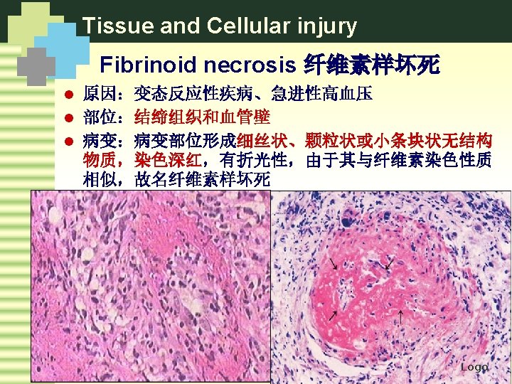
Tissue and Cellular injury Special types of necrosis u 干酪样坏死 Caseous necrosis u 坏疽 Gangrene u 脂肪坏死 Fat necrosis u 纤维素样坏死 Fibrinoid necrosis Logo
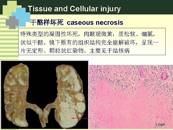
Tissue and Cellular injury 干酪样坏死 caseous necrosis 在结核病时,因病灶中 含脂质较多,坏死区呈 黄色,状似干酪 most often in TB Gross: white or light yellow, cheesy. LM: amorphous granular debris tissue, architecture is completely obliterated Logo
Tissue and Cellular injury Gangrene 坏疽 指继发有腐败菌感染的大块组织坏死。 Necrosis of big tissue with secondary putrefactive organisms infection Black and green appearance Dry gangrenes Gangrene Wet gangrene Gas gangrene Logo
Tissue and Cellular injury Dry gangrenes Occurs on the skin surface following arterial obstruction. It is particularly liable to affect the limbs, especially the toes. l 坏死组织水分少, 蒸发 l 干固皱缩,呈黑褐色,分界明显 l 四肢末端多见,动脉受阻,静脉 通畅 l 腐败菌感染一般较轻 frostbite injury Logo
Tissue and Cellular injury dry, black, clear border with surrounding normal tissue Logo
Tissue and Cellular injury Wet gangrene Conditions: occurs in naturally moist tissues and organs. Both arterial and venous obstruction; Character: wet swollen, foulsmelling, black or green. Location: small intestine, appendix, lung, uterus, limbs 与外界相通的内脏(如肺、肠、子宫、 阑尾、胆囊等)或四肢,动脉受阻, 静脉也不畅或受阻(淤血,水肿) l 坏死组织水分多,局部肿胀、暗绿 色、分界不清 l 腐败菌感染重,产生吲跺,粪臭素、 恶臭; Logo l 毒素吸收,毒血症 l
Tissue and Cellular injury Wet gangrene Soft, swollen, dark. Logo
Tissue and Cellular injury Gas gangrene Conditions: deep contaminated wounds in which there is considerable muscle damaged by gas forming bacteria. Character: swollen obviously, gas bubbles formation war wounds 深在的开放性创伤合并产气荚 膜杆菌感染 l 坏死组织含大量气体,呈蜂窝 状,污秽、暗棕色,捻发感; l 战伤,外伤,厌氧菌感染 l Logo
Tissue and Cellular injury Fat necrosis脂肪坏死 酶解性脂肪坏死 外伤性脂肪坏死 Logo
Tissue and Cellular injury Fat necrosis脂肪坏死 酶解性脂肪坏死 外伤性脂肪坏死 LM: shadowy outlines of necrotic fat cells, with basophilic calcium deposits and a surrounding inflammation reaction Logo
Tissue and Cellular injury 坏死的类型 Types of necrosis coagulative necrosis liquefaction necrosis caseous necrosis Specialized necrosis gangrene fibrnoid necrosis l Enzymatic digestion of the cells l Denaturation of proteins fat necrosis Logo
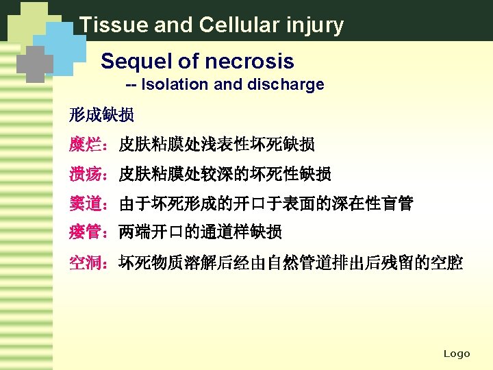
Tissue and Cellular injury 坏死结局 坏死对机体的影响 ü 生理上的重要性 ü 溶解和吸收 Physiologic importance Lysis and absorption ü 坏死细胞数量 ü 分离排出 Amount of necrotic cells Isolation and discharge ü 细胞再生能力 ü 机化和包裹 Ability of cells Organization ü 器官代偿能力 ü 钙化 Compensatory capacity Encapsulation calcification Logo
Tissue and Cellular injury Sequel of necrosis -- Lysis and absorption p 引起急性炎症(与死后自溶区别) 蛋白水解酶→溶解液化 p 淋巴管,血管 p 巨噬细胞 Logo
Tissue and Cellular injury Sequel of necrosis -- Isolation and discharge Ulcer & erosion ulcer cavity Logo
Tissue and Cellular injury Sequel of necrosis -- Isolation and discharge 腕关节窦道 瘘管(肛周围脓肿) Logo
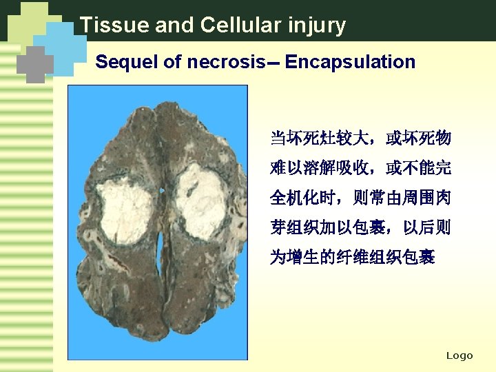
Tissue and Cellular injury Sequel of necrosis-- Organization necrosis Granulation tissue 机化:由肉芽组织取代坏死 组织、纤维素性渗出物、浓 缩的脓液、组织内血肿和血 栓等无生机物质的过程。 Logo
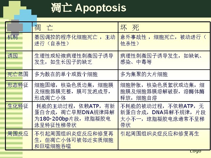
凋亡 Apoptosis 1972, “falling off” l A form of cell death l A way to eliminate unwanted host cells through activation of a coordinated, internally programmed series of events l 凋亡是活体内个别细胞程序性细胞死亡(programmed cell death)的表现形式,是由体内外因素触发细胞内 预存的死亡程序而导致的细胞主动性死亡方式,在形态 和生化特征上都有别于坏死。 是正常细胞群体中单个细胞的死亡。 A single cell death in living bodies controlled by the genes which is a energy-dependent suicide process. Logo
凋亡 Apoptosis Difference between apoptosis and necrosis Apoptosis üCell shrinkage üChromatin condensation nucleus fragments üFormation of cytoplasmic blebs and apoptotic bodies üPhagocytosis of apoptotic cells or bodies ü No inflammation Logo
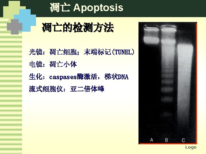
凋亡 Apoptosis Morphological changes LM: Single cells or small clusters of cells Round or oval mass of eosinophilic cytoplasm with dense nuclear chromatin fragments 凋亡小体多呈圆形或卵圆形,大小不等,胞浆浓缩,强嗜 酸性,核染色质浓集呈紫蓝色致密的球状 Logo
凋亡 Apoptosis Logo