Metabolism II and Glycolysis 5703 Organic reaction mechanisms

Metabolism II and Glycolysis 5/7/03

Organic reaction mechanisms Much can be learned by studying organic model reactions when compared to enzyme catalyzed reactions. 1. Group transfer reactions 2. Oxidations and reductions 3. Eliminations, isomerizations and rearrangements 4. Reactions that make or break carbon-carbon bonds

ATP is the energy carrier for most biological reactions ATP + H 2 O -> ADP + Pi ATP + H 2 O -> AMP + PPi

Coupled Reactions

Recycling ATP & ADP

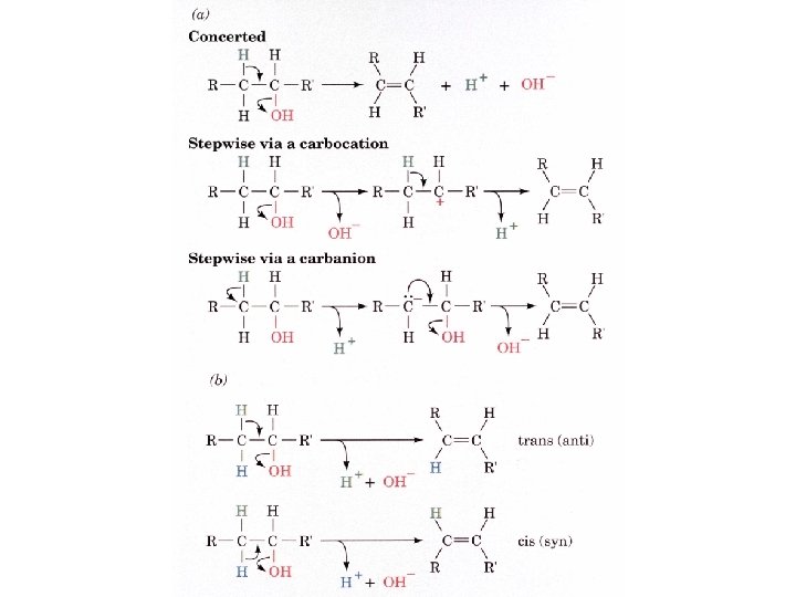
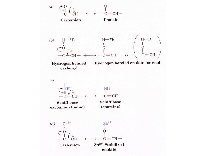
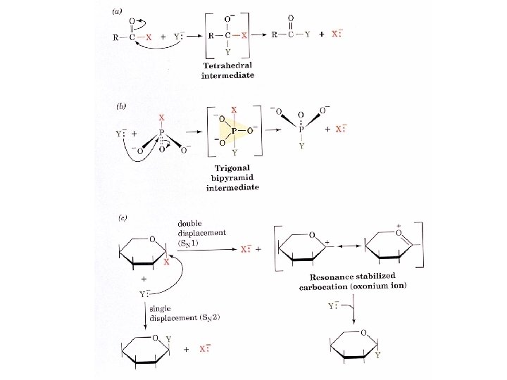
Heterolytic cleavage or bond formation is catalyzed using either nucleophiles or electrophiles.

Nucleophiles Basic reaction of amine Nucleophilic reaction of an amine

Biologically important nucleophiles

Amine Ketone or aldehyde Carbinolamine intermediate Imine Movement of an electron pair from a position and pointing to the electron deficient center attracting the pair.

Common biological electrophiles
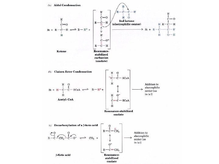

Group transfer reactions Acetyl group transfer Nucleophile attack on an acyl carbonyl to form a tetrahedral intermediate Peptide bond hydrolysis Phosphoryl group transfer nucleophile attack on a phosphate to yield a trigonal bipyramid intermediate Kinase reactions involving transfer of phosphate from ATP to organic alcohols Glycosyl group transfers substitution of one group at the C 1 carbon of a sugar for another
 High energy compound Carrier of acetyl and acyl groups Can be")
Thioesters (Acetyl-coenzyme A) High energy compound Carrier of acetyl and acyl groups Can be used to drive exogenic processes e. g. GTP from GDP

Oxidations and reductions Oxidation : Loss of Electrons Reduction: Gain of Electrons Many redox reactions involve the breaking of a C-H bond and the loss of two bonding electrons

Electron transfer reactions to oxygen undergo transfer of one electron at a time (Pauli exclusion principle) Oxidations to oxygen from NADH require two electron steps to be changed to one electron steps. Stable radical structures like FMN or FAD and cytochromes are involved.

Reduction of NAD+ to NADH

Electron transfer reactions
 Electron acceptor (oxidizing")
Half-cell reactions either donate or accept electrons Electron donor (reducing agent) Electron acceptor (oxidizing agent)

Nernst Equation- electromotive force -EMF- reduction potential Work is non -pressure volume work or DG = -w’ = -welec Welec = n. FDE or DG = -n. FDE F = Faraday constant = 96, 485 Coulombs per mole of electrons DE 0 = standard reduction potential or midpoint potential

Measuring potentials

However, there is no absolute potential to reference! At equilibrium and in contact with a platinum electrode and at 1 M H+ and STP this is defined as zero potential. At p. H of 7. 0 this is 0. 421 V = Eo´. Prime means that it is at p. H 7. 0. Every thing is referenced to this potential See Table in FOB pg 373 for standard potentials

Metabolic pathways are irreversible They have large negative free energy changes to prevent them running at equilibrium. 1 If two pathways are interconvertible (from 1 to 2 or 2 to 1), the two pathways must be different! Independent routes means independent control of A rates. 2 Y X The need to control the amounts of either 1 or 2 independent of each other.

Every pathway has a first committed step A committed step is an irreversible step that commits the pathway to the synthesis of the end product. This step is usually the regulated step in the pathway. All metabolic pathways are regulated The control of the flux through a pathway is regulated by regulatory enzymes at the committed step in the pathway. This control over metabolism allows the organism to make corrections and adjust to unforeseen changes.

Pathways in eukaryotic cells occur in separate organelles or cellular locations ATP is made in the mitochondria and used in the cytosol. Fatty acids are make in the cytosol and broken down in the mitochondria. Separation of pathways exerts a greater control over opposing pathways and the intermediates can be controlled by transport across the separating membranes.

Experimental approaches to study metabolism 1. Sequence of reactions by which a nutrient is converted to end products 2. Mechanism by which an intermediate is turned into its successor. 3. Regulation of the flow of metabolites in a pathway. Inhibitors and growth studies are used to see what is blocked. If a reaction pathway is inhibited products before the block increase and intermediates after the block decrease in concentration

Genetic Defects cause intermediates to accumulate

Genetic manipulations can cause a block to occur

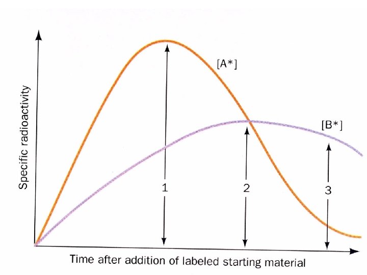
Radioactive tracers

Glycolysis The conversion of glucose to pyruvate to yield 2 ATP molecules • 10 enzymatic steps • Chemical interconversion steps • Mechanisms of enzyme conversion and intermediates • Energetics of conversions • Mechanisms controlling the Flux of metabolites through the pathway

Historical perspective Winemaking and baking industries 1854 -1865 Louis Pasture established that microorganisms were responsible for fermentation. 1897 Eduard Buchner- cell free extracts carried out fermentation no “vital force” and put fermentation in the province of chemistry 1905 - 1910 Arthur Harden and William Young • inorganic phosphate was required ie. fructose-1, 6 bisphosphate • zymase and cozymase fractions can be separated by diaylsis

Inhibitors were used. Reagents are found that inhibit the production of pathway products, thereby causing the buildup of metabolites that can be identified as pathway intermediates. Fluoride- leads to the buildup of 3 -phosphoglycerate and 2 -phosphoglycerate 1940 Gustav Embden, Otto Meyerhof, and Jacob Parnas put the pathway together.

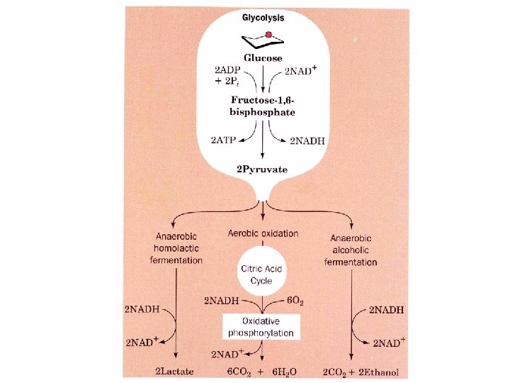
Pathway overview 1. Add phosphoryl groups to activate glucose. 2. Convert the phosphorylated intermediates into high energy phosphate compounds. 3. Couple the transfer of the phosphate to ADP to form ATP. Stage I A preparatory stage in which glucose is phosphorylated and cleaved to yield two molecules of glyceraldehyde-3 phosphate - uses two ATPs Stage II glyceraldehyde-3 -phosphate is converted to pyruvate with the concomitant generation of four ATPs-net profit is 2 ATPs per glucose. Glucose + 2 NAD+ + 2 ADP +2 Pi 2 NADH + 2 pyruvate + 2 ATP + 2 H 2 O + 4 H+

Oxidizing power of NAD+ must be recycled NADH produced must be converted back to NAD+ 1. Under anaerobic conditions in muscle NADH reduces pyruvate to lactate (homolactic fermentation). 2. Under anaerobic conditions in yeast, pyruvate is decarboxylated to yield CO 2 and acetaldehyde and the latter is reduced by NADH to ethanol and NAD+ is regenerated (alcoholic fermentation). 3. Under aerobic conditions, the mitochondrial oxidation of each NADH to NAD+ yields three ATPs
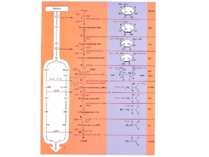

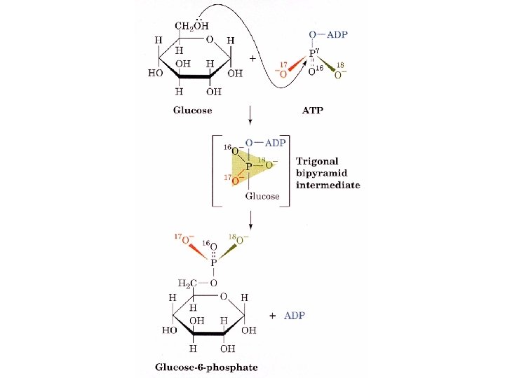
Hexokinase Mg++ + ATP Glucose + ADP + H+ Glucose-6 -phosphate Isozymes: Enzymes that catalyze the same reaction but are different in their kinetic behavior Tissue specific Glucokinase- Liver controls blood glucose levels. Hexokinase in muscle - allosteric inhibition by ATP Hexokinase in brain - NO allosteric inhibition by ATP

Hexokinase reaction mechanism is RANDOM Bi-Bi Glucose ATP ADP Glu-6 -PO 4 When ATP binds to hexokinase without glucose it does not hydrolyze ATP. WHY? The binding of glucose elicits a structural change that puts the enzyme in the correct position for hydrolysis of ATP.

The enzyme movement places the ATP in close proximity to C 6 H 2 OH group of glucose and excludes water from the active site. There is a 40, 000 fold increase in ATP hydrolysis upon binding xylose which cannot be phosphorylated! a-D-Xylose
 causes a")
Yeast hexokinase, two lobes are gray and green. Binding of glucose (purple) causes a large conformational change. A substrate induced conformational change that prevents the unwanted hydrolysis of ATP.
 Substrate binding 2) Acid attack")
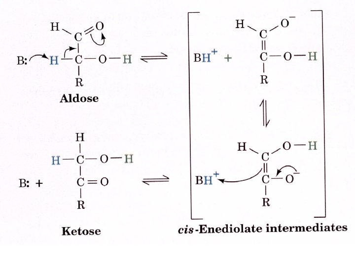
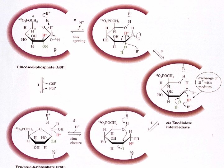
Phosphoglucose Isomerase Uses an “ ene dione intermediate 1) Substrate binding 2) Acid attack by H 2 N-Lys opens the ring 3) Base unprotonated Glu abstracts proton from C 2 4) Proton exchange 5) Ring closure

Uncatalyzed isomerization of Glucose
")
Phosphofructokinase + ATP Fructose-6 -PO 4 Mg++ + ADP Fructose-1, 6 -bisphosphate 1. ) Rate limiting step in glycolysis 2. ) Irreversible step, can not go the other way 3. ) The control point for glycolysis
 + Fructose -1, 6 -bisphosphate (FBP) Glyceraldehyde-3 phosphate (GAP) Aldol")
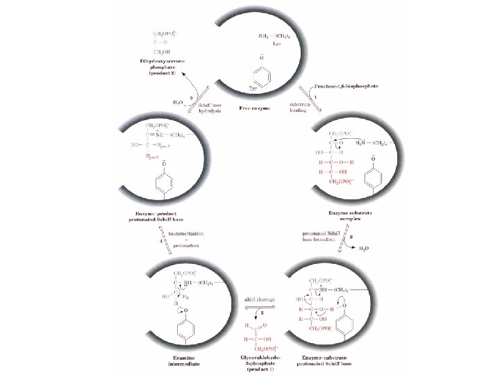
Aldolase Dihydroxyacetone phosphate (DHAP) + Fructose -1, 6 -bisphosphate (FBP) Glyceraldehyde-3 phosphate (GAP) Aldol cleavage (retro aldol condensation)

There are two classes of Aldolases Class I animals and plants - Schiff base intermediate Step 1 Substrate binding Step 2 FBP carbonyl groups reacts with amino LYS to form iminium cation (Schiff base) Step 3. C 3 -C 4 bond cleavage resulting enamine and release of GAP Step 4 protonation of the enamine to a iminium cation Step 5 Hydrolysis of iminium cation to release DHAP + Na. BH 4

Class II enzymes are found in fungi and algae and do not form a Schiff base. A divalent cation usually a Zn+2 polarizes the carbonyl intermediate. Probably the occurrence of two classes is a metabolic redundancy that many higher organisms replaced with the better mechanism.

Aldolase is very stereospecific When condensing DHAP with GAP four possible products can form depending on the whether the pro-S or pro R hydrogen is removed on the C 3 of DHAP and whether the re or si face of GAP is attacked.

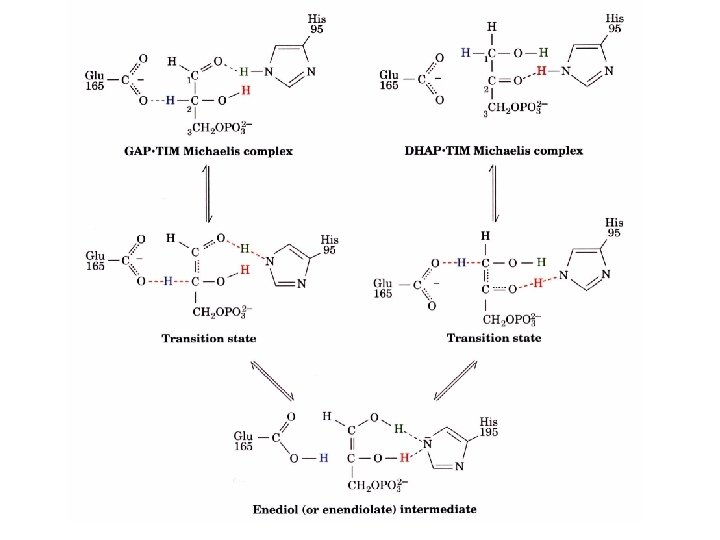
Triosephosphate isomerase DHAP GAP TIM is a perfect enzyme which its rate is diffusion controlled. A rapid equilibrium allows GAP to be used and DHAP to replace the used GAP.
 and")
TIM has an enediol intermediate GAP enediol DHAP Transition state analogues Phosphoglycohydroxamate (A) and 2 -phosphoglycolate (B) bind to TIM 155 and 100 times stronger than GAP of DHAP B. A.

TIM has an extended “low barrier” hydrogen bond transition state Hydrogen bonds have unusually strong interactions and have lead to p. K of Glu 165 to shift from 4. 1 to 6. 5 and the p. K of

Geometry of the eneolate intermediate prevents formation of methyl glyoxal Orbital symmetry prevents double bond formation needed for methyl glyoxal
- Slides: 59