Metabolism 102709 Introduction to metabolism Metabolism is the

Metabolism 10/27/09

Introduction to metabolism Metabolism is the overall process through which living systems acquire and utilize free energy to carry out their functions They couple exergonic reactions of nutrient breakdown to the endergonic processes required to maintain the living state Catabolism (degradation): nutrients and cell constituents broken down to salvage components and/or generate energy Anabolism (biosynthesis): biomolecules are synthesized from simpler components How do living things acquire the energy needed for these functions?

Autotrophs – self-feeders (synthesize their own cellular constituents from H 2 O, CO 2, NH 3, and H 2 S) Photoautotrophs - acquire free energy from sunlight Chemolithotrophs – obtain free energy from oxidation of inorganic compounds such as NH 3, H 2 S, or Fe 2+. Heterotrophs – oxidize organic compounds to make ATP is the energy carrier for most biological reactions

Organisms can be classified by the identity of the oxidizing agent. Obligate aerobes: must use O 2 Anaerobes: use sulfate or nitrate Facultative anaerobes: can grow in presence or absence of O 2 (e. g. E. coli) Obligate anaerobes: poisoned by O 2

Metabolic pathways are series of connected enzymatic reactions that produce specific products. Their reactants, intermediates, and products are called metabolites. There are over 2000 known metabolic reactions – see figure to the left.

Organizing metabolic reactions • See these useful sites below: • http: //www. genome. jp/kegg/m etabolism. html • http: //www. genome. jp/kegg/pa thway/map 01100. html • If you click on the “Carbohydrate Metabolism” button, you will get the clickable image on the next slide

Carbohydrate Metabolism • This figure shows most of the metabolic pathways that we will discuss in this half of the course, namely, the glycolysis pathway, gluconeogenesis, the citric acid cycle, and the pentose phosphate pathway. • If you click on the glycolysis/ gluconeogenesis node, you will get the map on the next slide that It also give the enzyme classification (EC) code that will help you search for structures, sequences, and other information about it.

Metabolic pathways • Metabolic pathways are compartmentalized. • Oxidative phosphorylation occurs in mitochondria while glycolysis and fatty acid biosynthesis occur in the cytosol. • Gluconeogenesis occurs in liver to maintain constant level glucose in the circulation but adipose tissue specializes in storage of triacylglycerols. • Isozymes: enzymes that catalyze the same reaction but are encoded by different genes and have different kinetic of regulatory properties. • Lactate dehydrogenase (LDH): type M [skeletal muscle and liver] participates in the reduction of pyruvate to lactate (using NADH) while type H [heart muscle] catalyzes the reverse reaction. • See Table 14 -3 in the book for more examples.

Pathways in eukaryotic cells occur in separate organelles or cellular locations ATP is made in the mitochondria and used in the cytosol. Fatty acids are made in the cytosol with the use of acetyl-Co. A (Co. A=coenzyme A) which is synthesized in the mitochondria. This exerts a greater control over opposing pathways and the intermediates can be controlled by transport across the separating membranes.

Roles of ATP and NADP+ in metabolism • In catabolic pathways, complex metabolites are exergonically broken down into simpler products, creating ATP or NADPH • In anabolic processes, simple molecules are converted into complex molecules at the expense of degradation of the energy storage molecules, ATP and/or NADPH.

Very Few metabolites are used to synthesize a large variety of biomolecules • Acetyl-Coenzyme A (acetyl-Co. A) • Pyruvate • Citrate cycle intermediates Three main pathways for energy production • Glycolysis • Citric acid cycle • Oxidative-Phosphorylation

Overview of catabolism • Complex metabolites are broken down into their monomeric units • Then to the common intermediate, acetyl-Co. A • The acetyl group is then oxidized to CO 2 via the citric acid cycle while NAD+ and FAD are reduced to NADH and FADH 2. • Reoxidation of NADH and FADH 2 by O 2 during oxidative phosphorylation yields H 2 O and ATP

Thermodynamic considerations Recall A + B C + D; DG = DGo’ + RT ln ([C][D]/[A][B]) When close to equilibrium, [C][D]/[A][B] Keq and DG 0. This is true for many metabolic reactions – near-equilibrium reactions When reactants are in excess, the reaction shifts toward products When product are in excess, the reaction shifts toward reactants However, some reactions are not near equilibrium are thus irreversible • • • – – This is true of highly exergonic reactions These metabolic reactions therefore control the flow of reactants through the pathway/cycle and they make pathways irreversible. 1. 2. 3. Metabolic pathways are irreversible Every metabolic pathway has a first committed step Catabolic and anabolic pathways must differ (so that they can be separately regulated)

Metabolic pathways are irreversible They have large negative free energy changes to prevent them running at equilibrium. 1 If two metabolites are interconvertible, the two interconversion pathways must be different Independent routes means independent control of A rates. 2 Y X The need to control the amounts of either 1 or 2 independent of each other.
 1. A 1. B C P 2. 2.")
Control of flux at committed step(s) 1. A 1. B C P 2. 2. 3. 3. 4. Allosteric control: by substrates, products, or coenzymes of the pathway (e. g. CTP in ATCase) Covalent modification: (de)phosphorylation by (phosphatases)kinases which are themselves regulated Substrate cycles: Fluxes through r and f can be separately regulated Genetic control: up or down regulated production or activation of an enzyme

Thermodynamics of Phosphate compounds Adenosine diphosphate, one phosphoester bond and one phosphoanhydride bond Adenosine monophosphate one phosphoester bond. Which bonds are exergonic?

Phosphoryl – coupled transfer reactions

These highly exergonic reactions are coupled to numerous endergonic biochemical processes so as to drive them to completion. ATP is generated by coupling its formation to more highly exergonic metabolic reactions. The bioenergetic utility of phosphoryl-transfers stems from their kinetic stability to hydrolysis combined with their capacity to transmit relatively large amounts of free energy. DG of ATP hydrolysis varies with p. H, divalent metal ion concentration, and ionic strength

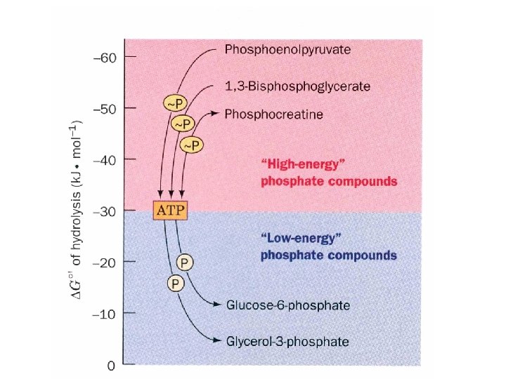
DG of ATP hydrolysis is in the middle of biological phosphate hydrolysis Compound Phosphoenol pyruvate 1, 3 -Bisphoglycerate Acetyl phosphate Phosphocreatine PPi ATP AMP + PPi ATP ADP + Pi Glucose-1 -phosphate Fructose-6 -phosphate Glucose-6 -phosphate Glycerol-3 -phosphate DGo' (k. J/mol) -61. 9 -49. 4 -43. 1 -33. 5 -32. 2 -30. 5 -20. 9 -13. 8 -9. 2

The P~P is a high energy bond Because of the concentrations of ATP, ADP, and Pi, the DG of a reaction is usually -50 k. J/mol. Usually anything over 25 k. J/mol is called a high energy bond. These bonds are sometimes designated as a ~, or a squiggle: AR-P~P~P (adenyl, ribosyl, phosphoryl). Why is the hydrolysis of ATP energetic? 1. Resonance stabilization of a phosphoanhydride bond is less than that of its hydrolysis products. 2. Electrostatic repulsion between three of four negative charges on the phosphate at neutral p. H. DG becomes even lower at higher p. H values which produces more charge. 3. Solvation energy of a phosphoanhydride bond is less than that of its hydrolysis products.

Resonance structures for phosphate bonds In phosphoanhydride, the P=O are each competing for the same anhydride oxygen lone pairs. In the separated phosphates, there is no competition so the resonance is better. Finally, there is electrostatic repulsion between adjacent O - atoms in the phosphoanhydride (see zigzag line). This repulsion leads to destabilization of this form, favoring hydrolysis.

Sample DG and K calculations Biochemical reactions are rarely at standard conditions. Temps. and concentrations vary from the standard state. • • DG = DGo’ + RT ln ([C][D]/[A][B]) For ATP ADP + Pi; [ATP]=3. 0 m. M, [ADP]=0. 8 m. M, [Pi]=4. 0 m. M DG = DGo’ + RT ln ([ADP][Pi]/[ATP]) at 310 K (37 o. C) DG = -30. 5 k. J/mol + (8. 3145 J/K)(310 K) ln [(0. 0008 M)(0. 0004 M)/ (0. 0003 M)] = -30. 5 k. J/mol – 17. 6 k. J/mol = -48. 1 k. J/mol • • K=? For hydrolysis of G-1 -P at 37 o. C Glucose-1 -phosphate + H 2 O glucose + Pi; DG 0’=-20. 9 k. J/mol DG 0’=-RTln. K; K=e-DG 0’/RT K=e-(-20, 900 J/mol)/(8. 3145 J/K-mol)(310 K) = 3. 3 x 10³

Other High-Energy Compounds Acyl phosphates Enol phosphates see previous page Phosphoguanidines

Compounds like a-D-glucose-6 - phosphate and l-Glycerol-3 -phosphate have smaller DG’s than ATP and have no significant resonance differences or charge repulsion.
 • Phosphate is and was originally scarce – thioesters are likely")
Thioesters (acetyl-Co. A) • Phosphate is and was originally scarce – thioesters are likely “highenergy” compounds • Thioesters are found today in Coenzyme A (Co. A) which links to various groups, most notably acetyl and is a common product of carbohydrate, fatty acid, and amino acid catabolism • Coenzyme A is sometimes written as Co. ASH since it has a reactive SH group • DG 0’ for hydrolysis of the thioester bond is – 31. 5 k. J/mol, 1 k. J/mol more then ATP hydrolysis!!

The role of ATP 1. Kinases: Early stages of nutrient breakdown transfers a phosphate to sugars 2. Interconversion of nucleoside triphosphates ATP, GTP, CTP, UTP Nucleoside diphosphate kinase

3. Physiological processes Muscle contraction Transport of ions against concentration gradients 4. Additional phosphoanhydride cleavage in highly endergonic reactions.

Formation of ATP 1. Substrate level phosphorylation - direct transfer of a phosphate group to ADP from a high energy compound. 2. Oxidative phosphorylation and photophosphorylationelectron transfer generates an ion gradient that is used to generate ATP. 3. Adenylate kinase reaction AMP + ATP 2 ADP About 1. 5 kg of ATP turnover per hour for the average person (about 3 moles) ATP + creatine phosphocreatine + ADP for ATP storage; ATP buffer in muscle and nerve cells.

Next Lecture Tuesday 10/29/09 Sugars
- Slides: 30