Mesotherapy for facial skin rejuvenation A clinical histologic


, 1. 5 mm long")
 and biopsy done 6")
 done before treatment and 6 months after. -evaluation by")

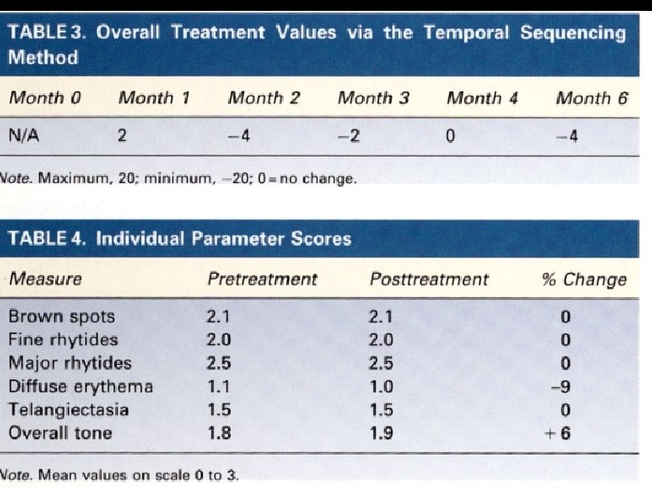
 (Table 3)")

 l Several adverse")


- Slides: 21
Mesotherapy for facial skin rejuvenation: A clinical, histologic and electronmicroscopic evaluation. Amin S. et al. Dermatol Surg 2006; 32: 14671472
Introduction described by Michel Pistor in 1952 l consists of the injection of hormones, enzymes, nutrients, pharmaceuticals, detergents, and other substances into the subcutaneous layer of the skin. l originally developed to treat vascular and lymphatic disorders. l
l l l is an intriguing yet poorly studied method for: -body contouring -cellulite treatment -lipolysis few clinical studies have been published showing the mechanism of action and active ingredients of mesotherapy for lipolysis. concern also raised for the potential adverse effects
l Although synonymous with injection induced lipolysis, it has also been used to treat a variety of rheumatologic and dermatologic disorders. l skin rejuvenation of the face is one of the latest applications -there are virtually no scientific peer-reviewed publications -rhytid reduction, increased elasticity, and improved pigmentation have been anecdoctally suggested, unthough umproven.
Objective first clinical evaluation on the effect of mesotherapy for facial skin rejuvenation. l evaluates clinical, histologic, and ultrastructural changes. Adverse effects also recorded l
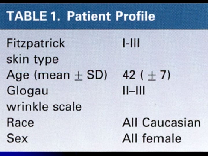
Methods l 10 subjects: exclusion: pregnancy, retinoid use, lasers in the previous 6 months…keloid. (Table 1 ). 4 treatments in a 6 month interval. l LMX (Ferndale Laboratories) topical anesthesia under occlusion one hour before procedure. injection of a 9: 1 suspension in hyaluronic acid gel (Hopewell pharmacy). Mixture immediately before injection. (Multivitamin Mixture, not specified) l
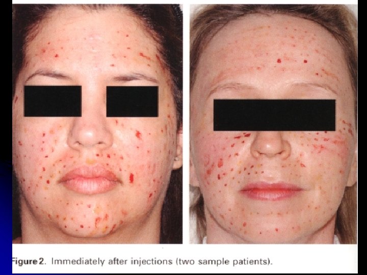
l l injections with needles with adjustable hubs (figure 1), 1. 5 mm long so that perpendicular injection feasible. 0. 01 ml injected at 1 cm intervals for a total of 200 -250 injections (2 -2. 5 ml)(figure 2) ice packs applied after 5 -10 min and observation for 30 minutes for adverse side effects. surveys performed monthly
l skin biopsies before first RX (angle of right mandible) and biopsy done 6 months later at 1 cm: -HE … stain -electron microscopy evaluation -blinded dermatopathologist: -epidermal thickness -vessel size and density -solar elastosis -elastin content -collagen fibre diameter by equal magnification electron microscope photographs -presence of sclerosis -overall appearance
l photography (6. 1 Mpixels) done before treatment and 6 months after. -evaluation by blinded physician using -temporal sequencing (Table 2): max 20, min – 20. -individual parameters (0 -3 points) (no statictical analysis because only 10 patients) -dyspigmentation -rhytid level -erythema
Results l l No significant improvement on the photograph assessment (figure 4) (Table 3) (Table 4) Microscopic evaluation showed no significant changes in epidermal thickness, vessel size and density, solar elastosis, elastin content, mucin content, dermal thickness, collagen fibre thickness, presence of sclerosis, and overall appearance.
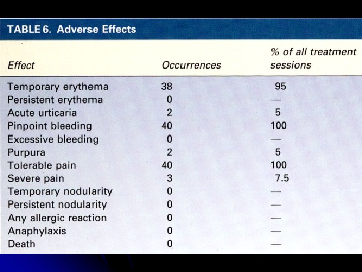
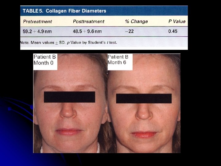
Electron microscopy showed a diminished diameter of collagen fibres (Table 5) l Several adverse effects noted, none serious (Table 6). l 4 out of 10 patients described increase smoothness to the skin and increased tone in the skin, which did not persist beyond one week after each session. l
Discussion temporal assessment: value close to 0 implies no effect. Slightly negative values could be explained by normal aging. l the transient effect on skin smoothness reported in 4/10 patients could be explained by a transient oedematous effect. l
l decrease in the diameter of collagen fibers can be explained by synthesis of new collagen with follows inflammation or thermal injury and is frequently associated with the presence of a repair zone.
Questions -evaluation at 1 year? -more treatments? -control group? -more effective than other non ablative treatments? Due to the popularity of mesotherapy, future studies will be required.