Mesoderm Mesodermal Regions Early mesodermal patterning buccopharyngeal membrane

Mesoderm

Mesodermal Regions
 Specific regions of the epiblast migrate through the streak")
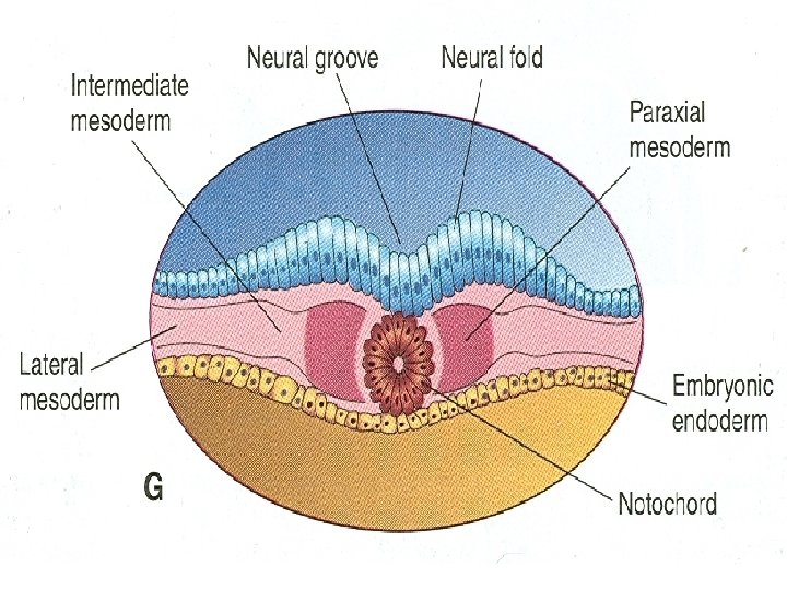
Early mesodermal patterning: (buccopharyngeal membrane) Specific regions of the epiblast migrate through the streak at different levels and assume different positions within the embryo: Cranial to caudal: Notochord (n) Paraxial mesoderm (pm) Intermediate mesoderm (im) *Lateral plate mesoderm (lpm) Extraembryonic mesoderm (eem) Langman’s fig 5 -07

MESODERMAL DERIVATIVES • • • Connective tissue Cartilage Bone Striated & smooth muscles Heart Blood & lymphatic vessels Kidneys, ovaries, testes & genital ducts Serous membrane lining the body cavities Spleen & cortex of the supra renal gland

Development of Somites • As the notochord and neural tube forms • Embryonic mesoderm on each side of them proliferates • Form a thick longitudinal columns of paraxial mesoderm • Each column is continuous with intermediate mesoderm

Head Amnion Amniotic cavity Neural plate Left Right Cut edge of amnion Primitive streak Tail Ectoderm Mesoderm Notochord Endoderm Yolk sac (a) 17 days. The flat three-layered embryo has completed gastrulation. Notochord and neural plate are present. Figure 28. 10 a

Neural groove Neural fold Neural crest Coelom Somite Intermediate mesoderm Lateral plate mesoderm • Somatic mesoderm • Splanchnic mesoderm (b) 20 days. The neural folds form by folding of the neural plate, which then deepens, producing the neural groove. Three mesodermal aggregates form on each side of the notochord (somite, intermediate mesoderm, and lateral plate mesoderm). Figure 28. 10 b
 22 days. The neural folds")
Surface ectoderm Neural crest Neural tube Somite Notochord (c) 22 days. The neural folds have closed, forming the neural tube which has detached from the surface ectoderm and lies between the surface ectoderm and the notochord. Embryonic body is beginning to undercut. Figure 28. 10 c
 Splanchnic mesoderm • Visceral serosa")
Somite Dermatome Myotome Sclerotome Kidney and gonads (intermediate mesoderm) Splanchnic mesoderm • Visceral serosa • Smooth muscle of gut Peritoneal cavity (coelom) Neural tube (ectoderm) Epidermis (ectoderm) Gut lining (endoderm) Somatic mesoderm • Limb bud • Parietal serosa • Dermis (d) End of week 4. Embryo undercutting is complete. Somites have subdivided into sclerotome, myotome, and dermatome, which form the vertebrae, skeletal muscles, and dermis respectively. Body coelom present. Figure 28. 10 d

Epiblast ECTODERM MESODERM Notochord • Epidermis, hair, nails, glands of skin • Brain and spinal cord • Neural crest and derivatives (sensory nerve cells, pigment cells, bones and blood vessels of the head) Nucleus pulposus of intervertebral discs Somite • Sclerotome: vertebrae and ribs • Dermatome: dermis of dorsal body region • Myotome: trunk and limb musculature Intermediate mesoderm • Kidneys • Gonads ENDODERM Lateral plate mesoderm Somatic mesoderm Splanchnic mesoderm • Parietal serosa • Dermis of ventral body region • Connective tissues of limbs (bones, joints, and ligaments) • Wall of digestive and respiratory tracts (except epithelial lining) • Visceral serosa • Heart • Blood vessels Epithelial lining and glands of digestive and respiratory tracts Figure 28. 13

Development of Somites • Intermediate mesoderm gradually thins into a layer of lateral mesoderm • Lateral mesoderm is continuous with the extraembryonic mesoderm • Extraembryonic mesoderm covers the yolk sac and amnion

Somites • Paraxial mesoderm differentiates and begins to divide into cuboidal bodies called somites by the end of 3 rd week • These blocks of mesoderm are located on each side of developing neural tube • About 38 pairs of somites form during the somite period of human development (20 -30 days)

Somites • About 42 -44 pairs of somites are present by the end of 5 th week • Are triangular in transverse section • Form distinct surface elevations on the embryo • Are used as one of the criteria to know the age of the embryo at this stage

Somites • First appear in the future occipital region • Soon develop craniocaudally • Gives rise to the axial skeleton and associated musculature • Also forms adjacent dermis of the skin • The first pair of somites appear at the end of 3 rd week

Somites • First appear at a short distance caudal to the cranial end of the notochord • Subsequent pairs form in a craniocaudal sequence

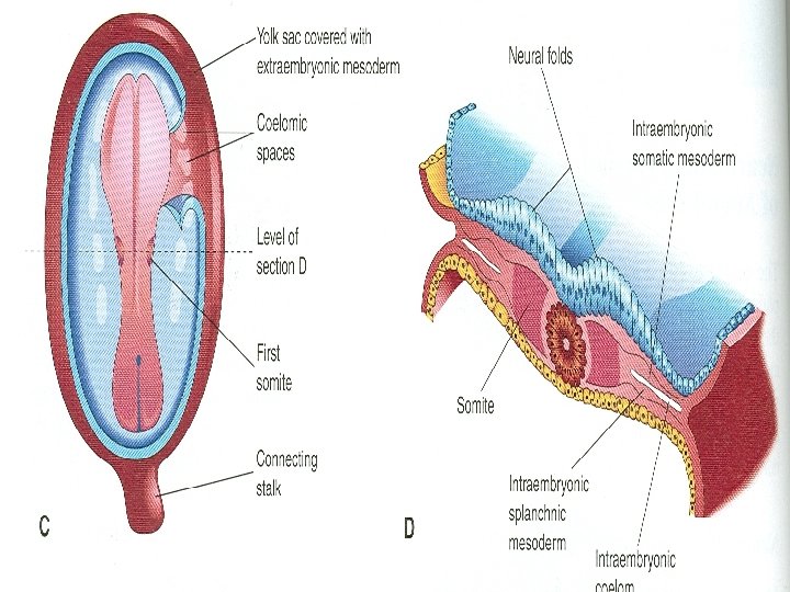
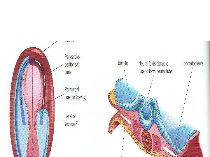
Intraembryonic Coelom • Also known as primordium of embryonic body cavity • Appears as isolated coelomic spaces in the lateral mesoderm and cardiogenic mesoderm • These spaces soon coalesce to form a single horseshoe shaped cavity called intraembryonic coelom


Parietal & Visceral Layers • Somatic or parietal layer continuous with the extraembryonic mesoderm covering the amnion • Splanchnic or visceral layer continuous with the extraembryonic mesoderm covering the yolk sac

Parietal & Visceral Layers • Somatic mesoderm with overlying embryonic ectoderm form the embryonic body wall or somatopleure • Splanchnic mesoderm with underlying embryonic endoderm form the embryonic gut or splanchnopleure

Fate of Intraembryonic Coelom During the 2 nd month, the intraembryonic coelom is divided into 3 body cavities: • Pericardial cavity • Pleural cavity • Peritoneal cavity

- Slides: 25