Merkezi grme mekanizmalar Merkezi grme yollar Ganglion hcrelerinin


: - Görsel (7) ve somatik duysal")


- Slides: 36
Merkezi görme mekanizmaları
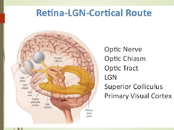
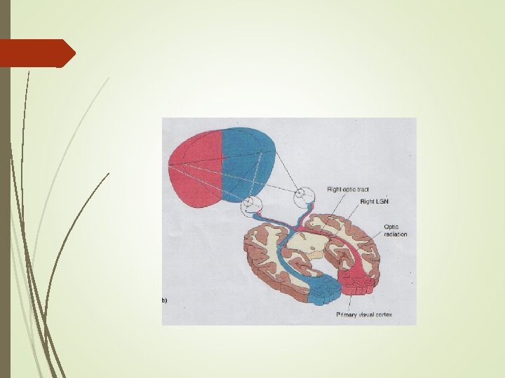
Merkezi görme yolları Ganglion hücrelerinin aksonları optik siniri oluşturur. Optik kiazmada her iki optik sinirin nazal retinadan gelen lifleri çaprazlaşır. Görme alanının sağ tarafı sol talamus ve sol kortekse, sol tarafı sağ talamus ve sağ kortekse projekte olur.
Nazal retina, temporal retina
Görme alanı: * Sağ ve sol görme alanı * Görme alanı kayıpları
Merkezi görme yolları Optik sinir Optik kiazma Optik traktus Lateral genikulat nukleus Optik radyasyo Görme korteksi
The Eye : III. Central Neurophysiology of Vision Hall, John E. , Ph. D, Guyton and Hall Textbook of Medical Physiology, Chapter 52, 661 -671 Principal visual pathways from the eyes to the visual cortex. (Modified from Polyak SL: The Retina. Chicago: University of Chicago, 1941. ) Copyright © 2016 by Elsevier, Inc. All rights reserved.
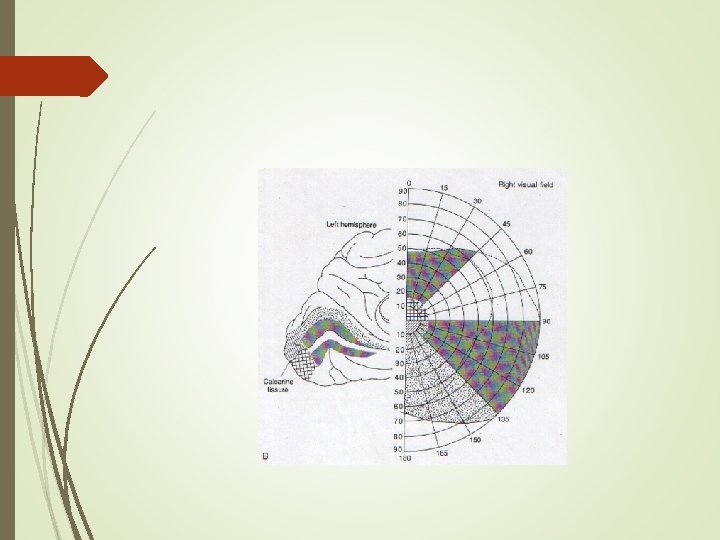
Circuits of the Central Nervous System Connors, Barry W. , Medical Physiology, Chapter 16, 390 -407. e 1 Visual fields and visual maps. A, The right sides of both retinas (which sense the left visual hemifield) project to the right lateral geniculate nucleus (LGN), which in turn projects to the right primary visual cortex (area V 1). B, The upper part. . . Copyright © 2017 by Elsevier, Inc. All rights reserved.
Optik traktus projeksiyonları Talamus LGN yolu ile Primer Görme Kort. Beyin sapı nukleusları: Gözlerin-başın hareketi, ışık refleksi Hipotalamus: Gündüz-gece ritmi, hormon salınımı düzenlenmesi
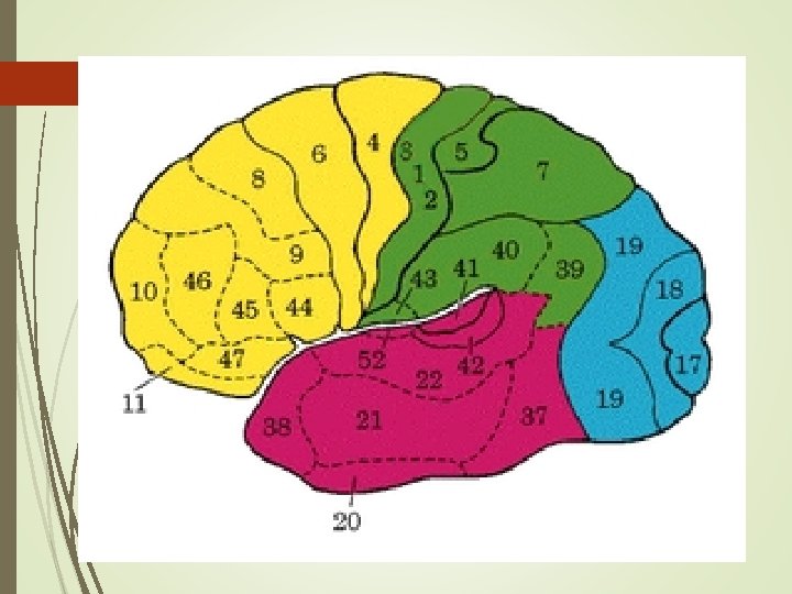
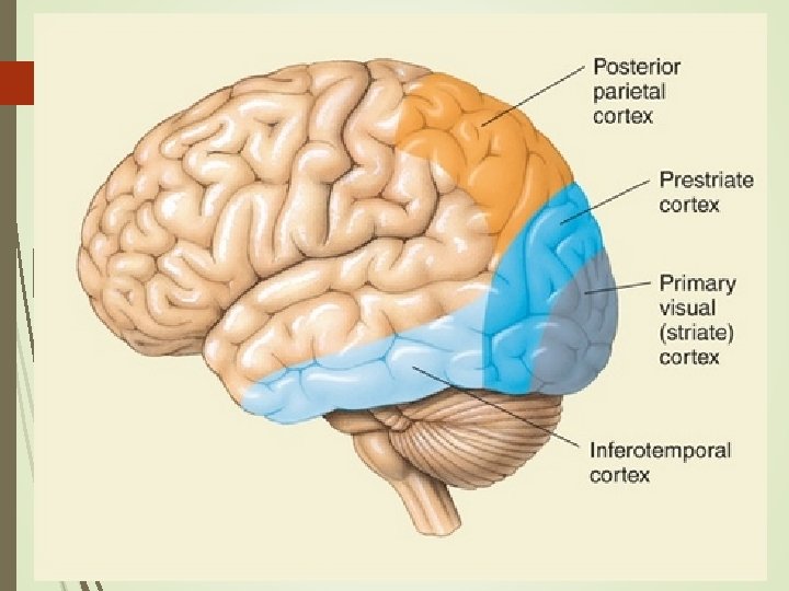
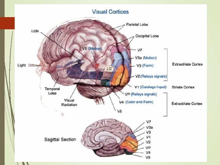
Kortikal Görme Alanları Tüm alanlarda retinotopi ve sütunsal organizasyon * Primer görme alanı (V 1 alanı, 17. alan) * Sekonder görme alanları (18. , 19. alanlar) * Üst düzey alanlar: - Posterior pariyetal korteks - İnferior temporal korteks
Circuits of the Central Nervous System Connors, Barry W. , Medical Physiology, Chapter 16, 390 -407. e 1 Ocular dominance columns and blobs in the primary visual cortex (area V 1). A, Ocular dominance columns are shown as alternating black (right eye) and gray (left eye) structures in layer IV. The alternating light and dark bands are visible in an au. . . Copyright © 2017 by Elsevier, Inc. All rights reserved.
* Posterior pariyetal korteks (5. , 7. alanlar): - Görsel (7) ve somatik duysal (5) uyarılar bütünleştirilir. - Kişi merkezli ve obje merkezli uzaysal konum bilgisi, dikkat, motor işlev * İnferior temporal korteks: - Obje tanıma - Bellek alanları yakınında yer alır ve bağlantılıdır.
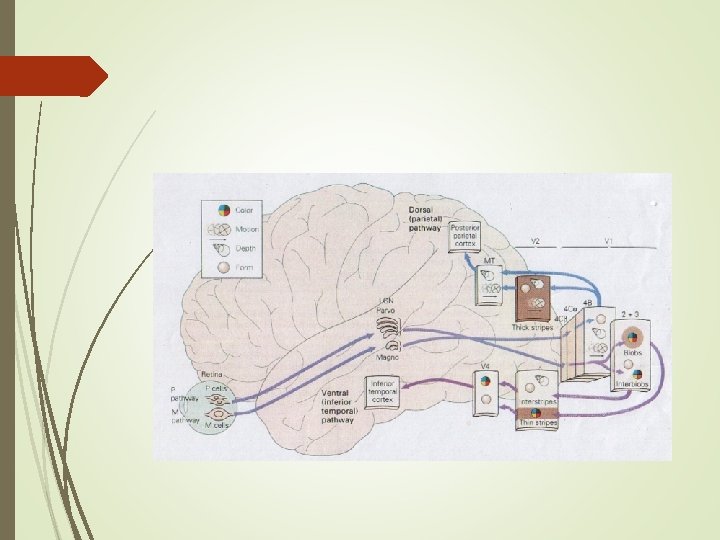
Paralel işlem Ganglion hücrelerinde başlar. Ganglion hücre tipleri: *M hücreleri: %10, büyük gövde-kalın akson, hızlı iletim, geçici yanıt, renk duy. yok *P hücreleri: %80, küçük gövde-ince akson, yavaş iletim, uyarı süresince yanıt, renk duyarlılığı var
M tip ganglion hücreleri: Hareketle ilgili enformasyon iletimi P tip ganglion hücreleri: Renkli görme, görüntünün ayrıntılı analizi, şekil görme ilgili enformasyon iletimi
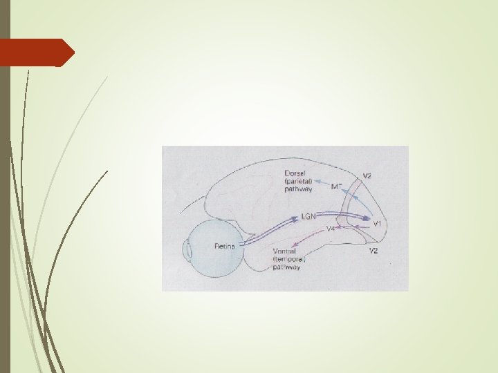
PARALEL İŞLEM Ventral yol: V 1 Sek. Alanlar İnf. temp. korteks *Görsel Tanıma-bellek alanları ile bağlantı Dorsal yol: V 1 Sek. Alanlar Post. par. korteks * Hareket analizi, uzaysal ilişkiler, motor alanlarla bağlantı
Retinadan kortekse paralel işlem Görüntüdeki hareket analizi Retina M ganglion hücreleri LGN tabakaları V 1 alanı Sekonder alanlar Posterior pariyetal korteks Görüntüdeki şekil ve renk analizi Retina P ganglion hücreleri LGN tabakaları V 1 alanı Sekonder alanlar İnferior temporal korteks
The Visual System Corbett, J. J. , Fundamental Neuroscience for Basic and Clinical Applications, Chapter 20, 286 -305. e 1 Overview of the visual fields ( A) and of the visual pathway as viewed from above ( B). Copyright © 2018 by Elsevier, Inc. All rights reserved.
The Visual System Corbett, J. J. , Fundamental Neuroscience for Basic and Clinical Applications, Chapter 20, 286 -305. e 1 Relationship of visual field diagrams to patient being examined. The observer draws the diagrams as if they were on the wall the patient is looking at. Copyright © 2018 by Elsevier, Inc. All rights reserved.
The Visual System Corbett, J. J. , Fundamental Neuroscience for Basic and Clinical Applications, Chapter 20, 286 -305. e 1 A visual field deficit, such as blindness in the left eye ( A), may result from a lesion of the left optic nerve ( B, seen from above). An aneurysm of the ophthalmic artery ( C, seen from below) may cause damage to the optic nerve on that side. Copyright © 2018 by Elsevier, Inc. All rights reserved.
The Visual System Corbett, J. J. , Fundamental Neuroscience for Basic and Clinical Applications, Chapter 20, 286 -305. e 1 Visual field deficits ( A) resulting from a lesion of the left optic nerve ( B and C) at its junction with the optic chiasm (junctional lesion). This combination of field defects is caused by destruction of fibers of the left optic nerve plus some. . . Copyright © 2018 by Elsevier, Inc. All rights reserved.
The Visual System Corbett, J. J. , Fundamental Neuroscience for Basic and Clinical Applications, Chapter 20, 286 -305. e 1 Visual field deficits ( A, right homonymous hemianopia) resulting from a lesion of the left optic tract ( B, seen from above). Interruption in the blood supply to the optic tract ( C, seen from below) may produce such deficits. Copyright © 2018 by Elsevier, Inc. All rights reserved.
The Visual System Corbett, J. J. , Fundamental Neuroscience for Basic and Clinical Applications, Chapter 20, 286 -305. e 1 Visual field defects produced by lesions at different places in the visual pathway. Visual fields are diagrammed as described in Fig. 20. 9. Regions of normal vision are indicated in white; regions of loss of vision are indicated in black. LGN, lat. . . Copyright © 2018 by Elsevier, Inc. All rights reserved.
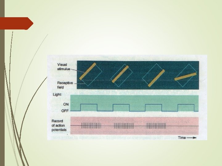
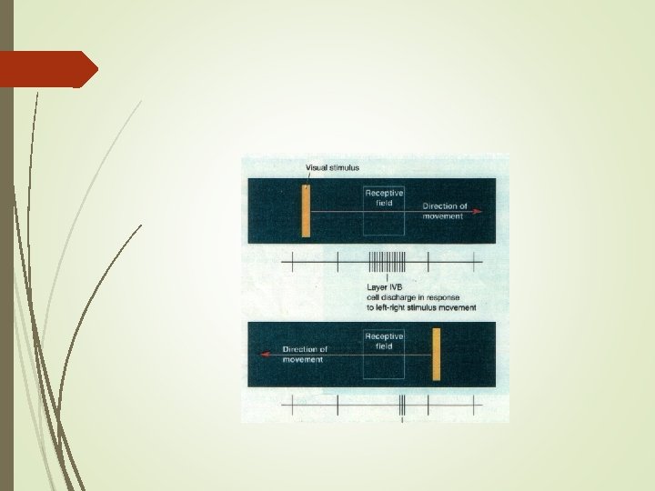
The Visual System Corbett, J. J. , Fundamental Neuroscience for Basic and Clinical Applications, Chapter 20, 286 -305. e 1 A to D, Representative receptive field organization of neurons in primary visual cortex. Copyright © 2018 by Elsevier, Inc. All rights reserved.
The Visual System Corbett, J. J. , Fundamental Neuroscience for Basic and Clinical Applications, Chapter 20, 286 -305. e 1 A, Diagram of how four stellate cells in layer IV with concentric receptive fields ( 1, on left) could converge onto a pyramidal cell ( 2) to produce simple receptive field characteristics ( 2, on right). B, Diagram of how several neurons, each ha. . . Copyright © 2018 by Elsevier, Inc. All rights reserved.
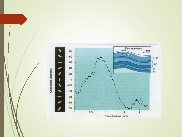
The Visual System Corbett, J. J. , Fundamental Neuroscience for Basic and Clinical Applications, Chapter 20, 286 -305. e 1 Ocular dominance columns and orientation columns in the cortex and their relationship to the layers of the lateral geniculate nucleus ( LGN). Copyright © 2018 by Elsevier, Inc. All rights reserved.
The Visual System Corbett, J. J. , Fundamental Neuroscience for Basic and Clinical Applications, Chapter 20, 286 -305. e 1 Simplified diagram of the cortical processing of visual information. Lateral ( A) and medial ( B) views of the cerebral cortex show some of the cortical areas (using Brodmann numbers) involved in the processing of visual input. C, The paths throug. . . Copyright © 2018 by Elsevier, Inc. All rights reserved.