MENTAL FORAMEN LOCATION AND DIMENSIONS OF THE MENTAL


- Slides: 22
MENTAL FORAMEN
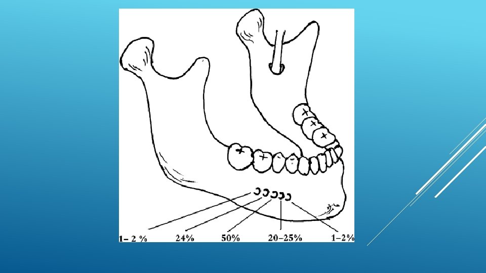
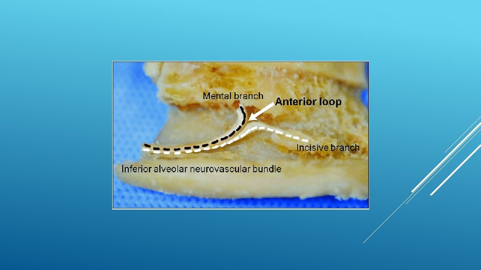
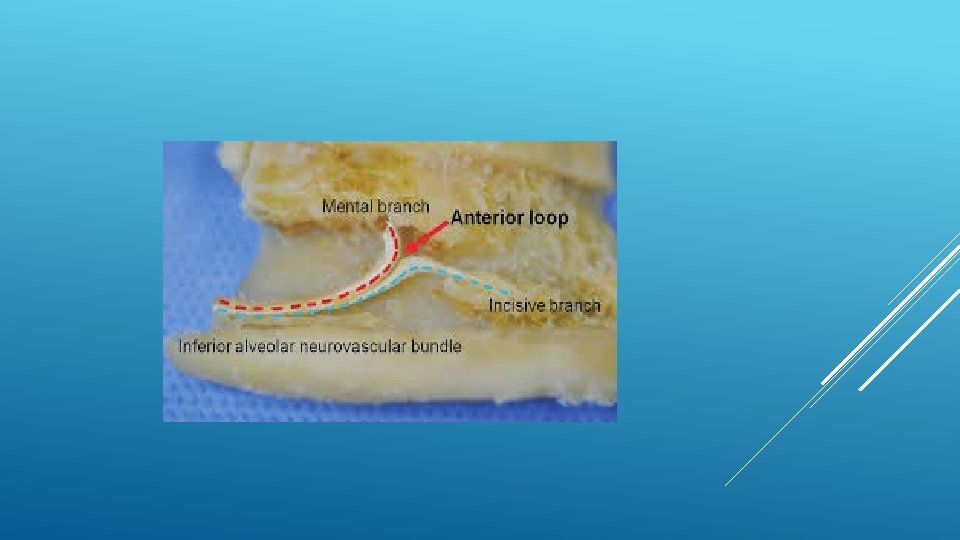
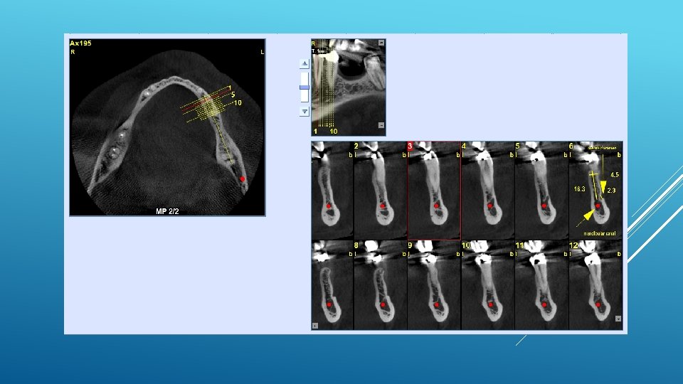
LOCATION AND DIMENSIONS OF THE MENTAL FORAMEN: A RADIOGRAPHIC ANALYSIS BY USING CONE-BEAM COMPUTED TOMOGRAPHY The majority of MF (56%) were located apically between the 2 premolars, and another 35. 7% of MF were positioned below the second premolar. On average, the MF was localized 5. 0 mm from the closest root of the adjacent tooth (range, 0. 3 -9. 8 mm). The mean size of the MF showed a height of 3. 0 mm and a length of 3. 2 mm; however, individual cases showed large differences in height (1. 8 -5. 1 mm) and in length (1. 8 -5. 5 mm). All mental canals exiting the MF demonstrated an upward course in the coronal plane, with 70. 1% of the mental canal presenting an anterior loop (AL) in the axial view. The mean extension of AL in cases with an AL was 2. 3 mm.
RELATIONSHIP BETWEEN THE POSITION OF THE MENTAL FORAMEN AND THE ANTERIOR LOOP OF THE INFERIOR ALVEOLAR NERVE AS DETERMINED BY CONE BEAM COMPUTED TOMOGRAPHY COMBINED WITH MIMICS The parameters were measured, and their values include mean (SD) anterior loop length, 1. 16 (1. 78) mm; anterior loop angle, 19. 13 (26. 89) degrees; inferior alveolar canal diameter, 3. 01 (0. 67) mm; height of the inferior alveolar canal, 10. 32 (1. 56) mm; 2 -dimensional mental foramen diameter, 2. 97 (0. 61) mm; 3 D mental foramen diameter, 2. 95 (0. 59) mm; 2 -dimensional vertical height of the mental foramen, 14. 67 (1. 67) mm; and 3 D vertical height of the mental foramen, 14. 61 (1. 69) mm. The mental foramen was located apically between the first and second premolars in 51. 67% and below the second premolar in 40. 83% of the cases.
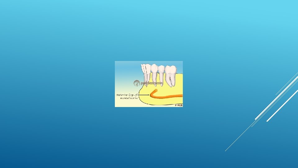
THE MENTAL FORAMEN OR "THE CROSSROADS OF THE MANDIBLE. " AN ANATOMIC AND CLINICAL OBSERVATION] [ARTICLE IN FRENCH, GERMAN] THOMAS VON ARX 1 This paper presents a clinical and anatomical review of the mental foramen (MF) based on recent publications (since 1990). Usually, the MF is located below the 2 nd premolar or between the two premolars, but it may also be positioned below the 1 st premolar or below the mesial root of the 1 st molar. At the level of the MF, lingual canals may join the mandibular canal (hence the term "crossroads"). Accessory MF are frequently described in the literature with large ethnic variations in incidence. The emergence pattern of the mental canal usually has an upward and posterior direction. The presence and extent of an "anterior loop" of the mental canal may be overestimated with panoramic radiography. Limited cone-beam computed tomography currently appears to be the most precise radiographic technique for assessment of the "anterior loop". The mental nerve exiting the MF usually has three to four branches for innervation of the soft tissues of the chin, lower lip, facial gingiva and mucosa in the anterior mandible. The clinician is advised to observe a safety distance when performing incisions and osteotomies in the vicinity of the MF.
ANATOMICAL RELATIONSHIP BETWEEN MENTAL FORAMEN, MANDIBULAR TEETH AND RISK OF NERVE INJURY WITH ENDODONTIC TREATMENT he root apex of the mandibular second premolar (70 %), followed by the first premolar (18 %) and then the first molar (12 %), was the closest to the MF. Ninetysix percent of root apices evaluated were >3 mm from the MF. An AL was present in 88 % of the cases. Conclusions: With regards to endodontic treatment, the risk of nerve injury in the vicinity of the MF would appear to be low. However, the high incidence of the AL highlights the need for clinicians to be aware and careful of this important anatomical feature.
ASSESSMENT OF MORPHOLOGICAL AND ANATOMICAL CHARACTERISTICS OF MENTAL FORAMEN USING CONE BEAM COMPUTED TOMOGRAPHY All mental foramina were visualized. Regarding location, 49. 2% of the MFs were located between first and second premolars, 7. 7 distal and 39. 7% coincident to the apex of the mandibular second premolar. The mean MF opening angle was 45. 4° on the right side, and 45. 9° on the left. There were no statistically differences between gender groups with regard to the opening angle degree