Meniscal Tears and Their Treatment Should I Refer


 red")


 Injury Results at 2 nd Look Lat Meniscus 69% healed")

 helps to stabilize")


![Observation: [ Valgus deformity ][ Varus deformity ][ Recurvatum deformity ][ no deformity ]](https://slidetodoc.com/presentation_image_h/c988502ab0c16e0bfb51ddd9f5efb26b/image-41.jpg "Observation: [ Valgus deformity ][ Varus deformity ][ Recurvatum deformity ][ no deformity ]")


- Slides: 48
Meniscal Tears and Their Treatment: Should I Refer? Robert S. Fawcett M. D. , M. S. , CAQ Sportsmedicine York Hospital Family Medicine Residency York, PA
Objectives n n n Discuss important teaching points in history, physical and testing leading diagnosis of damaged meniscus Understand the short and long term outcomes of meniscectomy Discuss the benefits and implications of surgical vs. conservative management of meniscal tear
Epidemiology n n Overall incidence unknown, but surgical incidence is 60 -70 per 100, 000 per year Most common orthopedic surgical procedure 1/3 of meniscal tears are sports-related (most of the rest from MVAs) 1/3 of meniscal tears associated with ACL injury
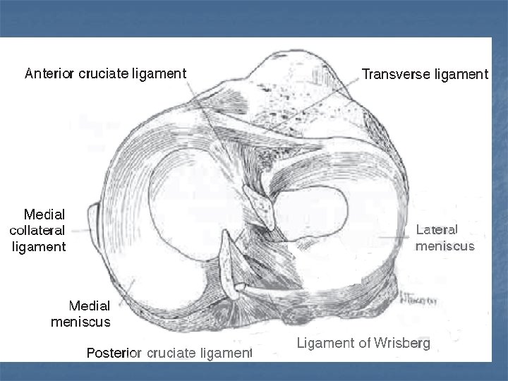
Structure of the Meniscus n Medial is semicircular Moves 2 -5 mm through full ROM n Lack of motion may promote tears n Fibers from the deep medial collateral n Covers 60% of articular cartilage n n Lateral almost a complete circle n n Moves ~1 cm through full ROM Both made of fibrocartilage 75% circumferential type 1 collagen fibers n 25% radial fibers n Covers 75% of articular cartilage n
Tears and Zones
Structure of the Menisci n Vascular supply good in the most peripheral 20% of the fibers n n Supplied by the geniculate arteries Inner 1/3 of the ring is avascular Relatively thin n Nourished through synovial fluid n n Middle 1/3 of the ring is combination
Function of the Menisci n Distribute load across the knee joint 2 -4 x body weight during walking n 6 -8 x body weight during running n n Axial compression is converted to “hoop stress”, or circumferential elongation in the meniscus Lateral meniscus distributes more load than medial meniscus, which contributes to greater degeneration if disrupted Menisci deepen the socket of the tibial plateau, contributing to stability
Function of the Menisci n n Wedge shape limits translation of femur on tibial plateau Menisci forced posteriorly in flexion, anteriorly in extension of the knee Menisci reduce stresses on the ACL Menisci force synovial fluid into articular cartilage (helping to nourish the white zone) during compression.
Pathophysiology n n In acute knee injuries with ACL intact, medial meniscal injury is 5 times more likely than lateral In acute knee injuries with ACL ruptured, lateral meniscus more likely to be involved In repetitive deep squatting, medial meniscus most likely to be injured (20: 1) In patients with arthritis in the knee, tears are present in the majority
History: the Key to Diagnosis n Twisting on planted foot n n n Inertial forces or external forces Acute effusion in acute injury Waxing and waning course with pain and effusion intermittently in chronic injury Locking or popping of knee, especially if followed by effusion However…
Meniscal Tears in Arthritis n In a random sample of 1000 people over age 50: Meniscal tears seen in 35% of sample group n Just as common in asymptomatic as symptomatic groups in those with OA on Xray n Became more common in older pts in study n Twice as common in medial as lateral meniscus n Slightly more common in overweight n
Physical Exam Finding/Test Sensitivity Specificity Joint Line Tenderness 71% 27% Mc. Murray 58. 5% Apley 58% Thessaly 5 o Thessaly 20 o 66%Me, 81%La 89%Me, 92%La 96%Me, 91%La 97%Me, 96%La MRI 75 -87% 87 -93% }80% }95% 93. 4% 80% *This test has undergone only one external validation study, but passed *
Thessaly Test? n n n Done with pt standing, first on normal leg Flex knee 5 degrees, rotate body on fixed leg back and forth 3 times, holding examiner’s hands for stability Flex further to 20 degrees and repeat Repeat on affected leg Positive is pain at joint line or feeling of locking or catching Validation results: 98% specific, 90% sensitive, PPV 98%, NPV 86%, and accuracy 89%
Value of MRI as Diagnostic Tool n n n Studies do NOT prove it superior to composite clinical exam Many false positives appear MRI has high NEGATIVE predictive value Sensitivity and specificity keep getting better as technology improves How will MRI result change treatment? No surgeon would touch a knee without one n Helps with planning procedure n
What About Ultrasound? n n n Compared to MRI, sensitivity of U/S = 85% Compared to MRI, specificity of U/S = 85% Compared to MRI, accuracy of U/S = 85% Compared to MRI, pos predictive value of U/S = 76% Compared to MRI, neg predictive value of U/S = 92%
Treatment Options n n n Total meniscectomy Partial meniscectomy Meniscal repair Inside out n Outside in n All inside n n Conservative (No operative intervention)
Consequences of Meniscectomy n n As early as 1948 Fairbanks noted increased osteophyte formation and femoral cartilage deterioration in meniscectomized knees Total meniscectomy remained a common procedure until the 1980’s In medial meniscectomy, load bearing surfaces are halved, doubling stress on tibial plateau If 15 -30% of meniscus is removed, forces between tibia and femur increase up to 350%
Bucket Handle Tear
Oblique Tear
Criteria for Meniscal Repair vs. Partial Meniscectomy Criterion Distance from rim Repair <3 mm Ptl. Meniscectomy >3 mm Mobility of fragment Stable Mobile Age of injury Recent Old Ret. To Play Later Sooner Age of patient Younger Older
Partial Meniscectomy n n Done when tear involves interior 70% May be done when athlete wants to resume activity ASAP Done with mobile fragments 10 -35 minute arthroscopic procedure under regional or general anesthetic Mobile areas removed n Edges contoured to “prevent further tears” n n n Immediate partial weight bearing allowed Crutches for 1 -2 days
Partial Meniscectomy n n Sedentary workers back to work in 1 week Laborers back in 2 -4 weeks Athletes back in 2 -6 weeks 88% “excellent” results at 15 years* *Burks RT, Metcalf MW, Metcalf RW; 15 yr f/u of arthroscopic partial meniscectomy; Arthroscopy 1997; 13: 673 -9.
Meniscus Repair n n Used in longitudinal tears Best results in (more vascular) red or pink zones Many fixation devices, none better than sutures, though some are faster Outside in, inside out, and all inside technique
Meniscus Repair Pts must wear brace with pwb for 2 weeks Sedentary workers back to work in 1 week Laborers back in 6 -8 weeks Athletes back in 12 -16 weeks 76% “excellent” results after 10 years* n n n *
Conservative Therapy n n n Not an option if knee locked, fragment not reduced Symptom relief with post-exercise RICE Symptom relief with NSAIDS, immobilization Physical therapy focusing on closed chain exercise of quadriceps and hamstrings Failure includes recurrent effusion, recurrent locking or pain that interferes with ADLs No randomized trials
Conservative Study Result n n n Retrospective review of 3612 arthroscopies Identified 80 “stable” tears (<3 mm movement) for whom nothing was done 70 were longitudinal, 10 were radial Only 6 needed subsequent surgery, 4 of which had additional trauma 32 patients had “second look” surgery 17/22 longitudinal tears, 0/6 radial tears healed completely Weiss CB, Lundberg M, De. Haven KD, Gillquist J; Non-operative treatment of meniscal tears. JBJS 1989 71 -A(6): 811 -22.
Conservative Study Results n n n Yagashita et al. Am J Spts Med 2004 32(8): 1953 “Stable” tears at ACL reconstruction left to heal and 2 nd look removing ACL hardware Lateral: 74% healed, 6% incompletely healed, 14% unhealed Medial: 56% healed, 6% incompletely healed, 24% unhealed Healing rate was “length dependent”
Conservative Study Results n n n 32 patients 30 lateral and 10 medial meniscal tears along with 25 ACL tears and 7 PCL tears Arthroscoped initially with repeat at 3 mo. Lateral meniscus: 69% completely healed and 18% incompletely healed Medial meniscus: 58% completely healed and 0% incompletely healed Ihara H, Miwa M, Takayanagi K, Nakayama A. Clin Orthop Relat Res. 1994 Oct; (307): 146 -54.
Results Without Surgery (Ihara) Injury Results at 2 nd Look Lat Meniscus 69% healed completely, 18% healed partially Medial Meniscus 58% healed completely 0% healed partially Ant. Cruc. Ligment 80% healed “satisfactorily” Post Cruc. Ligament 3/7 (40%) healed “satisfactorily”
Cochrane Review 2002 n n No evidence for comparing surgery to no treatment Partial is better than total meniscectomy: Less operative time n Enhance recovery rate n Improved long term stability n n Arthroscopic is better than open meniscectomy Less operative time n Quicker recovery post-op n n No long term advantages have been shown
Summary: What We Know n n n Meniscus (torn or intact) helps to stabilize and dissipate axial force in the knee Meniscectomy contributes to degenerative disease of the knee (Williams, others) When meniscal repairs fail, pts often engaging in same activity as initial injury Longitudinal tears heal better than radial tears, simple tears better than complex ones Peripheral tears (in vascularized area) heal more readily than central tears (Noyes, Krych)
Summary: What We Know n n Meniscal tears are accompanied by ligament tears in the majority of cases Ligamentous pathology with meniscal tears makes degenerative changes more likely Repairing both meniscus and ligaments (when both injured) improves outcomes (Noyes) Younger pts do better with meniscal repair than older patients (Mintzer) Less surgery is better than more surgery Arthroscopy better than open n Partial better than complete meniscectomy (Cochrane) n
Summary: What We Don’t Know n Is no surgery better than less surgery? Does operating on stable radial tears improve outcomes? n How do we tell (without surgery) that conservative treatment is a reasonable option n n Does immobilization help the acute tear? n n n If so, for how long? If a repair is undertaken, what timing and type of repair has the best outcomes? If no repair is done, should we do a “second look”? When?
Bibliography Karachalios T, Hantes M, Zibis AH, et al. Diagnostic accuracy of a new clinical test (the Thessaly test) for early detection of meniscal tears. J Bone Joint Surg 2005; 87: 955– 62. Krych AJ, Mc. Intosh AL, Voll, Michael AE, Stuart J, Dahm DL. Arthroscopic Repair of Isolated Meniscal Tears in Patients 18 Years and Younger. Am. J. Sports Med. 2008; 36; 1283 originally published online Mar 4, 2008. Manson TT, Cosgarea AJ. Meniscal injuries in active patients. Advanced Studies in Medicine November-December 2004, 4(10): 545 -552. Muellner T, Weinstabl R, Shabus R, Vecsei V, Kainberger F; The diagnosis of meniscal tears in athletes: a comparison of clinical and magnetic resonance imaging investigations. Am J Sports Med 1997; 25: 7 -12. Ihara H, Miwa M, Takayanagi K, Nakayama A. Clin Orthop Relat Res. 1994 (307): 146 -54. Johnson MJ, et al. (1999). Isolated arthroscopic meniscal repair: A long-term outcome study (more than 10 years). American Journal of Sports Medicine, 27(4): 44– 49. Mintzer CM, Richmond JC, Taylor J. Meniscal repair in the young athlete. Am J Sports Med 1998; 26: 630 -3. Noyes FR, Barber-Westin SD. Arthroscopic repair of meniscal tears extending into the avascular zone in patients younger than twenty years of age. Am J Sports Med 2002; 30: 589 -600. Weiss CB et al. Non-operative treatment of meniscal tears. JBJS 1989 71 -A(6): 811 -22. Howell JR Handoll HHG. Surgical treatment for meniscal injuries of the knee in adults (Cochrane Review). In: The Cochrane Library, Issue 3, 2002. Oxford: Update Software. Williams RJ et. al. MRI evaluation of isolated arthroscopic partial meniscectomy patients at a minimum five year follow-up. HSSJ 2007 3: 35 -43.
Harrison BK; Thessaly test for detection of meniscal tears: validation of a new physical examination technique for primary care medicine. Clin J Sport Med , 1/1/09; 19(1): 9 -12. Park GY; The value of ultrasonography in the detection of meniscal tears diagnosed by magnetic resonance imaging. Am J Phys Med Rehabil. Jan 1, 2008; 87(1): 14 -20. Englund M, Guermazi A, Gale D, et al. Incidental meniscal findings on knee MRI in middle-aged and elderly persons. N Engl J Med. 2008; 359: 1108 -1115.
Thank you
_______ is a ____ year old M/F presenting with knee symptoms as follows: Quality : [ pain ] [ ache ] [ burning ] [ other ] Location : [ right ][ left ][ bilateral ], [ diffuse ][ localized ] [ front (anterior) ][ back (posterior) ][ inside (medial) ][ outside (lateral) ] Associated signs and symptoms: [ swelling ] [ redness ] [ warmth ] [ fever ] [ rash ] Onset : [ 04/19/2007 ] after [ ][ at home ][ at work ] while playing [ sport ] Course : [ improving ][ stable ][ worsening ] Radiation : [ to thigh ] [ to hip ] [ to shin ] [ to foot ] Severity : [ 1 ] [ 2 ][ 3 ][ 4 ][ 5 ][ 6 ][ 7 ][ 8 ][ 9 ][ 10 ] out of 10 at its worst, and [ 1 ] [ 2 ][ 3 ][ 4 ][ 5 ][ 6 ][ 7 ][ 8 ][ 9 ][ 10 ] out of 10 now Exacerbating Factors: [ ] Remitting Factors: [ ] The patient [ does ][ does not ] have a history of locking or popping. [ think meniscus ] The patient [ does ][ does not ] have a history of prior knee injury. The patient [ does ][ does not ] have a history of other musculoskeletal problem. The patient [ does ][ does not ] have a history of weakness or knee giving away. [ think quads atrophy, anterior cruciate tear ] The patient [ does ][ does not ] have a history of pain with jumping. [ think tendonitis ] The patient [ does ][ does not ] have a history of pain after rest, needing to keep knee extended. [ patellofemoral syndrome ] The patient [ does ][ does not ] have a history of effusion immediately after trauma [ think internal derangement like AC tear, medial collateral or meniscus ] The patient [ does ][ does not ] have a feeling of friction or popping over lateral condyle. [ think IT band problem ]
Location of Pain/Tenderness Anterior n n n Quads tendinitis/tear Bipartite/fx patella Prepatellar bursitis Infrapatellar tendinitis Osgood Schlatter Housemaid’s knee Medial n n n Sprain/rupture MCL Medial meniscus tear Arthritis Pes anserine bursitis Pes anserine tendinitis Tibial plateau fracture
Location of Pain/Tenderness Lateral n n Posterior Iliotibial band friction n Torn medial meniscus Torn lateral meniscus n Bakers cyst Arthritis (female, n Arthritis obese) n Popliteal aneurysm Fracture of fibula. Generalized • Arthritis • Septic joint • Patellofemoral Syndrome
Observation: [ Valgus deformity ][ Varus deformity ][ Recurvatum deformity ][ no deformity ] The patient [ does ][ does not ] have quads atrophy. The patient [ does ][ does not ] have effusion. The patient [ does ][ does not ] have a limp. The patient [ does ][ does not ] have arthritis evidenced by [ osteophyte formation ][ crepitance ][ reduced ROM ][ effusion ][ other joint involvement ]. Range of motion is [ full. ] limited as follows: [ ] degrees extension to [ ] degrees flexion. Palpation: Tenderness of: [ tibial tubercle ][ inf. pole of patella ][ med. joint line ][ lat. joint line ][ med. collateral ligament ][ lat. femoral condyle ][ pes anserine insertion/bursa ]. No tenderness of: [ tibial tubercle ][ inf. pole of patella ][ med. joint line ][ lat. joint line ][ med. collateral ligament ][ lat. femoral condyle ][ pes anserine insertion/bursa ]. There [ is ][ is no ] tenderness with patellar motion. Provocative Testing: Lachman test is [ positive ][ negative ]. [ think ACL ] Drawer test is [ positive ][ negative ]. [ think ACL ] Medial collateral or valgus stress test is [ positive ][ negative ]. Lateral collateral or varus stress test is [ positive ][ negative ].
Is there effusion?
Patellar Palpation/Tilt/Apprehension
Lachman
Valgus/Varus Stress
Mc. Murray’s Test
Apley’s Test
Ober Test