
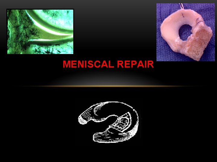
MENISCAL REPAIR Anatomy and Physiology of the Meniscus


- Slides: 49
MENISCAL REPAIR
Anatomy and Physiology of the Meniscus Articular cartilage acts as a friction reducer Meniscal cartilage acts as a cushion to absorb shock
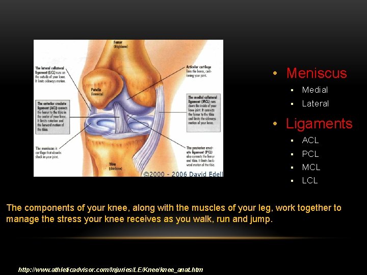
• Meniscus § Medial § Lateral • Ligaments § ACL § PCL § MCL § LCL The components of your knee, along with the muscles of your leg, work together to manage the stress your knee receives as you walk, run and jump. http: //www. athleticadvisor. com/Injuries/LE/Knee/knee_anat. htm
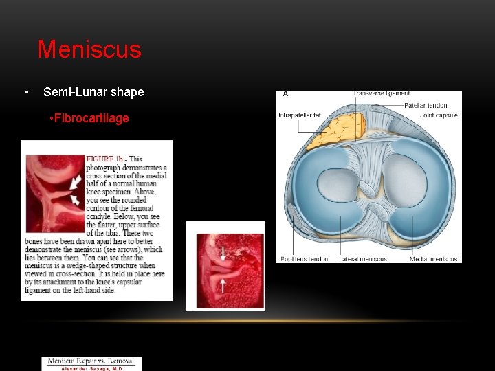
Meniscus • Semi-Lunar shape • Fibrocartilage
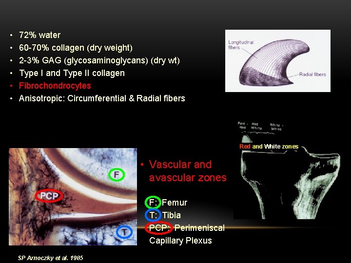
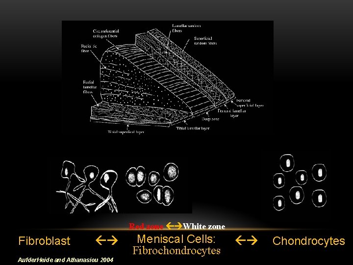
• • • 72% water 60 -70% collagen (dry weight) 2 -3% GAG (glycosaminoglycans) (dry wt) Type I and Type II collagen Fibrochondrocytes Anisotropic: Circumferential & Radial fibers Red and White zones • Vascular and avascular zones F: Femur T: Tibia PCP: Perimeniscal Capillary Plexus SP Arnoczky et al. 1985
Red zone White zone Fibroblast Aufder. Heide and Athanasiou 2004 Meniscal Cells: Fibrochondrocytes Chondrocytes
Damage of the Meniscus
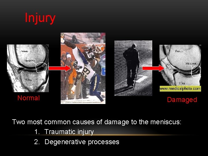
Injury Normal Damaged Two most common causes of damage to the meniscus: 1. Traumatic injury 2. Degenerative processes
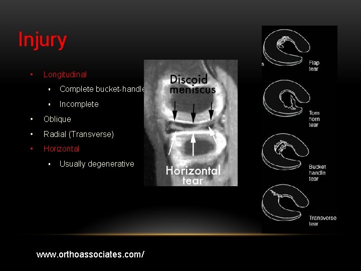
Injury • Longitudinal § Complete bucket-handle § Incomplete • Oblique • Radial (Transverse) • Horizontal § Usually degenerative www. orthoassociates. com/
Repair of the Meniscus
Repair • Meniscectomy: Partial and Total § Changes in contact area and stress distribution contribute to osteoarthritis (OA) • Meniscal repair: Sutures and implants • Does not heal in avascular zone • Allograft implantation: Transplant issues § Poor fixation technique § Difficulties with finding a donor of correct size • Future Options: SIS and CIS
Meniscectomy Partial Complete • Arthroscopic procedure • Minor trimming of a frayed edge to anything short of removing the rim • Evidence for partial regeneration? • Open or arthroscopic procedure • Removing a complete meniscus, that is a medial or a lateral meniscus, right to the meniscal rim • Arthritis
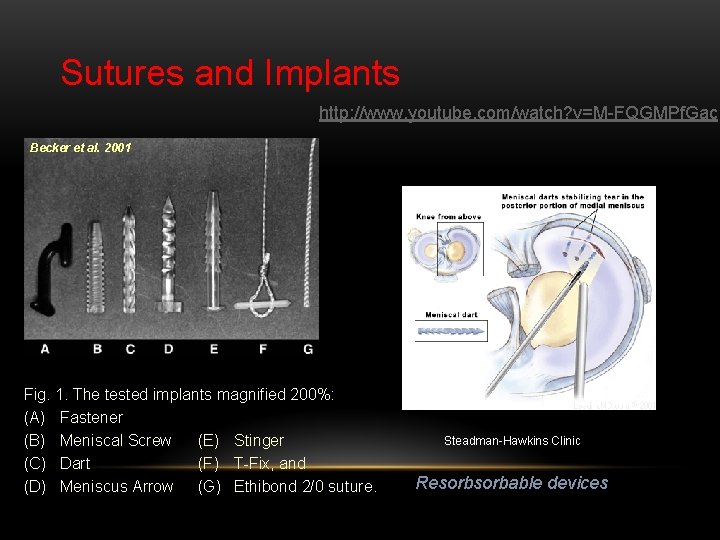
Sutures and Implants http: //www. youtube. com/watch? v=M-FQGMPf. Gac Becker et al. 2001 Fig. 1. The tested implants magnified 200%: (A) Fastener (B) Meniscal Screw (E) Stinger (C) Dart (F) T-Fix, and (D) Meniscus Arrow (G) Ethibond 2/0 suture. Steadman-Hawkins Clinic Resorbable devices
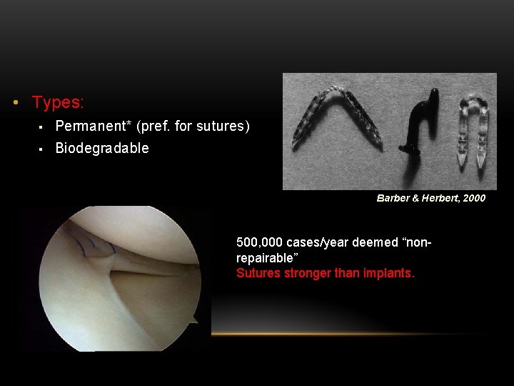
• Types: § Permanent* (pref. for sutures) § Biodegradable Barber & Herbert, 2000 500, 000 cases/year deemed “nonrepairable” Sutures stronger than implants.
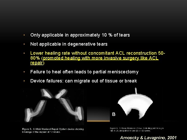
• Only applicable in approximately 10 % of tears • Not applicable in degenerative tears • Lower healing rate without concomitant ACL reconstruction 5080% (promoted healing with more invasive surgery like ACL repair) • Failure to heal often leads to partial meniscectomy • Device failures: can migrate out of tissue or break Arnoscky & Lavagnino, 2001
Meniscal Allograft Transplants (MAT) • Indications § Patients that have had meniscectomy § Patients with pain and discomfort associated with osteoarthritis § Patients undergoing ACL reconstruction for increased stability § Athletes for determent of osteoarthritis development • Preservation techniques § Fresh § Freeze-Dried § Cryo-preserved
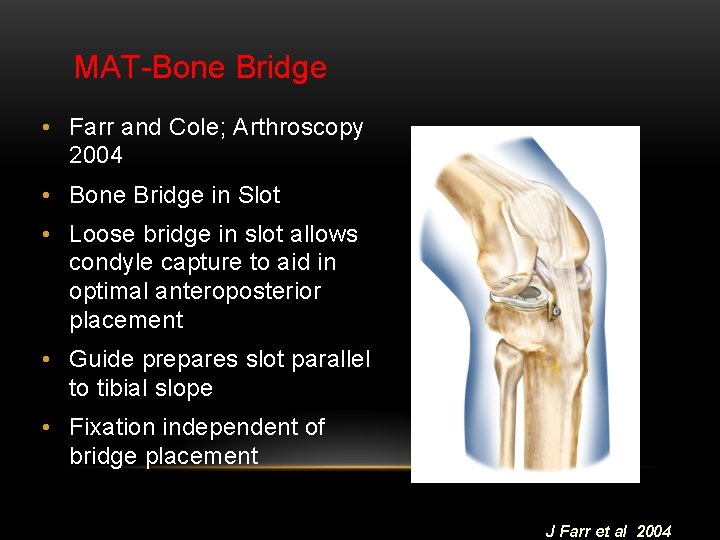
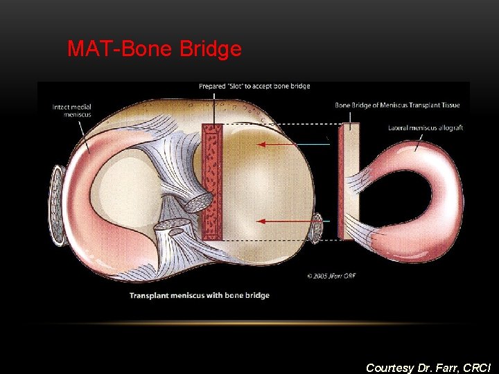
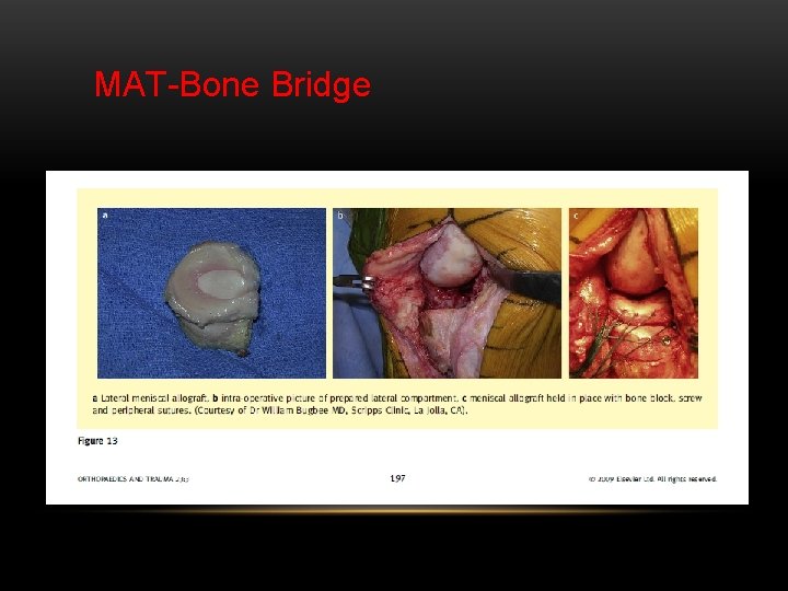
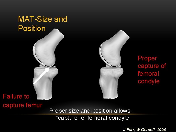
MAT-Bone Bridge • Farr and Cole; Arthroscopy 2004 • Bone Bridge in Slot • Loose bridge in slot allows condyle capture to aid in optimal anteroposterior placement • Guide prepares slot parallel to tibial slope • Fixation independent of bridge placement J Farr et al 2004
MAT-Bone Bridge Courtesy Dr. Farr, CRCI
MAT-Bone Bridge
MAT-Size and Position Proper capture of femoral condyle Failure to capture femur Proper size and position allows: “capture” of femoral condyle J Farr, W Gersoff 2004
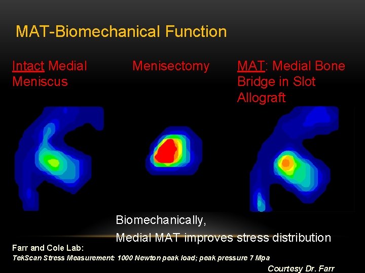
MAT-Biomechanical Function Intact Medial Meniscus Farr and Cole Lab: Menisectomy MAT: Medial Bone Bridge in Slot Allograft Biomechanically, Medial MAT improves stress distribution Tek. Scan Stress Measurement: 1000 Newton peak load; peak pressure 7 Mpa Courtesy Dr. Farr
MAT-issues • Availability • Reproducibility/Accuracy of Sizing • Lack of control over final cellular and extracellular matrix composition/ mechanical properties • Potential for disease transmission • Survivorship • Variation of Surgical Technique/ Placement
MATissues • Transplant is immunopriveliged -No immunosuppressants • Avascular meniscus has been frozen-Decreased viability • Inflammatory response • Disease transmission risk (eg. HIV risk 1/Million) • Meniscus may or may not be fully repopulated by patient § May be a variable duration on how long meniscus transplantation is going to last for enough repopulation § Degenerative types of tearing can happen § Need for additional surgery
• Current technique allows reproducible implantation of both medial and lateral menisci • MAT effective in the intermediate term for decreasing pain (medial and lateral) • Long-term effect on articular cartilage is under evaluation
Meniscal Substitues • Preserve biomechanical joint function • Prevent post meniscectomy chondrosis • Reestablish chondro-protective function • Regeneration of cell type producing extracellular environment duplicating original meniscus • Alleviate symptoms (primarily pain) • Durable • Summary: mimic the natural meniscus Courtesy Dr. Farr
Meniscal Substitutes • Manipulated Meniscal Allograft • * Collagen Meniscal Implant (CMI) * • * Small Intestine Submucosal (SIS) Implant * • Adult Mesenchymal Stem Cell Implants • * Tissue Engineered Meniscus: Scaffolds + Cells * • Tissue Engineered Meniscus: Cell-based Therapies • Tissue Engineered Meniscus: Gene Therapies Courtesy Dr. Farr
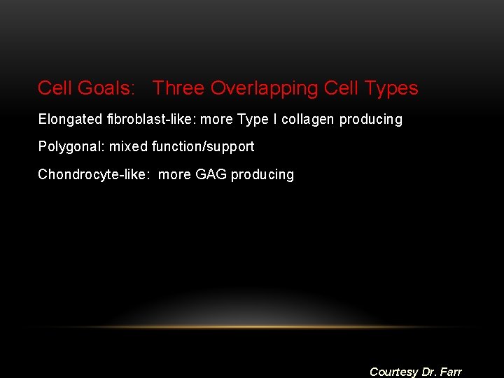
Cell Goals: Three Overlapping Cell Types Elongated fibroblast-like: more Type I collagen producing Polygonal: mixed function/support Chondrocyte-like: more GAG producing Courtesy Dr. Farr
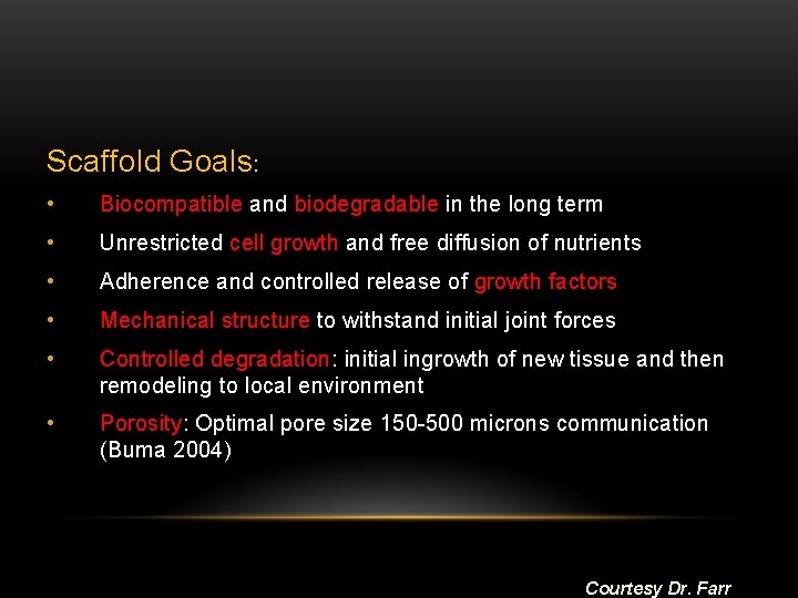
Scaffold Goals: • Biocompatible and biodegradable in the long term • Unrestricted cell growth and free diffusion of nutrients • Adherence and controlled release of growth factors • Mechanical structure to withstand initial joint forces • Controlled degradation: initial ingrowth of new tissue and then remodeling to local environment • Porosity: Optimal pore size 150 -500 microns communication (Buma 2004) Courtesy Dr. Farr
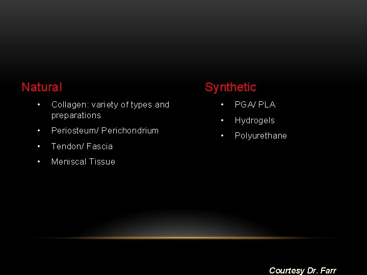
Natural • Collagen: variety of types and preparations • Periosteum/ Perichondrium • Tendon/ Fascia • Meniscal Tissue Synthetic • PGA/ PLA • Hydrogels • Polyurethane Courtesy Dr. Farr
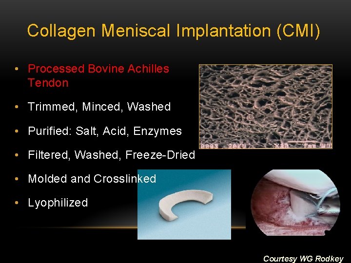
Collagen Menisca. I Implantation (CMI) • Processed Bovine Achilles Tendon • Trimmed, Minced, Washed • Purified: Salt, Acid, Enzymes • Filtered, Washed, Freeze-Dried • Molded and Crosslinked • Lyophilized Courtesy WG Rodkey
Appropriate Lesions • Partial meniscus loss (Medial only at present) • Need to have an intact rim of meniscus over entire length • No unstable segments • Lesion in red/red or red/white zone • Not suitable for total meniscus replacement • No uncorrected axial malalignment
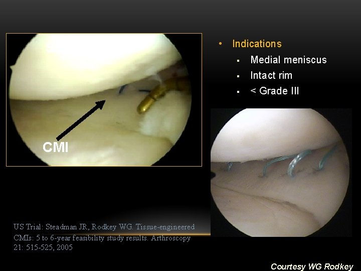
Surgery • Indications § Medial meniscus § Intact rim § < Grade III CMI US Trial: Steadman JR, Rodkey WG. Tissue-engineered CMIs: 5 to 6 -year feasibility study results. Arthroscopy 21: 515 -525, 2005 Courtesy WG Rodkey
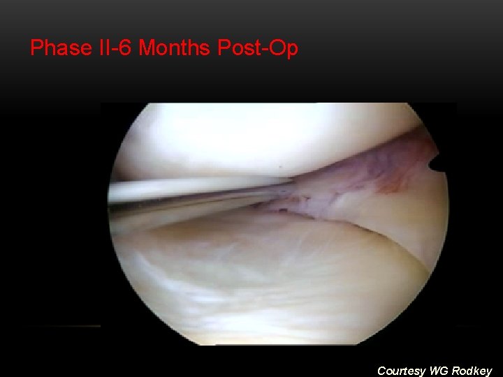
Phase II-6 Months Post-Op Courtesy WG Rodkey
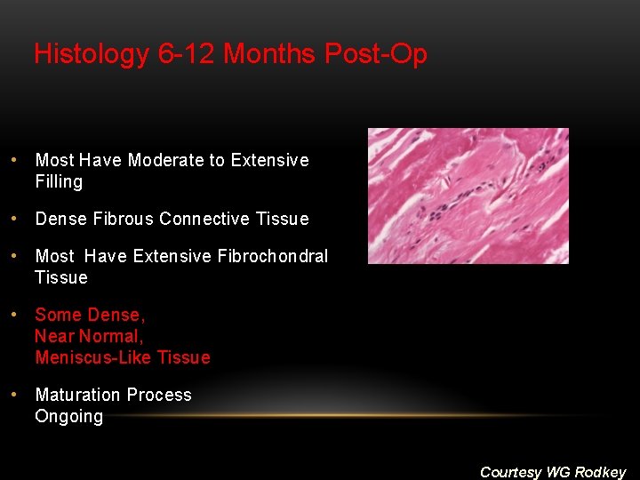
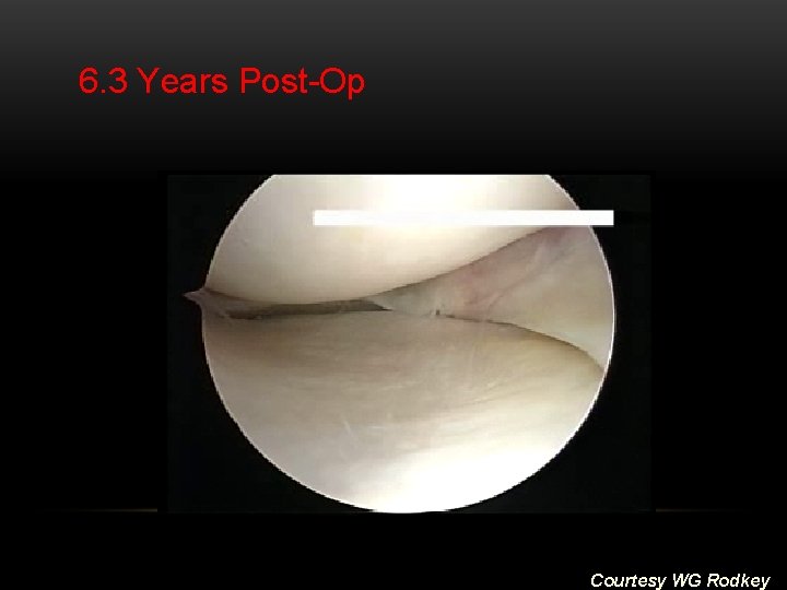
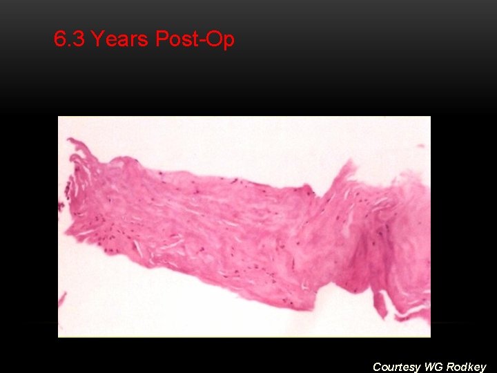
Histology 6 -12 Months Post-Op • Most Have Moderate to Extensive Filling • Dense Fibrous Connective Tissue • Most Have Extensive Fibrochondral Tissue • Some Dense, Near Normal, Meniscus-Like Tissue • Maturation Process Ongoing Courtesy WG Rodkey
6. 3 Years Post-Op Courtesy WG Rodkey
6. 3 Years Post-Op Courtesy WG Rodkey
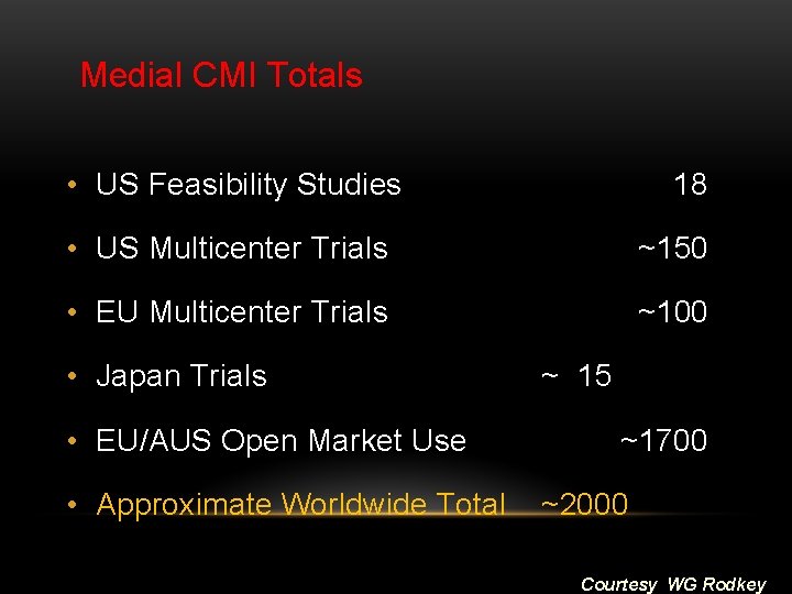
Medial CMI Totals • US Feasibility Studies 18 • US Multicenter Trials ~150 • EU Multicenter Trials • Japan Trials ~ 15 • EU/AUS Open Market Use ~100 ~1700 • Approximate Worldwide Total ~2000 Courtesy WG Rodkey
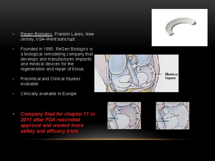
• Regen Biologics, Franklin Lakes, New Jersey, USA-Went bancrupt • Founded in 1990, Re. Gen Biologics is a biological remodeling company that develops and manufactures implants and medical devices for the regeneration and repair of tissue. • Preclinical and Clinical Studies Available • Clinically available in Europe • Company filed for chapter 11 in 2011 after FDA rescinded approval and wanted more safety and efficacy trials
• The data from the Rodkey study was used by the U. S. Food and Drug Administration (FDA) in the 510(k) application process for the Menaflex collagen meniscus implant. • An FDA executive summary of the Rodkey data indicated that patients who received the collagen meniscus implant followed a different post-op protocol than the control group and control patients were not required to undergo a planned second-look arthroscopy since it was assumed that there was no tissue regrowth in these patients. • The FDA also indicated that more meniscal tissue was removed from the collagen meniscus implant patients than in the control patients. • The FDA noted that the re-look arthroscopy results for collagen meniscus implant group showed that 16% of evaluated devices were not firmly attached to the host rim and 18% of knee compartments were determined to be worse than during the operative procedure at the time of the re-look arthroscopic procedure.
Issues • Clinically it has been shown that the collagen implant shrinks in time (Cummins et al. 1997, Ibarra et al. 1998) • Source for tissue ingrowth: native meniscus • The outer rim must be in place • Initial mechanical material properties poor • The outer rim must be in place (for fixation of scaffold)
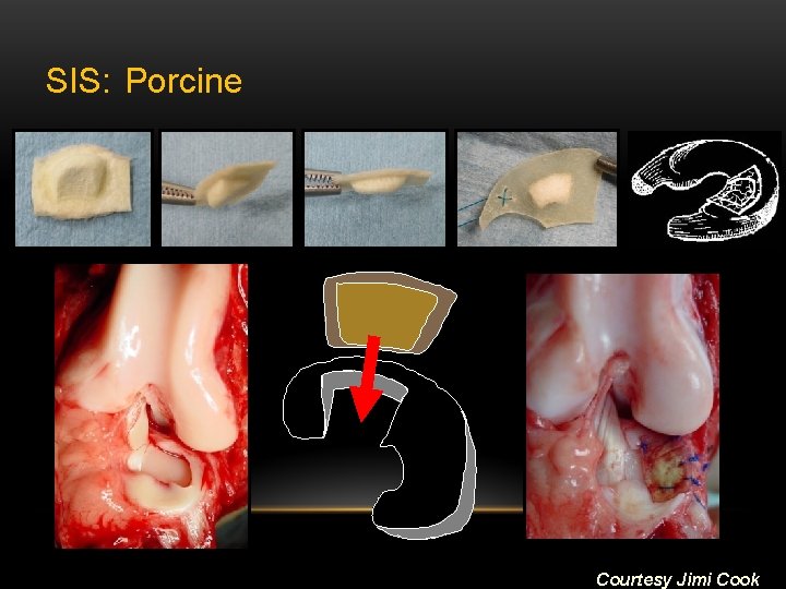
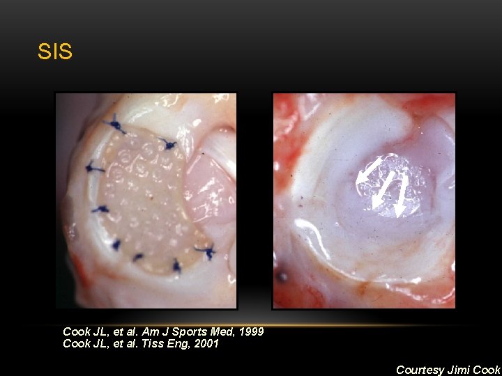
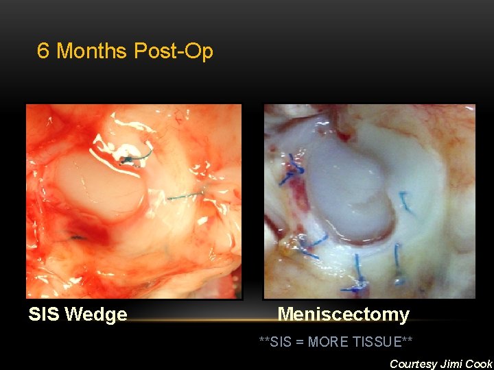
Small Intestine Submucosa (SIS) • De. Puy Biologics, Warsaw, Indiana, USA • Preclinical Studies; Phase 1/2 clinical research • Prepared and molded SIS • SIS composed of: § Collagen Types § Growth Factors • Segmental posterior horn, medial meniscus • Animal Studies, Cook et al
SIS: Porcine Courtesy Jimi Cook
SIS Cook JL, et al. Am J Sports Med, 1999 Cook JL, et al. Tiss Eng, 2001 Courtesy Jimi Cook
6 Months Post-Op SIS Wedge Meniscectomy **SIS = MORE TISSUE** Courtesy Jimi Cook
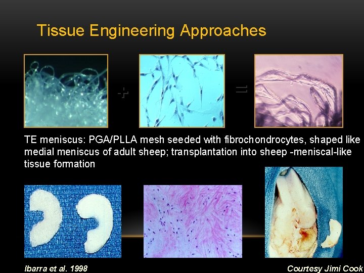
Tissue Engineering Approaches + = TE meniscus: PGA/PLLA mesh seeded with fibrochondrocytes, shaped like medial meniscus of adult sheep; transplantation into sheep -meniscal-like tissue formation Ibarra et al. 1998 Courtesy Jimi Cook
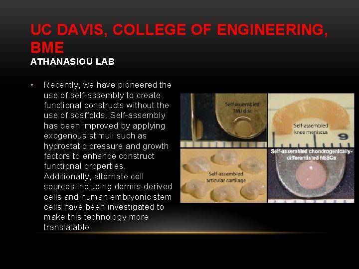
UC DAVIS, COLLEGE OF ENGINEERING, BME ATHANASIOU LAB • Recently, we have pioneered the use of self-assembly to create functional constructs without the use of scaffolds. Self-assembly has been improved by applying exogenous stimuli such as hydrostatic pressure and growth factors to enhance construct functional properties. Additionally, alternate cell sources including dermis-derived cells and human embryonic stem cells have been investigated to make this technology more translatable.
Chondroprotection is primary endpoint, through: • Reproduction of cell populations to reestablish stratified composite nature of natural meniscus • Long-term survivorship Additional Goals: • Ease of procedure to patient and surgeon • Affordable to health care system
The Bad and the Ugly • • • Anesthesia Infection (Superficial and Deep) Blood Clott (Deep vein thrombosis) Vascular Injury (Popliteal arteries) Nerve Injury (Peroneal nerve, saphenous nerve) Arthrofibrosis (Excessive scar tissue formation) Complex regional pain syndromes Inappropriate healing (Inadequate scar tissue formation) Possible Re-operation Improper alignment of implant Migration of implant With arthroscopic procedures, major complications are very rare (Small et al. 1988) Proper training of surgeons needed to ensure Byod & Myers, 2003
The Bad and the Ugly (Cont. ) • Degradation products accumulate at implant site § Delayed inflammatory response (Törmälä et al. ; Athanasiou et al. 1998) • Polyurethane & Teflon meniscus prosthesis § Fragentation of non-resorbable materials may induce macrophage & giant cell reactions, synovial irritation, cartilage softening (Buma et al. 2004) • PGA/PGLA relatively poor mechanical properties • Absorbable sutures: loss of mechanical strength