Meningitis Pathology CNS Infections Portals of entry of


 • Aseptic meningitis is a misnomer • it is a")
 • CSF: – increased number of lymphocytes (pleiocytosis) – protein")


- Slides: 26
Meningitis Pathology
CNS Infections • Portals of entry of infection into the CNS: – Hematogenous spread • the most common – Direct implantation • traumatic or in congenital CNS malformation – Local extension • occurs secondary to an established infection in a near by organ (air sinus, an infected tooth or middle ear) – Through the peripheral nervous system into the CNS • certain viruses, such as rabies and herpes zoster.
CNS Infections Meningitis An inflammatory process of the leptomeninges and CSF within the subarachnoid space. Meningoencephalitis?
CNS Infections Pyogenic meningitis • Medical emergency • The causative microorganisms (10 th edition, Robbins): – Neonates : Escherichia coli and group B streptococci – Adolescents and young adults: Neisseria meningitidis (Meningococcal meningitis) – Elderly: listeria monocytogenes and Streptococcus pneumoniae
CNS Infections Pyogenic meningitis • CSF Findings in spinal tap: – cloudy or frankly purulent CSF – as many as 90, 000 neutrophils /mm – raised protein level – markedly reduced glucose content – bacteria may be seen on a Gram stained smear or can be cultured, sometimes a few hours before the neutrophils appear
Acute meningitis
CNS Infections Meningitis Clinical Features • Systemic non-specific signs of infection • Meningeal irritation signs and neurologic impairment: – Headache, photophobia, irritability, clouding of consciousness and neck stiffness • Untreated, pyogenic meningitis can be fatal • Effective antimicrobial agents markedly reduce mortality associated with meningitis
CNS Infections Meningitis Complications • Phlebitis may venous occlusion hemorrhagic infarction of the underlying brain • Leptomeningeal fibrosis hydrocephalus • Septicemia hemorrhagic infarction of the adrenal glands and cutaneous petechiae (known as Waterhouse. Friderichsen syndrome, particularly common with meningococcal and pneumococcal meningitis) • Focal cerebritis & seizures • Cerebral abscess • Cognitive deficit • Deafness What is this complication
CNS Infections Brain abscess • Streptococci and staphylococci are the most common organisms identified in non-immunosuppressed populations • Predisposing conditions: – Acute bacterial endocarditis (usually give multiple microabscesses) – Cyanotic congenital heart disease in which there is a right-to-left shunt – Loss of pulmonary filtration of organisms ( e. g, bronchiectasis) • Most common on cerebral hemispheres
CNS Infections Brain abscess • Morphologically, – Liquefactive necrosis – The surrounding brain is edematous , congested & contains reactive astrocytes & perivascular inflammatory cells • Present clinically with progressive focal neurologic deficits in addition to the general signs of raised intracranial pressure • The CSF – Contains only scanty cells – ↑ protein – Normal level of glucose • Complications of Brain abscess: – Herniation – Rupture of abscess into subarachnoid space or ventricle
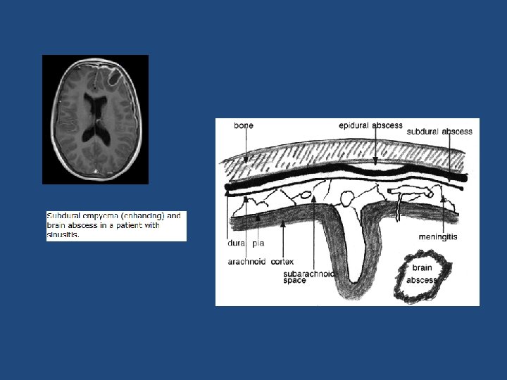
Epidural and Subdural Infections • These spaces can be involved with bacterial or fungal infections, usually as a consequence of direct local spread • Epidural abscess, commonly associated with osteomyelitis, arises from an adjacent focus of infection, such as sinusitis or a surgical procedure • When the process occurs in the spinal epidural space, it may cause spinal cord compression and constitute a neurosurgical emergency
Empyema • Infections of the skull or air sinuses may also spread to the subdural space, producing subdural empyema – The underlying arachnoid and subarachnoid spaces are usually unaffected, but a large subdural empyema may produce a mass effect – In addition, thrombophlebitis may develop in the bridging veins that cross the subdural space, resulting in venous occlusion and infarction of the brain
Empyema • Symptoms include those referable to the source of the infection. Most patients are febrile, with headache and neck stiffness, and if untreated may develop focal neurologic signs, lethargy, and coma • With treatment, including surgical drainage, resolution of the empyema occurs from the dural side; if resolution is complete, a thickened dura may be the only residual finding. With prompt treatment, complete recovery is usual
CNS Infections Tuberculosis • The subarachnoid space contains a fibrinous exudate, most often at the base of the brain • Tuberculoma is well-circumscribed intraparenchymal mass – Rupture of tuberculoma into subarachnoid space results in tuberculus meningitis – A tuberculoma may be up to several centimeters in diameter, causing significant mass effect – Always occurs after hematogenous dissemination of organism from primary pulmonary infection • On microscopic examination, there is usually a central core of caseous necrosis surrounded by a typical tuberculous granulomatous reaction
TB meningitis Exudate at the base of the brain
CNS Infections CSF in TB • There is only a moderate increase in cellularity of the CSF (pleiocytosis) made up of mononuclear cells, or a mixture of polymorphonuclear and mononuclear cells • The protein level is elevated, often strikingly so • The glucose content typically is moderately reduced or normal
Aseptic Meningitis (Viral Meningitis) • Aseptic meningitis is a misnomer • it is a clinical term for an illness comprising meningeal irritation, fever, and alterations of consciousness of relatively acute onset without recognizable organisms • The clinical course is less fulminant than in pyogenic meningitis, is usually self-limiting, and most often is treated symptomatically
Aseptic Meningitis (Viral Meningitis) • CSF: – increased number of lymphocytes (pleiocytosis) – protein elevation is only moderate – glucose content is nearly always normal • In approximately 70% of cases, a pathogen can eventually be identified, most commonly an enterovirus • There are no distinctive macroscopic characteristics except for brain swelling, seen in only some instances • On microscopic examination, there is either no recognizable abnormality or a mild to moderate infiltration of the leptomeninges with lymphocytes.
Homework • Create a table of CSF findings in Meningitis, aseptic meningitis, TB meningitis, Brain abscess and multiple sclerosis!