MENINGES OF THE BRAIN BY DR manjula Vastrad

MENINGES OF THE BRAIN BY: DR. manjula. Vastrad Asst. prof DEPT. OF RACHANA SHAREERA Smvvs rkm amc vijayapur Email : MANJULA. PRASAD 2010@GMAIL. COM

INTRODUCTION v Three meninges v Four dural septa v Two spaces

MENINGES �The Meninges are a system of membranes that envelop the complete central nervous system (brain and spinal cord).

MENINGES DURA MATER ARACHNOID MATER PIA MATER

4 DURAL SEPTA FALX CEREBRI TENTORIUM CEREBELLI FALX CEREBELLI DIAPHRAGMA SELLAE

TWO SPACES SUBDURAL SUBARACHNOID

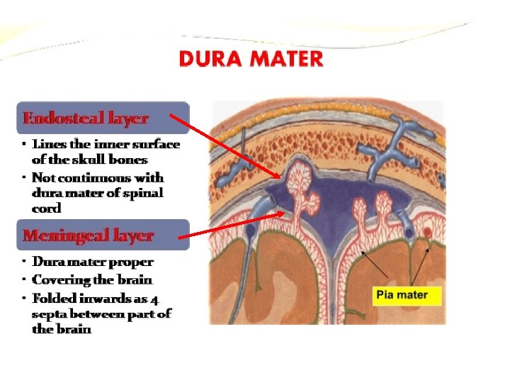
DURA MATER It is the outermost thickest and toughest membrane covering the brain. Also k/as Pachymeninx.

CEREBRAL DURA MATER ENDOSTEAL LAYER MENINGEAL LAYER

REFLECTIONS FALX CEREBRI TENTORIUM CEREBELLI FALX CEREBELLI DIAPHRAGMA SELLAE

FALX CEREBRI �Large sickle shaped fold of dura mater occupying the medial longitudinal fissure b/w two cerebral hemisphere. � 2 ENDS: 1. 2. Anterior end – narrow & attached to Crista Galli. Posterior end – broad & attached to Tentorium cerebelli.

FALX CEREBRI

FALX CEREBRI Ø 1. 2. 3. 2 MARGINS : Upper- convex & attached to lips of Sagittal Sulcus. Lower- concave & free. 3 VENOUS SINUSES: Superior sagittal sinus Inferior sagittal sinus Straight sinus

TENTORIUM CEREBELLI � It is a tent shaped fold of dura mater. � It separates the cerebellum from the occipital lobe of cerebrum. � It divides the cranial cavity into supra-tentorial & infra-tentorial compartments. q 1. 2. 2 MARGINS: ANTERIOR : ‘u’ shaped & free. Ends of ‘u’ are attached anteriorly to the anterior clinoid process. POSTERIOR : It is convex -Posterolaterally attached to transverse sulci on occipital bone. - Anterolaterally attached to superior border of petrous temporal bone & posterior clinoid process.

TENTORIUM CEREBELLI

TENTORIUM CEREBELLI q 1. 2. q 1. 2. 2 SURFACES: SUPERIOR – Convex - Gives attachment to falx cerebri in its midline - Related to occipital lobes of cerebrum INFERIOR – Concave - Gives attachment to falx cerebelli to its posterior part - It fits the upper superior surface of cerebellum. 2 VENOUS SINUSES: Transverse sinus Superior petrosal venous sinus
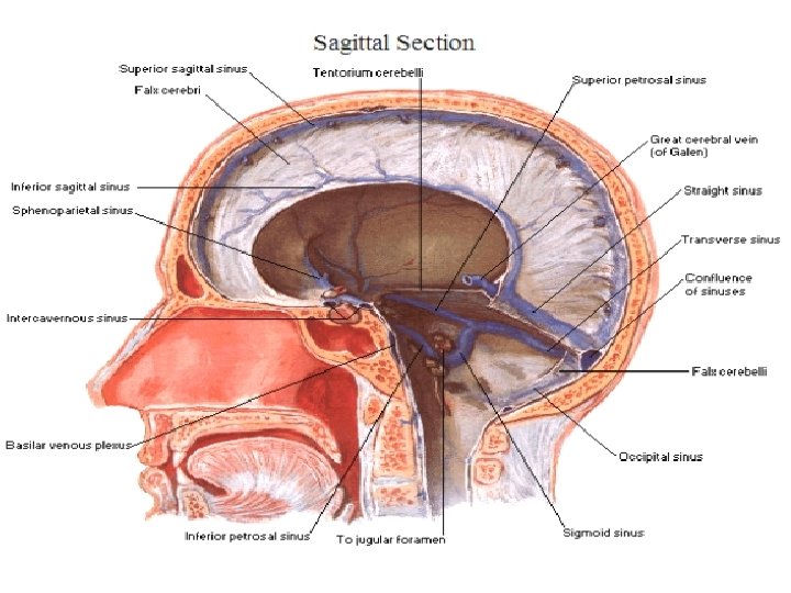

FALX CEREBELLI �Small sickle shaped fold of dura mater attached to internal occipital crest & project forwards b/w two cerebellar hemisphere. �Base of the sickle is attached to the posterior part of the inferior surface of tentorium cerebelli. � 2 MARGINS: 2. Anterior – concave & free Posterior – convex & attached to posteior occipital crest. � It encloses the Occipital Sinus. 1.

FALX CEREBELLI

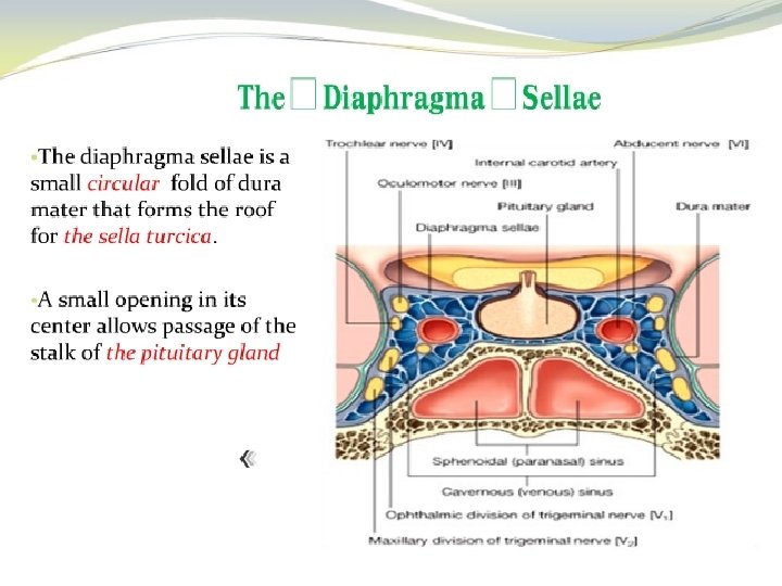
DIAPHRAGMA SELLAE �Small circular horizontal fold of dura mater, forming the roof of the hypophyseal fossa. q ATTACHMENTS: - Anteriorly attached to tuberculun sellae. - Posteriorly attached to dorsum sallae. - On each side, continuous with the dura mater of middle cranial fossa. - Having central aperture through which the stalk of pitutary passes.

Nerve supply and blood supply of duramater

ARACHNOID MATER �Thin transparent membrane that loosely surrounds the brain without dipping into sulci. �Bridges all the irregularities of the brain. �RELATIONS: 1) 2) Separated from Dura by subdural space. Separated from Pia by subarachnoid space containing CSF.

ARACHNOID MATER � PROLONGATIONS: 1. 2. Provide sheath for the cranial nerves as far as their exit from the skull. Arachnoid villi – small finger like projections of arachnoid tissue projecting into the cranial venous sinuses. - They absorb CSF.

PIA MATER � Vascular membrane that closely invests the brain, dipping into various sulci and other irregularities of the surface. � On the cerebellum , Pia mater dips and forms folds in relation to larger fissures of cerebellum. PROLONGAIONS: 1) Provides sheath for the cranial nerves 2) Also provide perivascular sheath for the minute vessels entering or leaving the brain 3) Folds of Pia mater enclosing tufts of capillaries, form the TELACHORIDEA.

PIA MATER

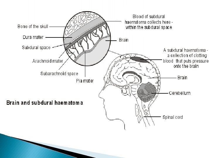
SUBDURAL SPACE Potential space between the dura & arachnoid mater. Becomes actual space in pathological conditions. Traversed by cerebral veins on their path for draining into dural venous sinuses.

SUBARACHNOID SPACE Space between arachnoid & pia mater. Traversed by a network of arachnoid trabeculae that give it a sponge like appearence. Contains CSF & large vessels of the brain Cranial nerves pass through the space.

APPLIED ANATOMY

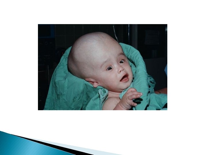
HYDROCEPHALUS Hydrocephalus also known as "water on the brain", is a medical condition in which there is an abnormal accumulation of cerebrospinal fluid (CSF) in the ventricles, or cavities, of the brain. This may cause increased intracranial pressure inside the skull and progressive enlargement of the head, convulsion, tunnel vision, and mental disability. Hydrocephalus can also cause death. It is more common in infants

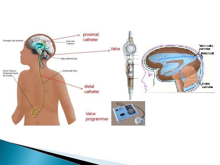
CEREBRAL SHUNTS Cerebral shunts are commonly used to treat hydrocephalus, the swelling of the brain due to excess build up of cerebrospinal fluid (CSF). The cerebral shunt can be used to alleviate or prevent these problems in patients who suffer from hydrocephalus or other related diseases. Shunts can come in a variety of forms but most of them consist of a valve housing connected to a catheter, the end of which is usually placed in the peritoneal cavity

MENINGITIS Meningitis is an acute inflammation of the protective membranes covering the brain and spinal cord, known collectively as the Meninges. The inflammation may be caused by infection with viruses, bacteria, or other microorganisms. The most common symptoms of meningitis are headache & neck stiffness associated with fever confusion or altered consciousness, vomiting, and an inability to tolerate light (photophobia) or loud noises (phonophobia).
 is a collection of blood accumulating")
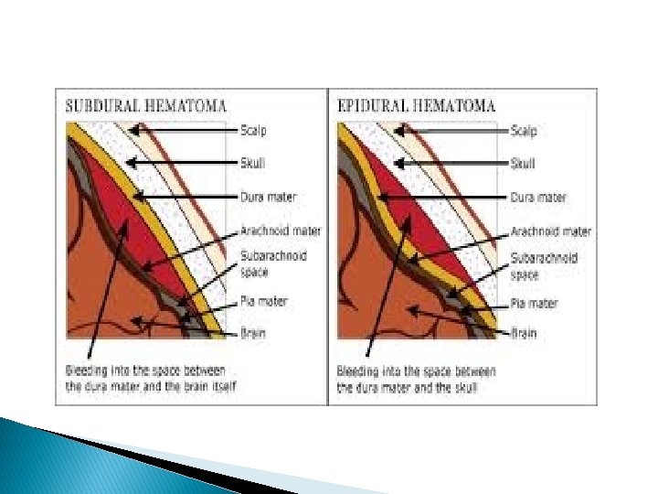
EXTRADURAL & SUBDURAL HAEMORRHAGE A subdural haemorrhage (SDH) is a collection of blood accumulating in the potential space between the dura and arachnoid mater of the meninges around the brain. The extradural haemorrhade is arterial due to injury to middle meningeal artery whereas subdural haemorrhage is venous in nature. In an extradural haemorrhage, there is no blood in CSF while it is acommom feature of subdural haemorrhage.

THANK YOU
- Slides: 38